. 2006 Sep 6;25(17):3975-85.
doi: 10.1038/sj.emboj.7601271. Epub 2006 Aug 10.

Functional cooperation between FACT and MCM helicase facilitates initiation of chromatin DNA replication

Affiliations

Functional cooperation between FACT and MCM helicase facilitates initiation of chromatin DNA replication

Bertrand Chin-Ming Tan et al. EMBO J. .

Abstract

Chromatin is suppressive in nature to cellular enzymes that metabolize DNA, mainly due to the inherent inaccessibility of the DNA template. Despite extensive understanding of the involvement of chromatin-modifying factors in transcription, roles of related activities in DNA replication remain largely elusive. Here, we show that the heterodimeric transcriptional elongation factor FACT (facilitates chromatin transcription) is functionally linked to DNA synthesis. Its involvement in DNA replication is partly mediated by the stable association with the replicative helicase complex, MCM, and further by the coexistence with MCM on replication origin. Furthermore, relying on its nucleosome-reorganizing activity, FACT can facilitate chromatin unwinding by the MCM complex, which is otherwise inert on the nucleosomal template. As a consequence, the physical and functional interaction between FACT and MCM is an important determinant in the proper initiation of DNA replication and S phase in vivo. Together, our findings identify FACT as an integral and conserved component of the endogenous replication machinery, and support a model in which the concerted action of helicase and chromatin-modifying activities promotes chromosome replication.


Figures
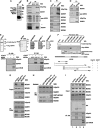
Figure 1
Figure 1
Replicative helicase MCM is a novel interacting protein of FACT. (A) FACT-associated complexes were immunoprecipitated from HeLa nuclear extracts (derived from 2 × 10⁷ cells) using αSSRP1 mAb 10D1, resolved by and shown on a silver-stained gel (also see Tan and Lee, 2004). Specific SSRP1-interacting protein bands that are absent in the control immunoprecipitates (not shown) were identified as MCM4 and MCM6 (∼100 kDa), and MCM7 (80 kDa). The 120-kDa band represents a previously identified associated protein, Nek9 (Tan and Lee, 2004). (B) Western blot analysis of the different immunocomplexes targeted by control (2B12) and αSSRP1 (10D1) mAbs. Immunoblotting was carried out using the indicated antibodies against pan-MCM or individual subunits. The positions of MCM2, MCM4/6 and MCM7 in the IP are indicated by the black, gray and white arrowheads (middle panel), respectively. The amount of the input (Input) is equivalent to 1/40 the IP. The identity of the protein band, marked by the asterisk, is unknown. (C) Western blot analysis of the immunoprecipitates targeted by αSSRP1 polyclonal antibodies (PI, preimmune serum). Immunoblotting was carried out using antibodies against the indicated MCM subunits. The amount of the Input is equivalent to 1/30 of the IP. (D) Western blot analysis of the immunoprecipitates targeted by preimmune serum (PI) or αMCM4 antibodies. Immunoblotting was carried out using the specified antibodies. The amount of the Input is equivalent to 1/50 of the IP. (E) Recombinant FACT heterodimer (FLAG-hSpt16/His6-SSRP1, left panel) and MCM subcomplexes (MCM2/4/6/7, MCM2/3/5, MCM2–7 and MCM4/6/7, as indicated) were isolated as described in Materials and methods and visualized by silver-stained gel. In vitro pull-down assay was carried out using the anti-FLAG M2 agarose, with the indicated combinations of protein complexes. The presence of specifically bound proteins was detected by the indicated antisera (bottom two panels). (F) Bacterially expressed GST and GST-fused MCMs were used as baits in the in vitro pull-down assay. The presence and purity of the immobilized proteins were seen on Coomassie blue-stained gels. The presence of bound FLAG-hSpt16 or His6-SSRP1 protein was detected by immunoblotting with M2 or αSSRP1 antibodies, respectively. The schematic representation shown below the panels depicts the domain organization of MCM4 as well as the presence or absence of interaction between various deleted constructs of MCM4 and the FACT subunits (input is 1/15 of IP). (G) Ablation of endogenous MCM3 and MCM4 expression was achieved by RNAi. Equal loadings of whole-cell extracts (‘Input') derived from HeLa cells transiently harboring control-, MCM3- or MCM4-targeting dsRNA (for 54 h) were blotted with the indicated antibodies. Lysates (30 × of input) were immunoprecipitated with 10D1 (bottom four panels, ‘IP: 10D1') and subsequently probed with the indicated antibodies. (H) In vitro pull-down assay was performed as in (E), using FACT (10 nM) and MCM2–7 (10 nM). GST (15 nM) or GST-fused full-length or deletion constructs of MCM4 (GST-MCM4120–250 GST-MCM4250–380) was added to the binding mixture. Precipitated rFACT as well as co-precipitated MCMs were probed with the indicated antibodies. (I) Extracts were prepared from HeLa cells ectopically expressing empty vector, HA-MCM4120–250, HA-MCM4250–380 or HA-MCM4-FL (full-length), and subsequently subjected to immunoprecipitation using the HA antibody. Immunoprecipitates (IP: HA) and lysate input (Input) were probed for HA, MCM or FACT, as indicated.
Figure 2
Figure 2
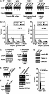
FACT coexists with MCM on the chromosomal replication origin. (A) ChIP was performed as described in Materials and methods. Chromatin fragments were prepared from cells at different stages: asynchronous (lanes 1, 4, 7 and 10), G1/S (lanes 2, 5, 8 abd 11) and G2/M (lanes 3, 6, 9 and 12). Immunoprecipitation was performed with either control (IgG, lanes 4–6) or 10D1 (lanes 1–3, 7–9 and 10–12) antibody. Products (∼160 bp) from final PCR analysis using primers specific to lamin B2 origin (lanes 1–6), to a non-transcribed region (lanes 7–9), or to the γ-actin gene, were resolved by 1.5% agarose gel (positions of the size marker are denoted on the right). Precipitated products are shown in the upper panels, DNA input in the lower. (B) Chromatin samples were immunoprecipitated with 10D1 (FACT, left) or anti-MCM4 (MCM4, right) antibody. The charts represent the quantitative determination of bound DNA, as determined by quantitative RT–PCR. Regions correspond to the lamin B2 origin, a non-origin region, a region of no annotated genes (background), and γ-actin ORF were analyzed (indicated on the x-axis). Data presented are normalized to input DNA (shown in percentage), and the mean±s.d. values from at least four experiments are shown. (C) HeLa cells were treated with various concentrations of α-amanitin for 12 h to inhibit RNA polII transcription. Binding of FACT to the lamin B2 origin (left), or to the non-origin, transcribed region of the γ-actin gene (right), under such treatment was monitored by the ChIP assay using the 10D1 antibody, as in (A). (D) The mRNA level of the lamin B2 (upper panel) and TIMM3 (middle) genes in asynchronous or double thymidine-arrested (G1/S) HeLa cells were examined by RT–PCR. Expression level of GAPDH (lower panel) was used as a loading control. (E) Coexistence of FACT and MCM on origin was demonstrated by a sequential ChIP experiment. Chromatin was first immunoprecipitated by MCM4 antibody (lane 3). A second round of ChIP was performed using control (lane 1) or 10D1 (lane 2) antibody. The presence of DNA fragments corresponding to the lamin B2 origin in the second ChIP was assessed by PCR. (F) Chromatin was prepared from asynchronous cells. ChIP was performed using the preimmune (IgG) or MCM4-specific (αMCM4) antibodies, and was subsequently probed for the presence of the non-origin, transcribed region of the γ-actin gene fragment. (G) Chromatin was prepared from the control and MCM4RNAi cells. ChIP was performed as above to probe for the presence of lamin B2 origin DNA fragment in the FACT immunocomplexes (left). Chart on the right represents quantification of bound lamin B2 origin region and shows the percent of starting material immunoprecipitated by 10D1 or control IgG (noted on the x-axis), as determined by RT–PCR. The mean±s.d. values from at least three experiments are shown.
Figure 3
Figure 3
FACT promotes the DNA unwinding activity of the MCM helicase on nucleosomal template. (A) Schematic representation of the strategy for generating a linear, tailed DNA template by PCR. The presence of 5′ dT (15-mer) tail is known to increase the helicase activity of MCM (You et al, 2003). Asterisk marks the site of labeling. See Materials and methods for details. (B) Reconstitution of nucleosome cores onto the ∼200-bp, end-labeled DNA fragment. Aliquot of the transfer reactions were analyzed and bands corresponding to reconstituted nucleosomes and free DNA are indicated. (C) Helicase assay was performed with the indicated amounts of isolated MCM4/6/7 (lanes 2–5) or MCM2/3/5 (lanes 7 and 8), or in combination with purified recombinant FACT (lane 4). Reactions were performed on the free DNA substrate. Lane 5 represents unwinding reactions done in the absence of ATP. Deprotienated reaction products were resolved by native gel electrophoresis. M, 100 bp ladder DNA marker. Denaturation of the DNA substrate by heat is also shown (‘Boiled', lane 9). Degree of DNA unwinding/displacement for each reaction was calculated based on the quantified signal ratio of ss- to ds-DNA bands. It is expressed in percentage (%) and shown on the bottom of the figure. (D) DNA helicase assay was carried out as in (C), except the use of the reconstituted nucleosomal template. Unwinding of the nucleosomal DNA by various combinations of MCM4/6/7 and/or FACT complexes (amounts indicated on top) was monitored by autoradiography. Lane 5 represents unwinding reaction carried out in the absence of ATP. Degree of DNA unwinding/displacement is determined as above. (E) DNA helicase activity of the FACT–MCMs complex was assayed on the naked DNA (lanes 1 and 2) or the nucleosomal (lanes 3–7) template, as described above. MCM4/6/7 (36 nM) (lanes 1–2 and 5–7) was assayed in combination with 200 nM of purified recombinant FACT. Before reaction, the complexes were preincubated with GST (50 nM, lanes 1 and 6), GST-fused deletion construct of MCM4 (GST-MCM4120–250, 50 nM, lanes 2 and 7) or buffer (lane 5). Percentage of displaced DNA is calculated as above and shown below the panel. M, DNA marker (lane 4). Heat denaturation of the DNA is also shown (‘Boiled', lane 3). (F) DNA helicase activity was assayed on the nucleosomal template, as described above. Preincubation experiments were performed with the indicated combinations of proteins or template (‘Preincubation:'), for a reaction length of 20 min, before adding the remaining components. Reactions were also performed without (lanes 1–4) or with (lanes 5 and 6) GST-MCM120–250. M, MCM4/6/7; F, rFACT; Nuc, nuclesomal DNA; G, GST-MCM120–250; all, all components without any preincubation. Lane 1: MCM4/6/7 alone. In (C–F), the positions of the double- and single-stranded fragments are indicated by arrowheads and arrows, respectively.
Figure 4
Figure 4
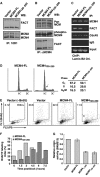
Disruption of the FACT–MCM complex triggers delayed DNA replication initiation. Stable clones of HeLa cells ectopically expressing empty vector, HA-MCM4-FL (full-length) or HA-MCM4120–250 were established as described in the Materials and methods. Mixed clones of each line were subjected to analysis. (A) Extracts were subjected to immunoprecipitation using the 10D1 antibody. Immunoprecipitates were probed for FACT or MCM2/4, as indicated. (B) Anti-MCM4 immunocomplexes isolated from these extracts were subjected to immunoblotting using the pan MCM, phospho-MCM4 (Thr110) and FACT antibodies, as indicated. (C) Using the indicated antibodies, ChIP was performed as in Figure 2 to probe for the presence of lamin B2 origin DNA fragment in the corresponding immunocomplexes. DNA input is shown on the bottom. (D) Cell cycle profiles of the MCM4-FL and MCM4120–250 cells. Cells were subjected to FACS for measurement of DNA content. Cells in the G0/G1, S and G2/M phases were defined by gating. Percentages of gated events are summarized on the right. Arrowhead indicates a major difference in phase distribution (see text). (E) BrdU-incorporation of the indicated cell lines at the G1/S junction. Upon release from thymidine treatment (at time 0), cells were pulse-labeled with BrdUTP. Analog incorporation (FL1) was compared with DNA content (FL2-PI, y-axis). BrdU-stained cells were defined by electronic gating (‘BrdU+'). Percentages of gated events are shown (in the upper left corner of each panel). The ‘Vector (−BrdU)' panel represents the distribution of cells in the absence of BrdU labeling, and thus serves as a control for the gating of stained cells. The arrow indicates the distribution of BrdU+ cells of the MCM4120–250 line (see text). (F) MCM4-FL, MCM41–250 and MCM4120–250 cells were synchronized and released at the G1/S junction and, at the indicated time points, pulse-labeled with BrdU (30 min) before being harvested. Cell cycle profiles and degree of BrdU incorporation were monitored by FACS. The labeling index quantitatively corresponds to the percentage (%) of BrdU-positive cells in a particular cell population. (G) Origin function of the lamin B2 replicator in the three indicated cell lines was assessed as described in Materials and methods (also see Supplementary Figure S5). The bar graph is a quantitative representation of the origin activity, with the activity in the control cells represented as 1. Data are averaged±s.d. of three independent experiments.

Similar articles

Cited by

References

    1. Abdurashidova G, Danailov MB, Ochem A, Triolo G, Djeliova V, Radulescu S, Vindigni A, Riva S, Falaschi A (2003) Localization of proteins bound to a replication origin of human DNA along the cell cycle. EMBO J 22: 4294–4303 - PMC - PubMed
    1. Abdurashidova G, Deganuto M, Klima R, Riva S, Biamonti G, Giacca M, Falaschi A (2000) Start sites of bidirectional DNA synthesis at the human lamin B2 origin. Science 287: 2023–2026 - PubMed
    1. Aggarwal BD, Calvi BR (2004) Chromatin regulates origin activity in Drosophila follicle cells. Nature 430: 372–376 - PubMed
    1. Alexiadis V, Varga-Weisz PD, Bonte E, Becker PB, Gruss C (1998) In vitro chromatin remodelling by chromatin accessibility complex (CHRAC) at the SV40 origin of DNA replication. Embo J 17: 3428–3438 - PMC - PubMed
    1. Aparicio OM, Weinstein DM, Bell SP (1997) Components and dynamics of DNA replication complexes in S. cerevisiae: redistribution of MCM proteins and Cdc45p during S phase. Cell 91: 59–69 - PubMed

Publication types