. 2006 Aug 11;126(3):597-609.
doi: 10.1016/j.cell.2006.06.048.

Blimp1 defines a progenitor population that governs cellular input to the sebaceous gland

Affiliations

Blimp1 defines a progenitor population that governs cellular input to the sebaceous gland

Valerie Horsley et al. Cell. .

Abstract

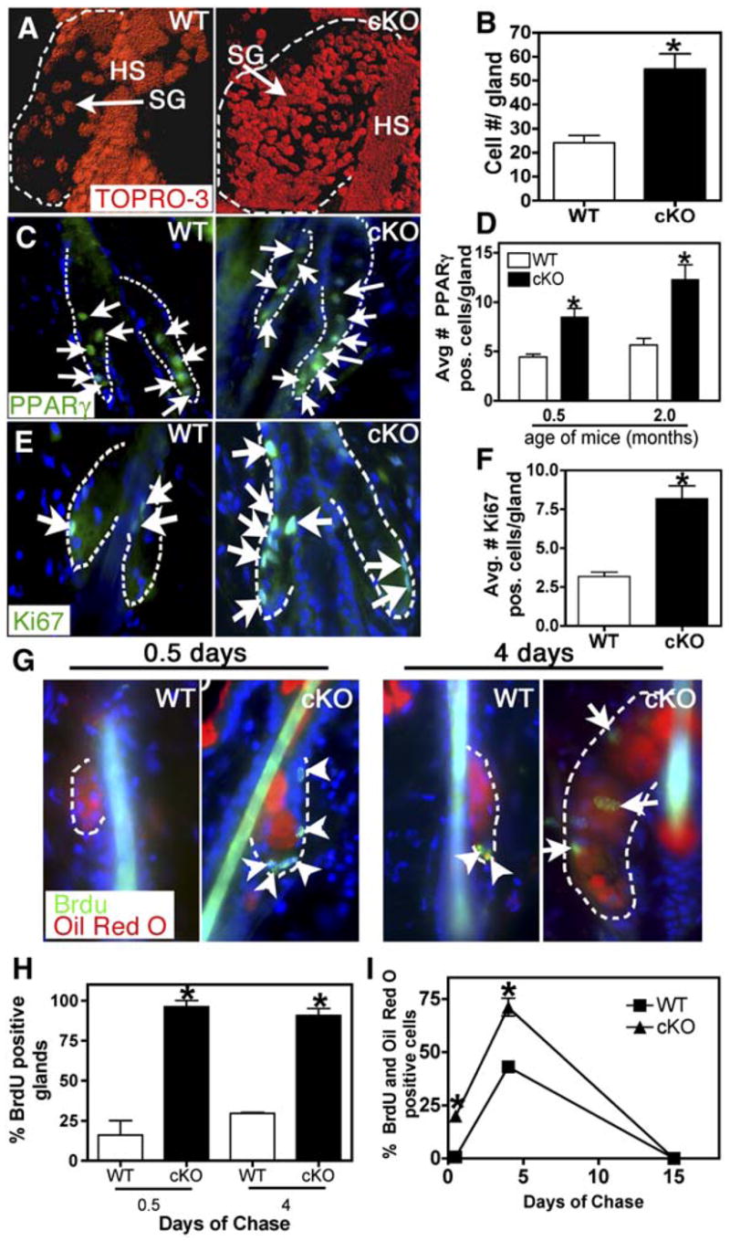
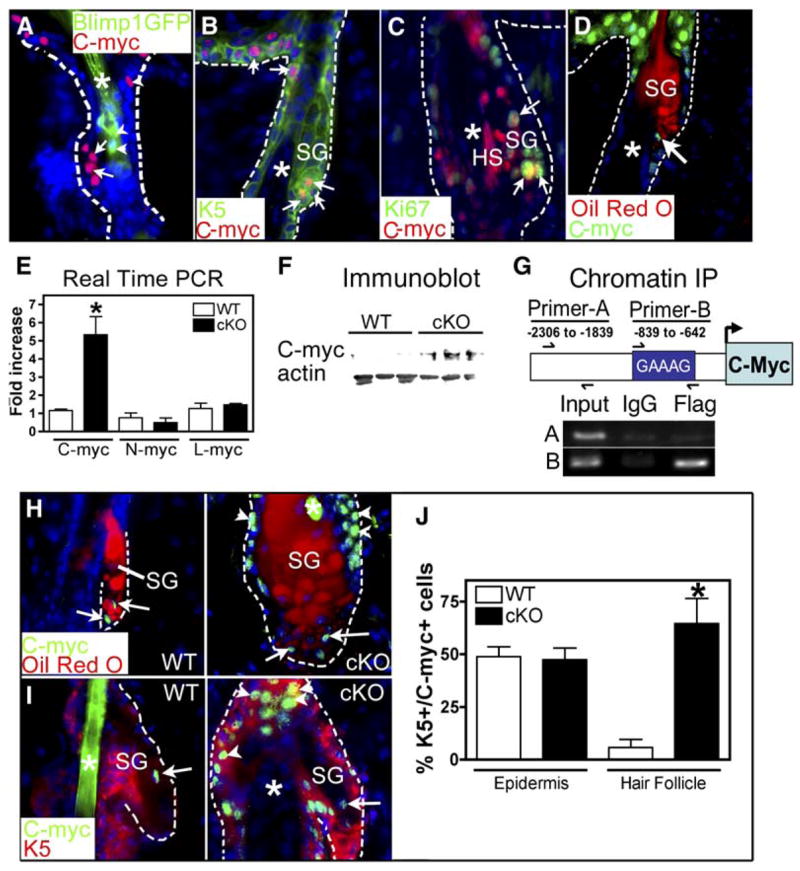
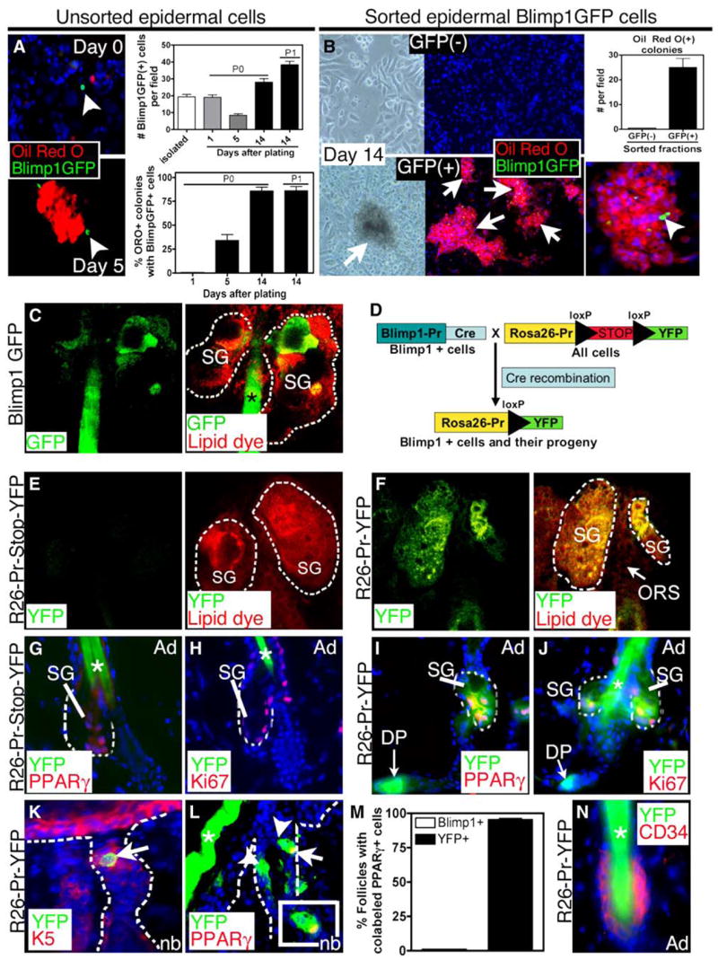
Epidermal lineage commitment occurs when multipotent stem cells are specified to three lineages: the epidermis, the hair follicle, and the sebaceous gland (SG). How and when a lineage becomes specified remains unknown. Here, we report the existence of a population of unipotent progenitor cells that reside in the SG and express the transcriptional repressor Blimp1. Using cell-culture studies and genetic lineage tracing, we demonstrate that Blimp1-expressing cells are upstream from other cells of the SG lineage. Blimp1 appears to govern cellular input into the gland since its loss leads to elevated c-myc expression, augmented cell proliferation, and SG hyperplasia. Finally, BrdU labeling experiments demonstrate that the SG defects associated with loss of Blimp1 lead to enhanced bulge stem cell activity, suggesting that when normal SG homeostasis is perturbed, multipotent stem cells in the bulge can be mobilized to correct this imbalance.


Figures

Figure 1
Figure 1. Blimp1 Expression in the Sebaceous Gland
Immunofluorescence and/or epifluorescence microscopy was performed on skin sections of wild-type or Blimp1GFP transgenic mice at ages indicated. Color-coding is according to secondary Abs or GFP and is marked on each frame. (H), (K), and (M) are confocal images. Schematic in (N) summarizes the localization of Blimp1-expressing cells in the SG. Abbreviations: β4, β4 integrin; SG, sebaceous gland; ep, epidermis; DP, dermal papilla; IRS, inner root sheath; HS, hair shaft, positive for Blimp1GFP (*); NB, newborn; Ad, adult; Bu, bulge (bracketed). Arrows denoted Blimp1+ or Blimp1GFP+ cells in or near the orifice to the sebaceous gland. Note that patterns of anti-Blimp1 Ab and Blimp1GFP expression were comparable. Blimp1 was first observed at E17.5, in the suprabasal epidermis and dermal papillae (DP). By birth, Blimp1 expression expanded to the IRS and to a site in upper outer root sheath (ORS) where the sebaceous gland (SG) buds. Blimp1-expressing SG cells were K14/K5+, Ki67, ORO, PPARγ , and CD34.
Figure 2
Figure 2. Blimp1 Regulates Sebocyte-Containing Gland Size
K14-Cre:Blimp1fl/fl conditional knockout (cKO) mice and organs are compared to wild-type (WT) littermate controls. (A and B) Images of hair coats of 2-month-old mice. (C) PCR genotype analyses. (D) Anti-Blimp1 immunostaining of skin sections from 6 day WT and Blimp1 cKO mice. Sections were counterstained with hematoxylin. (E–L) Data from 2-month-old WT and Blimp1 cKO mice. All SG analyses were performed in telogen phase of the hair cycle. (E and F) Brightfield images of Oil Red O and hematoxylin-stained skin sections (E) and whole mounts of Oil Red O-stained tail skins (F). (G) Sebaceous gland measurements. The average area of 50 individual WT and cKO sebaceous glands was determined. n = 3 mice for each genotype. (H) Thin-layer chromatography of skin lipids. CE, cholesterol esters; TG, triglycerides; C, cholesterol. (I) Real-time PCR of skin mRNAs tested for sebocyte markers (Mc5R, PPARγ, and SCD3). n = 4 for each genotype. (J) Brightfield images of hematoxylin and eosin staining of eyelid skin showing meibomian glands. (K) Brightfield images of preputial glands from male mice. Abbreviations: Meibomian glands (meib), epidermis (ep), and muscle (m). (L) Measurements of meibomian and preputial gland area. n = 4–5 for each genotype. Asterisks indicate significance, p < 0.05.
Figure 3
Figure 3. Increased Cell number and Proliferation in Blimp1 Null Sebaceous Glands
All analyses of SG cell number and proliferation were performed on telogen phase hair cycles in 2-month-old mice. (A) Confocal images of tail whole-mount skin sections stained with the nuclear dye TOPRO-3. Images were edited with Imaris software. (B) Quantification of cell number in SGs. n = 3 for each genotype. (C and D) Immunostaining with anti-PPARγ Abs of skin sections from 2-month-old mice. Nuclei are counterstained with DAPI. Graph shows quantification of PPARγ-positive nuclei. n = 3 for each genotype. (E and F) Immunostaining with anti-Ki67 Abs of skin sections from 2-month-old mice. Graph shows quantification of Ki67-positive nuclei in individual SGs. n = 3 for each genotype. (G) Epifluorescence of Oil Red O and anti-BrdU immunofluorescence of 2-month-old mice pulsed with BrdU and chased for either 0.5 or 4 days. Arrows indicate nuclei within the Oil Red O+ cells, while arrowheads indicate nuclei outside of these cells. (H) Quantification of SGs containing BrdU+ nuclei at indicated time points. (I) Quantification of the percentage of BrdU+/Oil Red O+ cells after 0.5, 4, and 15 days of chase. Asterisks indicate signifcance, p < 0.05.
Figure 4
Figure 4. c-Myc Expression Is Elevated in Blimp1 cKO Skin
(A–D) Localization of c-Myc within SGs of P6 Blimp1GFP (A) or WT (B–D) mice. Skin sections were labeled with Abs against c-Myc, K5, and/or Ki67 or costained with Oil Red O as indicated. Nuclei were counterstained with DAPI. Arrowheads, Blimp1GFP-expressing cells; arrows, c-Myc+ nuclei; asterisks, hair shaft (HS). (E) Real-time PCR analysis of c-myc, L-myc, and N-myc mRNAs in backskins of 2-month-old mice. Asterisks indicate significance, p < 0.05. (F) Anti-c-Myc immunoblot analysis of 2-month-old backskin proteins. (G) Chromatin immunoprecipitation (IP) assays were performed on keratinocytes expressing a flag epitope-tagged, recombinant Blimp1 protein with anti-FLAG or control IgG antisera. PCR analyses on the input or immunoprecipitated fragments were performed using primers encompassing a Blimp1 binding site previously shown to bind Blimp1 (Primer-B) or primers spanning an upstream region of the c-myc promoter lacking Blimp1 binding sites (Primer-A). (H and I) Immunostaining with anti-c-Myc Abs and Oil Red O counterstain (H) or anti-K5 antibodies (I). Arrows indicate c-Myc+ nuclei within the SG. Arrowheads indicate c-Myc-positive nuclei outside the gland. Asterisks denote hair shaft. (J) Quantification of the number of K5+ c-Myc+ cells in epidermis and hair follicles of 2-month-old mice. Asterisks indicate significance, p < 0.05.
Figure 5
Figure 5. Blimp1+ SG Cells Are a Resident Population of Progenitor Cells with Properties of Unipotent Progenitor Cells
(A and B) Cell culture. Isolated Blimp1GFP keratinocytes are initially distinct from Oil Red O+ cells, but after day 5, sebocyte colonies composed of GFP+ and Oil Red O+ cells were detected. Numbers of Blimp1GFP+ cells increased with passage. (B) FACS isolated Blimp1GFP+ and (−) cells were cultured for 14 days and stained for Oil Red O. Oil Red O colonies were only found in cultures of GFP+ cells. (C) Confocal projection of GFP and lipid dye (FM 4-64) epifluorescence from a whole mount of Blimp1GFP Bac transgenic tail skin. (D) Schematic illustrating the genetic strategy to lineage-trace Blimp1+ SG cells in the skin. (E and F) YFP epifluorence of tail skin whole mounts (left) stained a membrane dye (merge, right). Note YFP expression in the entire sebaceous gland but exclusion of signal in the ORS and bulge. (G–L) Backskin sections of R26-Pr-Stop-YFP or R26-Pr-YFP mice were immunostained with Abs against GFP and either PPARγ (G, H, and L), Ki67 (H and J), or K5 (K). (M) Quantification of the percentage of hair follicles containing either Blimp1+ (Blimp1GFP skin sections) or YFP+ (R26-Pr-YFP skin sections) cells that colocalize with PPARγ. (N) Backskin sections of R26-Pr-YFP mice were immunostained with Abs against GFP and CD34. Note the colocalization of YFP with PPARγ- and Ki67-positive sebocytes and in the DP in R26-Pr-YFP mice. SG, Sebaceous gland; HS, hair shaft; DP, dermal papillae.
Figure 6
Figure 6. Enhanced Activity of Bulge Stem Cells in Blimp1 cKO Skin
(A) Increased Anti-Ki67 along anti-β4 ORS above the bulge (Bu). (B) After a 12 hr BrdU pulse, 2-month-old back-skin samples were analyzed for anti-BrdU (arrows in the SG, arrowhead nucleus in ORS above the bulge) and anti-K5 immunofluorescence. Note: The anti-K5 chicken Ab used displayed atypically reduced staining in the bulge. Asterisks denote hair shaft. (C and D) After a 36 hr pulse, 2-month-old backskin samples were analyzed for BrdU and CD34. For quantification, n = 3 animals each. (E–I) At P6, matched WT and Blimp1 cKO litter-mates were given a 72 hr pulse of BrdU and were analyzed after a 28 day chase. (E) FACS analysis of backskin cells shows that the proportions of α6+/CD34+ bulge cells are similar. (F and G) Representative histogram (F) and quantification (G) of the number of α6+/CD34+ cells with BrdU incorporation after BrdU pulse-chase. n = 3 for each genotype. Asterisks indicate significance, p < 0.05. (H and I) Immunofluorescence was used to analyze the numbers of BrdU (green, arrowheads) label-retaining bulge cells (CD34+, red). For quantification, n = 3 mice each. (J and J′) GFP+ epifluorescence and anti-Blimp1 immunofluorescence colocalize at the SG base of sections of skin grafts derived from a single K14-actinGFP bulge stem cell, after clonal expansion in vitro. (J′) shows DIC phase-contrast. (K–N) Samples from (H) revealed BrdU+CD34 LRCs above the CD34+ (red) cKO bulge (K). This population at the SG base is positive for K5 (L and N) and shows partial colocalization with anti-c-Myc Abs (M). (N) shows quantification of K5- and BrdU-positive cells at the SG base in WT and cKO mice. n = 3 for each genotype. Asterisks indicate significance, p < 0.05.
Figure 7
Figure 7. Model for the Role of Blimp1 in Sebocyte Development
Blimp1 is expressed in a population of cells that act as unipotent precursors for the proliferative sebocytes, which in turn differentiate into lipid-containing sebocytes. Blimp1 represses c-Myc expression in these sebocyte progenitors and regulates the size and number of cells within the gland. Upstream, the multipotent bulge stem cells are sensitive to the Blimp1-positive progenitors at the base of the gland and can replenish them as necessary. Blimp1 null glands may resemble a wound situation, as without Blimp1, the resident progenitor cells are pushed down a distinct differentiation pathway.

Similar articles

Cited by

References

    1. Allen M, Grachtchouk M, Sheng H, Grachtchouk V, Wang A, Wei L, Liu J, Ramirez A, Metzger D, Chambon P, et al. Hedgehog signaling regulates sebaceous gland development. Am J Pathol. 2003;163:2173–2178. - PMC - PubMed
    1. Alonso LC, Rosenfield RL. Molecular genetic and endocrine mechanisms of hair growth. Horm Res. 2003;60:1–13. - PubMed
    1. Arnold I, Watt FM. c-Myc activation in transgenic mouse epidermis results in mobilization of stem cells and differentiation of their progeny. Curr Biol. 2001;11:558–568. - PubMed
    1. Blanpain C, Lowry WE, Geoghegan A, Polak L, Fuchs E. Self-renewal, multipotency, and the existence of two cell populations within an epithelial stem cell niche. Cell. 2004;118:635–648. - PubMed
    1. Bull JJ, Pelengaris S, Hendrix S, Chronnell CM, Khan M, Philpott MP. Ectopic expression of c-Myc in the skin affects the hair growth cycle and causes an enlargement of the sebaceous gland. Br J Dermatol. 2005;152:1125–1133. - PubMed

Publication types

MeSH terms