

Early diabetes-induced biochemical changes in the retina: comparison of rat and mouse models
- PMID: 16896942
- PMCID: PMC2228251
- DOI: 10.1007/s00125-006-0356-7
Early diabetes-induced biochemical changes in the retina: comparison of rat and mouse models
Abstract
Aims/hypothesis: Recently, various transgenic and knock-out mouse models have become available for studying the pathogenesis of diabetic retinopathy. At the same time, diabetes-induced retinal changes in the wild-type mice remain poorly characterised. The present study compared retinal biochemical changes in rats and mice with similar (6-week) durations of streptozotocin-induced diabetes.
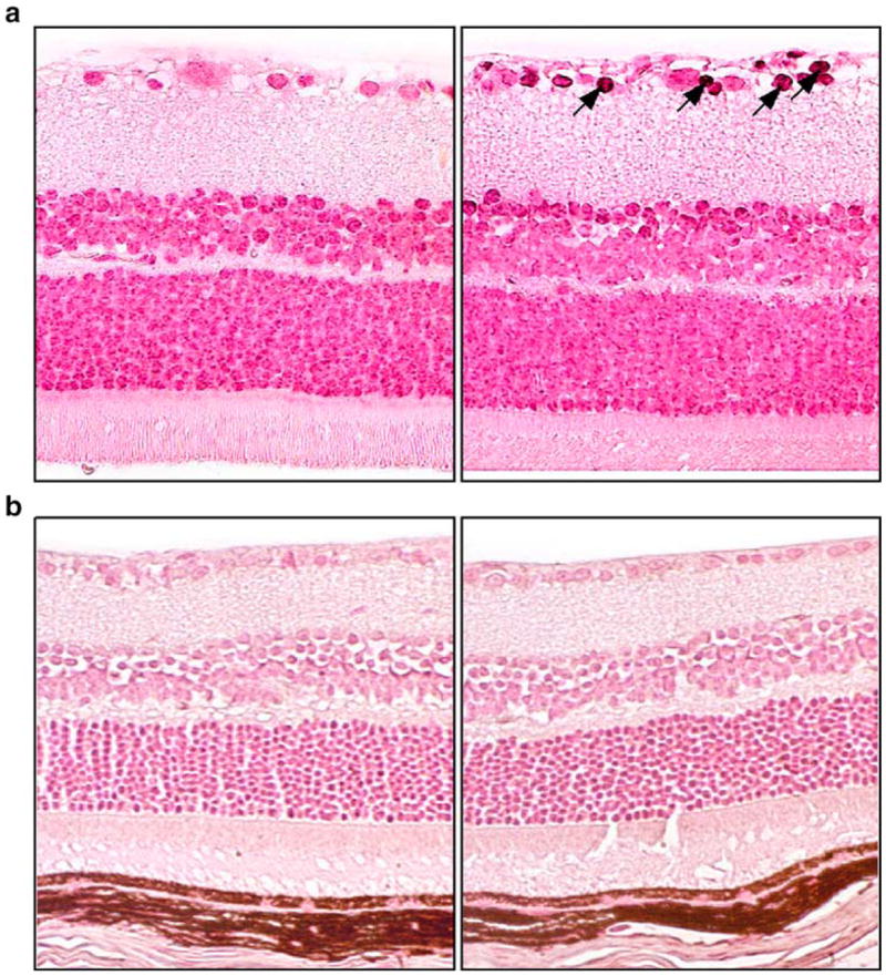
Materials and methods: The experiments were performed on Wistar rats and C57Bl6/J mice. Retinal glucose, sorbitol, fructose, lactate, pyruvate, glutamate, alpha-ketoglutarate and ammonia were measured spectrofluorometrically by enzymatic methods. Vascular endothelial growth factor (VEGF) protein was assessed by ELISA, and poly(ADP-ribosyl)ation by immunohistochemistry and western blot analysis. Free mitochondrial and cytosolic NAD(+)/NADH ratios were calculated from the glutamate and lactate dehydrogenase systems.
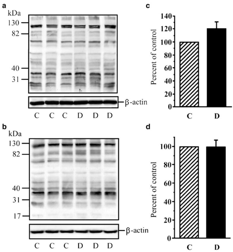
Results: Retinal glucose concentrations were similarly increased in diabetic rats and mice, vs controls. Diabetic rats manifested approximately 26- and 5-fold accumulation of retinal sorbitol and fructose, respectively, whereas elevation of both metabolites in diabetic mice was quite modest. Correspondingly, diabetic rats had (1) increased retinal malondialdehyde plus 4-hydroxyalkenal concentrations, (2) reduced superoxide dismutase (SOD), glutathione peroxidase, glutathione reductase and glutathione transferase activities, (3) slightly increased poly(ADP-ribose) immunoreactivity and poly(ADP-ribosyl)ated protein abundance, and (4) VEGF protein overexpression. Diabetic mice lacked these changes. SOD activity was 21-fold higher in murine than in rat retinas (the difference increased to 54-fold under diabetic conditions), whereas other antioxidative enzyme activities were 3- to 10-fold lower. With the exception of catalase, the key antioxidant defence enzyme activities were increased, rather than reduced, in diabetic mice. Diabetic rats had decreased free mitochondrial and cytosolic NAD(+)/NADH ratios, consistent with retinal hypoxia, whereas both ratios remained in the normal range in diabetic mice.
Conclusions/interpretation: Mice with short-term streptozotocin-induced diabetes lack many biochemical changes that are clearly manifest in the retina of streptozotocin-diabetic rats. This should be considered when selecting animal models for studying early retinal pathology associated with diabetes.
Figures
Similar articles
-
Antioxidants attenuate early up regulation of retinal vascular endothelial growth factor in streptozotocin-diabetic rats.Diabetologia. 2001 Sep;44(9):1102-10. doi: 10.1007/s001250100631. Diabetologia. 2001. PMID: 11596663
-
Aldose reductase inhibitor fidarestat prevents retinal oxidative stress and vascular endothelial growth factor overexpression in streptozotocin-diabetic rats.Diabetes. 2003 Mar;52(3):864-71. doi: 10.2337/diabetes.52.3.864. Diabetes. 2003. PMID: 12606532
-
Poly(ADP-ribose) polymerase inhibitors counteract diabetes- and hypoxia-induced retinal vascular endothelial growth factor overexpression.Int J Mol Med. 2004 Jul;14(1):55-64. Int J Mol Med. 2004. PMID: 15202016
-
Oxidative stress in diabetic retina.EXS. 1992;62:299-307. doi: 10.1007/978-3-0348-7460-1_30. EXS. 1992. PMID: 1450593 Review.
-
Biochemical manifestations of diabetes mellitus in microscopic layers of the cornea and retina.Diabetes Metab Rev. 1989 Feb;5(1):1-15. doi: 10.1002/dmr.5610050102. Diabetes Metab Rev. 1989. PMID: 2649333 Review.
Cited by
-
Novel diabetic mouse models as tools for investigating diabetic retinopathy.PLoS One. 2012;7(12):e49422. doi: 10.1371/journal.pone.0049422. Epub 2012 Dec 12. PLoS One. 2012. PMID: 23251343 Free PMC article.
-
Therapeutic applications of PARP inhibitors: anticancer therapy and beyond.Mol Aspects Med. 2013 Dec;34(6):1217-56. doi: 10.1016/j.mam.2013.01.006. Epub 2013 Jan 29. Mol Aspects Med. 2013. PMID: 23370117 Free PMC article. Review.
-
Retinal neurodegeneration may precede microvascular changes characteristic of diabetic retinopathy in diabetes mellitus.Proc Natl Acad Sci U S A. 2016 May 10;113(19):E2655-64. doi: 10.1073/pnas.1522014113. Epub 2016 Apr 25. Proc Natl Acad Sci U S A. 2016. PMID: 27114552 Free PMC article.
-
The Norway rat, from an obnoxious pest to a laboratory pet.Elife. 2020 Jan 17;9:e50651. doi: 10.7554/eLife.50651. Elife. 2020. PMID: 31948542 Free PMC article.
-
Levels of selected oxidative stress markers in the vitreous and serum of diabetic retinopathy patients.Mol Vis. 2015 Jun 12;21:649-64. eCollection 2015. Mol Vis. 2015. PMID: 26120270 Free PMC article.
References
-
- Frank RN. Diabetic retinopathy. N Engl J Med. 2004;350:48–58. - PubMed
-
- Frank RN, Amin R, Kennedy A, Hohman TC. An aldose reductase inhibitor and aminoguanidine prevent vascular endothelial growth factor expression in rats with long-term galactosemia. Arch Ophthalmol. 1997;115:1036–1047. - PubMed
-
- Kato N, Yashima S, Suzuki T, Nakayama Y, Jomori T. Long-term treatment with fidarestat suppresses the development of diabetic retinopathy in STZ-induced diabetic rats. J Diabetes Complications. 2003;17:374–379. - PubMed
-
- Dagher Z, Park YS, Asnaghi V, Hoehn T, Gerhardinger C, Lorenzi M. Studies of rat and human retinas predict a role for the polyol pathway in human diabetic retinopathy. Diabetes. 2004;53:2404–2411. - PubMed
-
- Kern TS, Tang J, Mizutani M, et al. Response of capillary cell death to aminoguanidine predicts the development of retinopathy: comparison of diabetes and galactosemia. Invest Ophthalmol Vis Sci. 2000;41:3972–3978. - PubMed
Publication types
MeSH terms
Substances
Grants and funding
LinkOut - more resources
Full Text Sources
Other Literature Sources
Medical
