Review
. 2006:81:179-229.
doi: 10.1016/S0079-6603(06)81005-6.

Repair of topoisomerase I-mediated DNA damage

Affiliations
Review

Repair of topoisomerase I-mediated DNA damage

Yves Pommier et al. Prog Nucleic Acid Res Mol Biol. 2006.

Abstract

Topoisomerase I (Top1) is an abundant and essential enzyme. Top1 is the selective target of camptothecins, which are effective anticancer agents. Top1-DNA cleavage complexes can also be trapped by various endogenous and exogenous DNA lesions including mismatches, abasic sites and carcinogenic adducts. Tyrosyl-DNA phosphodiesterase (Tdp1) is one of the repair enzymes for Top1-DNA covalent complexes. Tdp1 forms a multiprotein complex that includes poly(ADP) ribose polymerase (PARP). PARP-deficient cells are hypersensitive to camptothecins and functionally deficient for Tdp1. We will review recent developments in several pathways involved in the repair of Top1 cleavage complexes and the role of Chk1 and Chk2 checkpoint kinases in the cellular responses to Top1 inhibitors. The genes conferring camptothecin hypersensitivity are compiled for humans, budding yeast and fission yeast.


Figures

Fig. 1
Fig. 1. Topoisomerase cleavage complexes
Topoisomerases (abbreviated Topo in panel A) utilize a catalytic tyrosine residue for nucleophilic attack and breakage of a DNA phosphoester bond. A. The polarity depends on the Topo (only human enzymes are considered here). B. Topoisomerases I (nuclear and mitochondrial Top1) form a covalent bond with the 3′-DNA end and generate a 5′-hydroxyl-end. This cleavage intermediate allow controlled rotation of the 5′-end around the intact DNA strand (see Fig. 3B). Under normal conditions, the reaction is reversible. Religation (back arrow from B -> A) is favored over cleavage and requires the alignment of the 5′-hydroxyl-end with the phosphoester tyrosyl-DNA bond for nucleophilic attack. C. All other human Topo enzymes (Top2 and Top3) have an opposite polarity compared to Top1 (see Fig. 2). They form covalent bonds with the 5′-end of the break and generate 3′-hydroxyl ends.
Fig. 2
Fig. 2. Schematic architecture of the topoisomerase cleavage complexes
A. Topoisomerases I (Top1 nuclear and Top1mt) bind to double-stranded DNA and form covalent complexes at the 3′-end of the breaks. All other topoisomerases form covalent complexes at the 5′-end of the breaks. Top1 cleavage complexes are selectively stabilized by the natural alkaloid camptothecin (CPT). B. Topoisomerase II homodimers (Top2α and Top2β) bind to double-stranded DNA and form cleavage complexes with a canonical 4-base pair overhang. Top2 binds and hydrolyze ATP during catalysis. Top2 inhibitors stabilize the Top2 cleavage complexes and are potent anticancer drugs. C. Topoisomerases III (Top3α and Top3β) bind as monomers to non-canonical DNA structures (single-stranded DNA) [194] in association with a RecQ helicase (BLM in humans, Sgs-1 in budding yeast, Rhq1 in fission yeast). Top3 has been proposed to resolve double-holiday junctions arising from stalled replication forks (see Fig. 5A and corresponding text). Top3 inhibitors have not been reported.
Fig. 3
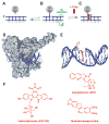
Fig. 3. Trapping of Top1 cleavage complexes by camptothecin and non-camptothecin inhibitors
A. Under physiological conditions, Top1 is associated with chromatin in non-covalent complexes. B. A small fraction of Top1 forms cleavage complexes that relax DNA supercoiling by controlled rotation of the cleaved strand around the intact strand (green curved arrow). C. Anticancer drugs such as those shown in panel F reversibly trap the Top1 cleavage complex by inhibiting religation. D. Crystal structure of camptothecin bound to the Top1-DNA cleavage complex [from [29]] showing “interfacial inhibition” [26, 27] of the Top1 cleavage complex by camptothecin. Interfacial inhibition also applies to non-camptothecin Top1 inhibitors shown in panel F [29, 30]. E. Same structure as in panel D. The Top1 has been removed except for the catalytic tyrosine (in orange). Camptothecin is shown intercalated between the base pairs flanking the Top1 cleavage site. F. Structures of three Top1 inhibitors.
Fig. 4
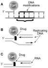
Fig. 4. Conversion of reversible Top1 cleavage complexes into DNA damage
A. Irreversible (“suicide”) Top1 cleavage complexes are produced when Top1 cleaves previously damaged DNA (DNA modifications that trap Top1 are detailed in Table 1). B. Top1 cleavage complexes can be converted to irreversible complexes upon replication fork collision when the Top1 cleavage complex is on the leading strand for DNA synthesis. The drug is shown as the initiating event for the collision. However, once the replication double-strand break (Rep-DSB) is formed, dissociation of the drug has no impact on the irreversible covalent complex. C. Conversion of Top1 cleavage complexes into irreversible covalent complexes by transcription complexes.
Fig. 5
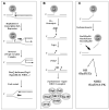
Fig. 5. Schematic representation of three main pathways for the repair of Top1 covalent complexes
A. 5′-end-religation requires realignment of the 5′-hydroxyl with the end of the broken DNA bonded to Top1. This would require a “pull-back” (“regression”) of the replication or transcription complexes (see Figs. 4B and 4C). Fork regression and restart require helicase activities (in particular RecQ helicases such as Sgs-1 in budding yeast and BLM or WRN in humans) in association with Top3. B. Top1 excision by Tdp1 requires prior proteolysis of Top1 [106] or denaturation of Top1 [105] to expose the phosphotyrosyl bond to be attacked (see Fig. 6A–C). Tdp1 generates a 3′-phosphate DNA end, which needs to be hydrolyzed by polynucleotide kinase phosphatase (PNKP = hPNK). PNKP also catalyzes the phosphorylation of the 5′-end of the DNA. Tdp1 and PNKP are part of the XRCC1 complex (shown at the bottom). C. Excision of the Top1-DNA covalent complex by 3′-endonucleases. Studies in budding yeast have implicated at least 3 endonuclease families. The human orthologs are listed: Rad1/Rad10, Mus81/Eme1 and Mre11/Rad50. The resulting DNA lesion is probably processed by homologous recombination initiated by the Rad51, Rad52 complexes and by non-homologous end joining (Ku-DNA-PK pathway).
Fig. 6
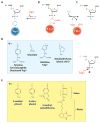
Fig. 6. Tyrosyl-DNA phosphodiesterase (Tdp1)-mediated reactions and substrates
A. Structure of the Top1-DNA covalent intermediate (see Fig. 1B). B. Tdp1 releases the Top1 by forming a covalent bond between its active histidine 263 and the DNA end. C. Histidine 493 from the second HKD motif of Tdp1 (and which is mutated H493R in SCAN1) promotes the hydrolysis of the Tdp1-DNA intermediate, and frees the 3′-phosphate DNA end. Tdp1 is regenerated for another catalytic cycle. D. Physiological substrates for Tdp1. E. Tdp1 substrates used for biochemical assays.
Fig. 7
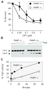
Fig. 7. Hypersensitivity of PARP-1−/− cells to camptothecin and functional Tdp1 deficiency in PARP-1 −/− cells
A. Mouse fibroblasts were exposed to camptothecin (concentrations indicated on X axis) for 1 hour, washed, kept in culture for 4 days and counted. The curves represent the averages and standard deviations from five independent experiments. B. A 14-mer single-stranded oligonucleotide with a phosphotyrosine at the 3′-end (see Fig. 6) (14-Y) [106] was incubated with nuclear extracts obtained from PARP-1−/− and PARP-1 +/+ cells [195] in the presence of 50 mM EDTA and absence of MgCl2 to eliminate PNKP activity, which requires MgCl2. A representative experiment is shown. TDP1 activity was determined as a shift in band position (from 14-Y to 14-P). C. Quantitation of the results shown in panel B using ImageQuant (Molecular Dynamics, Sunnyvale, CA).
Fig. 8
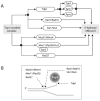
Fig. 8. Repair involved in the repair of Top1 covalent complexes in budding yeast
A. Schematic representation of the genetic pathways implicated in the removal of the Top1-DNA covalent complexes. Tdp1 appear to function in alternative pathways with Rad1/Rad10 and Slx1/Slx4. The other endonucleases (Mus81/Mms4; Mre11/Rad50/Xrs2 and Rad27) appear to function in parallel. Sae2 is a cofactor for the endonuclease activity of Mre11. Tpp1, Apn1 and Apn2 are 3′-phosphatases that remove the 3′-phosphate left after Tdp1 hydrolyzes the tyrosyl-DNA adduct (see Figs. 5B and 6C). B. Shematic representation of the sites of attack for Tdp1 and the endonucleases described in panel A. Srs2 helicase is also shown.

Similar articles

Cited by

References

    1. Wang JC. Cellular roles of DNA topoisomerases: a molecular perspective. Nat Rev Mol Cell Biol. 2002;3:430–440. - PubMed
    1. Zhang H, Barcelo JM, Lee B, Kohlhagen G, Zimonjic DB, Popescu NC, Pommier Y. Human mitochondrial topoisomerase I. Proc Natl Acad Sci U S A. 2001;98:10608–10613. - PMC - PubMed
    1. Zhang H, Meng LH, Zimonjic DB, Popescu NC, Pommier Y. Thirteen-exon-motif signature for vertebrate nuclear and mitochondrial type IB topoisomerases. Nucleic Acids Res. 2004;32:2087–2092. - PMC - PubMed
    1. Hickson ID. RecQ helicases: caretakers of the genome. Nat Rev Cancer. 2003;3:169–178. - PubMed
    1. Harmon FG, Brockman JP, Kowalczykowski SC. RecQ helicase stimulates both DNA catenation and changes in DNA topology by topoisomerase III. J Biol Chem. 2003;278:42668–42678. - PubMed

Publication types

Substances