. 2006 Jul 26;26(30):7826-38.
doi: 10.1523/JNEUROSCI.1866-06.2006.

Expression and function of SNAP-25 as a universal SNARE component in GABAergic neurons

Affiliations

Expression and function of SNAP-25 as a universal SNARE component in GABAergic neurons

Lawrence C R Tafoya et al. J Neurosci. .

Erratum in

  • J Neurosci. 2006 Aug 23;26(34):8875

Abstract

Intracellular vesicular trafficking and membrane fusion are important processes for nervous system development and for the function of neural circuits. Synaptosomal-associated protein 25 kDa (SNAP-25) is a component of neural soluble N-ethylmaleimide-sensitive factor attachment protein receptor (SNARE) core complexes that mediate the exocytotic release of neurotransmitters at chemical synapses. Previous results from mouse mutant models and pharmacological/neurotoxin blockades have demonstrated a critical role for SNAP-25-containing SNARE complexes in action potential (AP)-dependent release at cholinergic and glutamatergic synapses and for calcium-triggered catecholamine release from chromaffin cells. To examine whether SNAP-25 participates in the evoked release of other neurotransmitters, we investigated the expression and function of SNAP-25 in GABAergic terminals. Patch-clamp recordings in fetal Snap25-null mutant cortex demonstrated that ablation of SNAP-25 eliminated evoked GABA(A) receptor-mediated postsynaptic responses while leaving a low level of spontaneous AP-independent events intact, supporting the involvement of SNAP-25 in the regulated synaptic transmission of early developing GABAergic neurons. In hippocampal cell cultures of wild-type mice, punctate staining of SNAP-25 colocalized with both GABAergic and glutamatergic synaptic markers, whereas stimulus-evoked vesicular recycling was abolished at terminals of both transmitter phenotypes in Snap25-/- neurons. Moreover, immunohistochemistry and fluorescence in situ hybridization revealed coexpression of SNAP-25, VGAT (vesicular GABA transporter), and GAD65/67 (glutamic acid decarboxylase 65/67) in interneurons within several regions of the adult brain. Our results thus provide evidence that SNAP-25 is critical for evoked GABA release during development and is expressed in the presynaptic terminals of mature GABAergic neurons, consistent with its function as a component of a fundamental core SNARE complex required for stimulus-driven neurotransmission.


Figures
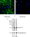
Figure 1.
Figure 1.
SMI81 monoclonal antibody shows robust and specific immunoreactivity in both cultured neurons and Western blots. A, Wild-type E17.5 cultured hippocampal neurons immunostained at 12 DIV using SMI81 monoclonal antibody and viewed by confocal microscopy with a 63× objective (NA, 1.4; optical slice, 0.81 μm). Probing for SNAP-25 (green; Alexa 488) resulted in staining that was both continuous throughout the neurites with regular punctate accumulations, but not appreciably within the soma or around the nucleus (blue; ToPro3). B, SMI81 showed no apparent immunoreactivity in Snap25-null mutant neuronal cultures. C, Protein fractions prepared from the cortices of Snap25−/− and wild-type animals at E17.5 (mutant, lane 1; wild type, lane 2; 30 μg) and P24 (wild type, lane 3; 1.0 μg) fractionated on a 12% SDS-PAGE gel, blotted, and probed for SNAP-25. SMI81 immunoreactivity was evident in wild-type lanes as a single band at ∼25 kDa with no cross-reactivity in the mutant lane. The synaptic vesicle protein synaptophysin was used as a loading control.
Figure 2.
Figure 2.
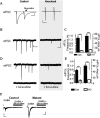
GABAergic transmission could not be evoked in SNAP-25-deficient mutant neurons, although spontaneous currents persist. A, Whole-cell patch-clamp recordings of field stimulation ePSCs obtained from cortical slices of E17.5 control and Snap25-null mutant fetuses. GABAergic responses were isolated by recording in the presence of NBQX (10 μm) plus APV (100 μm) to block glutamatergic transmission. Representative tracings are presented on the left with respective calibrations. The trace (dotted) obtained after application of 20 μm bicuculline to control slices is superimposed over the recording in the absence of GABAA receptor inhibition and indicates GABAergic origin of the response. Mutant slices did not show any detectable response to stimulation. B, Recordings of sPSCs in the absence of TTX. C, sPSCs from Snap25-null mutant slices (n = 9) were decreased in frequency (26.1-fold) and amplitude (6.0-fold) compared with controls (n = 5; ***p < 0.001). Error bars represent SEM. D, mPSCs recorded in the presence of TTX (0.5 μm). E, Both the amplitude (5.2-fold) and frequency (12.9-fold) of TTX-resistant mPSCs were decreased in Snap25-null mutants (n = 3) compared with controls (n = 3; ***p < 0.001). TTX treatment, however, did not significantly reduce the frequency or amplitude of mPSCs recorded from SNAP-25-deficient slices (see Results) indicating that action potential-dependent responses that contribute to the sPSCs of control slices are completely absent in Snap25-null mutants. Error bars represent SEM. F, Response of Snap25-null mutant (n = 5) and control slices (n = 5) to bath application of GABA (50 μm). Note that the response to exogenous GABA in Snap25-null mutant slices was more robust than control (8.4-fold; p < 0.001; n = 8), suggesting that the decreased amplitude for mIPSCs recorded from mutant slices was not attributable to inherent receptor defects and that GABAA receptors may be upregulated in SNAP-25-deficient fetal brain.
Figure 3.
Figure 3.
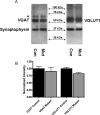
Similar levels of synaptic vesicle proteins expressed in SNAP-25-deficient and control mice. A, Crude synaptic vesicle fraction (LP2; 2.5 μg) prepared from cortex and hippocampus of E17.5 Snap25/ and control littermates were fractionated on a 10% SDS-PAGE gel, blotted, and probed with antibodies to synaptophysin and VGAT, or VGLUT1, as indicated. B, Three animals per genotype were assayed in duplicate with the mean of each animal’s repeated values being normalized to the average synaptophysin levels of the control group. Error bars indicate SEM. One-way ANOVA analysis showed no difference between levels of immunoreactive VGAT, VGLUT1, and synaptophysin in mutant and control fractions, indicating that neither the total vesicular content nor specific GABA- or glutamate-containing vesicles are significantly decreased in SNAP-25-deficient neurons. The slower migrating species (indicated by an asterisk) recognized by VGAT antibodies likely reflects phosphorylated VGAT (see Results).
Figure 4.
Figure 4.
Vesicular recycling within both glutamatergic and GABAergic terminals requires SNAP-25. Hippocampal neurons prepared from both Snap25−/− (KO) and control (WT) E17.5 mice grown for 12 d in culture (DIV 12) were loaded with in the presence of 15 μm FM1-43FX by application of either high K+ or hypertonic sucrose buffer as described in Materials and Methods; control wild-type neurons were also destained by applying a subsequent 90 s exposure to 90 mm K+ to demonstrate exocytotic release and washout of the endocytosed FM dye. After fixation, and immunostaining with either VGLUT1 or VGAT to distinguish dye uptake at glutamatergic or GABAergic synapses, the neurons were viewed by laser confocal microscopy. Representative confocal fluorescent images are shown in A–H, with A, C, E, and G on the left depicting merged images of FM1-43 dye fluorescence and transporter immunostaining taken at 63× (optical slice, 0.81 μm; scale bar, 50 μm). The panels on the right (B1–B3, D1–D3, F1–F3, H1–H3) are series of separated color and merged images of the areas outlined in the low-power images (white box) that were digitally magnified 7× (Adobe Photoshop; scale bar, 7 μm). Insets in the far right merged panels are further digitally enlarged images of the puncta indicated by white arrowheads. Note that FM1-43 dye (green in all panels) is taken up readily by wild-type neurons with equal and consistent colocalization in both glutamatergic (red, B1–B3) and GABAergic (red, F1–F3); whereas in SNAP-25-deficient terminals, there is no detectable FM1-43 dye uptake regardless of their neurotransmitter phenotype (VGLUT, red, D1–D3; VGAT, red H1–H3), consistent with of the lack of stimulus-evoked vesicular recycling in these mutant neurons. I shows the quantitative data of FM1-43 dye intensity over immunoreactive puncta obtained for each transporter after high K+ stimulation. Note that levels of FM1-43 dye fluorescence in loaded wild-type terminals were highly significant (p < 0.001) compared with both destained control synapses and to knock-out mutant synapses, which did not differ from background fluorescence. J compares the relative amount of FM1-43 fluorescence between wild-type and control neurons in a similar series of experiments using hyperosmotic sucrose to promote exocytosis. As in I, although wild-type neurons showed robust vesicular recycling, no detectable fluorescence was observed over VGLUT- or VGAT-immunoreactive puncta of Snap25−/− mutant neurons, indicating that there was no loading of a readily releasable pool of synaptic vesicles at either glutamatergic or GABAergic terminals in these neurons. Error bars indicate SEM.
Figure 5.
Figure 5.
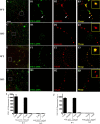
Immunostaining of cultured hippocampal neurons reveals SNAP-25 colocalization with GAD65/67, VGAT, and VGLUT1 in presynaptic terminals. Hippocampal neurons prepared from E17.5 mouse fetuses were cultured, probed with antibodies to the indicated proteins, and viewed by confocal microscopy as described in Materials and Methods. A–H are representative confocal fluorescent images of stained neurons at DIV 15. A, C, E, and G are merged images of dual staining taken at 63× (optical slice, 0.81 μm; scale bar, 50 μm). B1–B3, D1–D3, F1–F3, and H1–H3 are series of separated color and merged images at higher digital magnification of the areas outlined in the low-power images (scale bar, 20 μm). Insets in the far right merged panels are digitally enlarged images of the puncta indicated by white arrows. Note that dual immunostaining for SNAP-25 (green), and GAD65/67 (red; A, B1–B3), VGAT (red; C, D1–D3), and VGLUT1 (red; E, F1–F3) show marked colocalization (yellow in merged images) of punctate fluorescence for transmitter-specific proteins with SNAP-25, consistent with expression of the SNARE protein in GABAergic and glutamatergic terminals (and see I). Arrowheads indicate punctate staining of SNAP-25 that is not colocalized with transmitter-specific antibodies indicating accumulation of SNAP-25 outside terminals of the indicated transmitter phenotype. G, H, Dual staining for transporters VGLUT1 (green) and VGAT (red) show little or no colocalization. I summarizes quantitative data of colocalized punctate staining from dual-stained cultures at DIV 9, 12, 15, and 21 obtained using MetaMorph software (∗∗∗p < 0.001). Error bars indicate SEM.
Figure 6.
Figure 6.
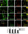
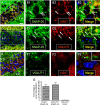
SNAP-25 immunoreactivity colocalizes with VGAT in the CA1 pyramidal layer of the adult hippocampus. Thirty micrometer coronal sections of adult mice (>60 d of age) were coimmunostained with antibodies to SNAP-25 and either vesicular transporters VGAT or VGLUT1, and as a control for specificity coimmunostained with anti-VGLUT1 and VGAT antibodies. After nuclear counterstaining with ToPro3 (blue), the sections were imaged by laser confocal microscopy as described in Materials and Methods. A, C, and E on the right are images taken using a 63× objective (optical slice, 0.81 μm), and the series of panels on the left (B1–B3, D1–D3, F1–F3) show separate channel and merged images of the boxed areas with overlapping red and green pixels depicted as yellow after digital magnification to 420×. A, B, SNAP-25 (green) colocalizes with VGAT (red) within the stratum pyramidale (S.P.) layer of the CA1 region. C, D, VGLUT1 immunoreactivity (red) localized to the stratum oriens (S.O.) and stratum radiatum (S. R.), also colocalizes with SNAP-25 (green). E, F, Nonoverlapping immunoreactivity for VGLUT1 (green) and VGAT (red) seen throughout the hippocampus. Note that, although the patterns of immunostaining for the two transporters are predominantly located in different layers of the hippocampus (E), in regions in which VGAT- and VGLUT1-immunoreactive puncta are interspersed, such as the border of the stratum oriens and pyramidale (boxed area in E; magnified in F), there is virtually no colocalization of these transporters. G, VGLUT1 and VGAT colocalize to a similar extent with SNAP-25. The proportion of pixel overlap in representative immunofluorescent images for transporters VGLUT1 and VGAT with SNAP-25 (VGLUT1/SNAP-25 and VGAT/SNAP-25, respectively) and between the two transporters (VGLUT1 and VGAT) was quantified using MetaMorph software. The histogram represents average values (error bars indicate SEM) obtained from 12 images taken from three animals (∗∗∗p < 0.001). Scale bar: (in A) A, C, E, 20 μm; B, D, F, 3.0 μm.
Figure 7.
Figure 7.
Colocalization of SNAP-25 and VGAT immunoreactivity occurs in the thalamus. Confocal images of immunofluorescent staining for SNAP-25 (green), VGAT (red), and VGLUT1 (red) in 30 μm sections of >P60 wild-type mice were obtained as described in Materials and Methods. A, B1–B4, SNAP-25 and VGAT immunostaining within the VPL of the thalamus. The separate color channel and merged images illustrate the extensively overlapping punctate pattern of robust immunoreactivity observed for VGAT and SNAP-25 (arrows) in this region; consistent cocompartmentalization of SNAP-25 and the GABA transporter within presynaptic terminals of GABAergic neurons. The arrowhead indicates an example of colocalized punctate stained structure after 5× digital enlargement in far upper right corner. C, D1–D4, In contrast, VGLUT1 staining was scarce within the VPL, although small regions with prominent immunoreactivity were evident. Immunofluorescent staining for the glutamate transporter within these patches also appeared punctate and overlapped with SNAP-25 immunostaining, again reflecting the expression of SNAP-25 in glutamatergic presynaptic terminals. A, C, Magnification, 20×; scale bar, 50 μm. B, D, Magnification, 63×; scale bar, 20 μm.
Figure 8.
Figure 8.
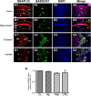
Adult GABAergic neurons express both SNAP-25 and GAD65/67 mRNA in several brain regions. FISH of SNAP-25 and GAD65/67 cRNA probes, followed by nuclear counterstaining with DAPI (blue), was performed on 30 μm coronal brain sections of >P60 wild-type mice as described in Materials and Methods. Sections were visualized using a wide-field fluorescent microscope and imaged with MetaMorph software. A–D, Fluorescent images of hybridization for SNAP-25 (red) and GAD65/67 (green), and ToPro3 staining in selected brain regions in separate color channels, and a color merged micrograph (far right). A, Primary motor cortex (layers I–V). B, Hippocampal CA1 region (SO, stratums oriens; SP, stratums pyramidale; SR, stratums radiatum, and SL, lacunosum molecular). C, Thalamus (RT, reticular nucleus; ic, internal capsule). D, Caudate–putamen. GAD65/67-positive nuclei throughout these selected anatomical regions display robust expression and colocalization with SNAP-25 mRNA (white arrows, digitally enlarged in white box in upper far right corner of the merged image). Images were taken with a 20× objective. Scale bar: A–D, 50 μm. E, Quantitation of colocalized GAD65/67 and SNAP-25 hybridization with 20× magnification. The fraction of GAD65/67-positive cells with overlapping SNAP-25 hybridization, determined using ImageJ software, demonstrates that nearly all cells expressing GAD65/67 mRNA coexpressed SNAP-25 mRNA in each brain region assayed. Error bars indicate SEM. Statistical analysis was performed by one-way ANOVA with Bonferroni’s post hoc comparisons.

Similar articles

Cited by

References

    1. Augustin I, Rosenmund C, Sudhof TC, Brose N (1999). Munc13-1 is essential for fusion competence of glutamatergic synaptic vesicles. Nature 400:457–461. - PubMed
    1. Bark C, Bellinger FP, Kaushal A, Mathews JR, Partridge LD, Wilson MC (2004). Developmentally regulated switch in alternatively spliced SNAP-25 isoforms alters facilitation of synaptic transmission. J Neurosci 24:8796–8805. - PMC - PubMed
    1. Bark IC, Wilson MC (1994). Regulated vesicular fusion in neurons: snapping together the details. Proc Natl Acad Sci USA 91:4621–4624. - PMC - PubMed
    1. Bark IC, Hahn KM, Ryabinin AE, Wilson MC (1995). Differential expression of SNAP-25 protein isoforms during divergent vesicle fusion events of neural development. Proc Natl Acad Sci USA 92:1510–1514. - PMC - PubMed
    1. Bedet C, Isambert MF, Henry JP, Gasnier B (2000). Constitutive phosphorylation of the vesicular inhibitory amino acid transporter in rat central nervous system. J Neurochem 75:1654–1663. - PubMed

Publication types

MeSH terms

Substances

LinkOut - more resources