Optimization and functional effects of stable short hairpin RNA expression in primary human lymphocytes via lentiviral vectors
- PMID: 16844419
- PMCID: PMC2562632
- DOI: 10.1016/j.ymthe.2006.05.015
Optimization and functional effects of stable short hairpin RNA expression in primary human lymphocytes via lentiviral vectors
Abstract
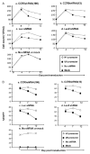
Specific, potent, and sustained short hairpin RNA (shRNA)-mediated gene silencing is crucial for the successful application of RNA interference technology to therapeutic interventions. We examined the effects of shRNA expression in primary human lymphocytes (PBLs) using lentiviral vectors bearing different RNA polymerase III promoters. We found that the U6 promoter is more efficient than the H1 promoter for shRNA expression and for reducing expression of CCR5 in PBLs. However, shRNA expression from the U6 promoter resulted in a gradual decline of the transduced cell populations. With one CCR5 shRNA this decline could be attributed to elevated apoptosis but another CCR5 shRNA that caused cytotoxicity did not show evidence of apoptosis, suggesting sequence-specific mechanisms for cytotoxicity. In contrast to the U6 promoter, PBLs transduced by vectors expressing shRNAs from the H1 promoter could be maintained without major cytotoxic effects. Since a lower level of shRNA expression appears to be advantageous to maintaining the shRNA-transduced population, lentiviral vectors bearing the H1 promoter are more suitable for stable transduction and expression of shRNA in primary human T lymphocytes. Our results suggest that functional shRNA screens should include tests for both potency and adverse metabolic effects upon primary cells.
Figures


Similar articles
-
Determinants of interferon-stimulated gene induction by RNAi vectors.Differentiation. 2004 Mar;72(2-3):103-11. doi: 10.1111/j.1432-0436.2004.07202001.x. Differentiation. 2004. PMID: 15066190
-
Short-term cytotoxic effects and long-term instability of RNAi delivered using lentiviral vectors.BMC Mol Biol. 2004 Aug 3;5:9. doi: 10.1186/1471-2199-5-9. BMC Mol Biol. 2004. PMID: 15291968 Free PMC article.
-
Of mice and men: human RNA polymerase III promoter U6 is more efficient than its murine homologue for shRNA expression from a lentiviral vector in both human and murine progenitor cells.Exp Hematol. 2010 Sep;38(9):792-7. doi: 10.1016/j.exphem.2010.05.005. Epub 2010 May 26. Exp Hematol. 2010. PMID: 20685233
-
Lentiviral vector design for multiple shRNA expression and durable HIV-1 inhibition.Mol Ther. 2008 Mar;16(3):557-64. doi: 10.1038/sj.mt.6300382. Epub 2008 Jan 15. Mol Ther. 2008. PMID: 18180777 Free PMC article.
-
In search of the most suitable lentiviral shRNA system.Curr Gene Ther. 2009 Jun;9(3):192-211. doi: 10.2174/156652309788488578. Curr Gene Ther. 2009. PMID: 19519364 Review.
Cited by
-
Gene Editing of HIV-1 Co-receptors to Prevent and/or Cure Virus Infection.Front Microbiol. 2018 Dec 17;9:2940. doi: 10.3389/fmicb.2018.02940. eCollection 2018. Front Microbiol. 2018. PMID: 30619107 Free PMC article. Review.
-
Optimization and characterization of tRNA-shRNA expression constructs.Nucleic Acids Res. 2007;35(8):2620-8. doi: 10.1093/nar/gkm103. Epub 2007 Apr 10. Nucleic Acids Res. 2007. PMID: 17426139 Free PMC article.
-
Stem cell-based anti-HIV gene therapy.Virology. 2011 Mar 15;411(2):260-72. doi: 10.1016/j.virol.2010.12.039. Epub 2011 Jan 17. Virology. 2011. PMID: 21247612 Free PMC article. Review.
-
Stem cell-based therapies for HIV/AIDS.Adv Drug Deliv Rev. 2016 Aug 1;103:187-201. doi: 10.1016/j.addr.2016.04.027. Epub 2016 May 2. Adv Drug Deliv Rev. 2016. PMID: 27151309 Free PMC article. Review.
-
Silencing of endogenous envelope genes in human choriocarcinoma cells shows that envPb1 is involved in heterotypic cell fusions.J Gen Virol. 2012 Aug;93(Pt 8):1696-1699. doi: 10.1099/vir.0.041764-0. Epub 2012 May 9. J Gen Virol. 2012. PMID: 22573740 Free PMC article.
References
-
- Hannon GJ. RNA interference. Nature. 2002;418:244–251. - PubMed
-
- Elbashir SM, Harborth J, Lendeckel W, Yalcin A, Weber K, Tuschl T. Duplexes of 21-nucleotide RNAs mediate RNA interference in cultured mammalian cells. Nature. 2001;411:494–498. - PubMed
-
- Lee NS, et al. Expression of small interfering RNAs targeted against HIV-1 rev transcripts in human cells. Nat Biotechnol. 2002;20:500–505. - PubMed
-
- Miyagishi M, Taira K. U6 promoter driven siRNAs with four uridine 3′ overhangs efficiently suppress targeted gene expression in mammalian cells. Nat Biotechnol. 2002;20:497–500. - PubMed
-
- Brummelkamp TR, Bernards R, Agami R. A system for stable expression of short interfering RNAs in mammalian cells. Science. 2002;296:550–553. - PubMed
Publication types
MeSH terms
Substances
Grants and funding
LinkOut - more resources
Full Text Sources
Other Literature Sources

