. 2006 Aug;80(15):7405-15.
doi: 10.1128/JVI.02533-05.

Intramolecular and intermolecular uridylylation by poliovirus RNA-dependent RNA polymerase

Affiliations

Intramolecular and intermolecular uridylylation by poliovirus RNA-dependent RNA polymerase

Oliver C Richards et al. J Virol. 2006 Aug.

Abstract

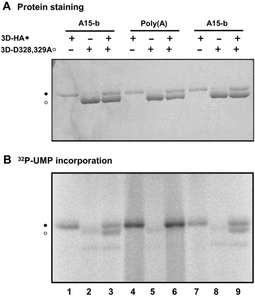
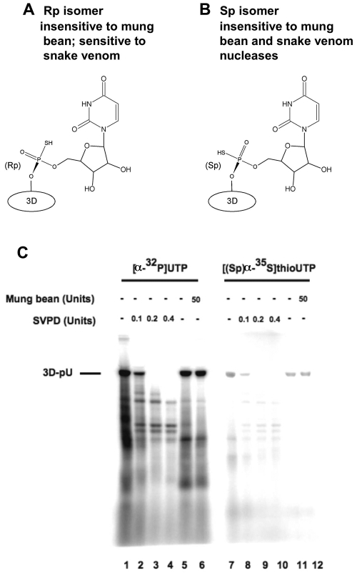
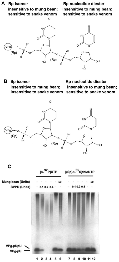
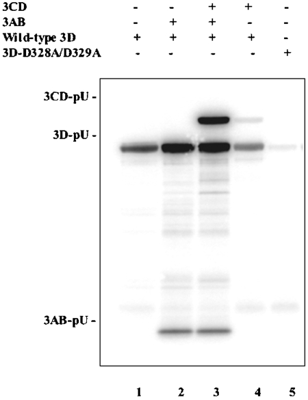
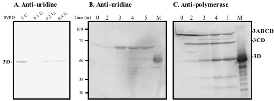
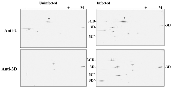
The 22-amino-acid protein VPg can be uridylylated in solution by purified poliovirus 3D polymerase in a template-dependent reaction thought to mimic primer formation during RNA amplification in infected cells. In the cell, the template used for the reaction is a hairpin RNA termed 2C-cre and, possibly, the poly(A) at the 3' end of the viral genome. Here, we identify several additional substrates for uridylylation by poliovirus 3D polymerase. In the presence of a 15-nucleotide (nt) RNA template, the poliovirus polymerase uridylylates other polymerase molecules in an intermolecular reaction that occurs in a single step, as judged by the chirality of the resulting phosphodiester linkage. Phosphate chirality experiments also showed that VPg uridylylation can occur by a single step; therefore, there is no obligatory uridylylated intermediate in the formation of uridylylated VPg. Other poliovirus proteins that could be uridylylated by 3D polymerase in solution were viral 3CD and 3AB proteins. Strong effects of both RNA and protein ligands on the efficiency and the specificity of the uridylylation reaction were observed: uridylylation of 3D polymerase and 3CD protein was stimulated by the addition of viral protein 3AB, and, when the template was poly(A) instead of the 15-nt RNA, the uridylylation of 3D polymerase itself became intramolecular instead of intermolecular. Finally, an antiuridine antibody identified uridylylated viral 3D polymerase and 3CD protein, as well as a 65- to 70-kDa host protein, in lysates of virus-infected human cells.


Figures

FIG. 1.
FIG. 1.
(A) Template requirements for uridylylation of VPg and 3D polymerase. The incorporation of [32P]UTP into 3D polymerase and VPg, using template poly(A) (lanes 1 to 4), A15-b (lanes 5 to 8), or A30-b (lanes 9 to 12), is illustrated by autoradiography in reactions described in Materials and Methods. Reaction mixtures contained 3D polymerase (2 μM), 3AB (4 μM), and VPg (100 μM) as indicated. Products were digested with RNase A and resolved in 12% polyacrylamide-Tris-Tricine gels. The positions of migration of the 2.5-kDa uridylylated VPg and the 52-kDa uridylylated 3D polymerase are indicated. These assignments were made by the known mobilities of VPg and 3D polymerase in comparison to markers, by the absence of other proteins in the purified preparations, and by immunoblotting (not shown). (B) Basic requirements for uridylylation of 3D polymerase. The incorporation of [32P]UTP into 3D polymerase and VPg is shown, as described in Materials and Methods. Where indicated, A15-b was present at 10 μM, 3AB at 3.6 μM, VPg at 100 μM, [32P]UTP at 0.12 μM (2 μCi/30 μl), and 3D polymerase at 2 μM. Products were resolved in 12% polyacrylamide-Tris-Tricine gels. The positions of uridylylated 3D polymerase and VPg are as in panel A. (C) 3D polymerase uridylylation as a function of 3AB concentration and incubation time. Reaction mixtures (see Materials and Methods) included template A15-b and 3D polymerase (2 μM). Products were digested with RNase A and resolved in 12% polyacrylamide-Tris-Tricine gels, as above. The position of uridylylated 3D polymerase is as in panel A. Unmarked labeled species most likely result from incomplete RNase digestion.
FIG. 2.
FIG. 2.
Intra- and intermolecular uridylylation of 3D polymerase. Enzymatically active HA-tagged 3D polymerase (lanes 1, 4, and 7), enzymatically inactive 3D-D328/329A (lanes 2, 5, and 8), and mixtures of these two molecules (lanes 3, 6, and 9) were incubated under 3D uridylylation conditions with [32P]UTP but without 3AB. Products were resolved in 10% polyacrylamide-SDS gels and analyzed (A) by protein staining, to detect both HA-tagged 3D and 3D-D328/329A, and (B) by autoradiography, to identify the uridylylated 3D species. The templates used for uridylylation reactions are indicated.
FIG. 3.
FIG. 3.
Phosphate chirality can be used to determine whether uridylylation reactions are single-step reactions or proceed through covalent intermediates. (A) Expected chirality if the uridylylation of 3D polymerase and VPg uridylylation are single-step reactions. (B) Expected chirality if uridylylated 3D polymerase is an obligate intermediate in VPg uridylylation. Chirality is defined based on the priority of the groups in the chiral [(Sp)α-35S]UTP.
FIG. 4.
FIG. 4.
Chirality of uridylylation of 3D. (A) Formation of a 3D uridylylation product containing a chiral phosphate diester linkage (3D polymerase to UMP) of the Rp configuration is susceptible to SVPD cleavage (specific for Rp chiral phosphate). (B) Formation of a 3D polymerase uridylylation product with a chiral phosphate diester linkage of the Sp configuration is not cleavable by SVPD. (C) Uridylylation of 3D polymerase was performed as described in Materials and Methods with 10 μM A15-b template, 3 μM 3AB, 2 μM [32P]UTP, and 2 μM 3D at 30°C for 60 min (lanes 1 to 6) or with A15-b, 3AB, 3D, and 0.9 μM [(Sp)α-35S]UTP at 30°C for 180 min (lanes 7 to 12). Products were treated with SVPD or mung bean nuclease (Sp-specific cleavage) at the indicated concentrations, followed by RNase A digestion, as described in Materials and Methods. Products were resolved in 12% polyacrylamide-Tris-Tricine gels and analyzed by autoradiography.
FIG. 5.
FIG. 5.
Chirality of uridylylation of VPg. (A) Formation of a VPg uridylylation product may produce VPg-pU or VPg-pUpU. If the diester linkage between VPg and UMP is in the Rp configuration, it is susceptible to cleavage by Rp-specific SVPD. It is predicted that the internucleotide linkage would be a single-step reaction, thus of the Rp configuration and, therefore, susceptible to cleavage by SVPD. In both cases the bonds would not be susceptible to mung bean nuclease cleavage (Sp-specific enzyme). (B) If formation of the linkage between VPg and UMP goes through an intermediate, that chiral phosphate would have the Sp configuration and hence not be susceptible to SVPD cleavage but still not cleavable by mung bean nuclease (requires internucleotide linkage; resistant to a mixed diester linkage). However, the internucleotide linkage remains, as in panel A, and hence is susceptible to SVPD only. (C) Uridylylation of VPg was performed as described in Materials and Methods and includes 1 μM poly(A) template, 50 μM VPg, 2 μM [32P]UTP, and 2 μM 3D polymerase, incubated at 30°C for 60 min (lanes 1 to 6), or with poly(A), VPg, 3D polymerase, and 0.9 μM [(Sp)α-35S]UTP at 30°C for 180 min (lanes 7 to 12). Products were treated as described in the legend to Fig. 4 but without RNase A digestion, resolved in 12% polyacrylamide-Tris-Tricine gels, and analyzed by autoradiography. The labeled material at the top of the lanes is end-labeled poly(A) template used in these VPg uridylylation reactions; 3D polymerase is known to catalyze terminal uridylylation of unblocked RNA molecules (45).
FIG. 6.
FIG. 6.
Intermolecular uridylylation of 3CD and 3AB by 3D polymerase. Uridylylation reactions were as described in Materials and Methods with 10 μM A15-b, 5 μM UTP (5 μCi/30 μl), 1 μM 3D polymerase, and 1 μM 3CD and/or 5 μM 3AB, as indicated. Incubations were at 30°C for 2 h; products were digested with RNase A, heated at 90°C in sample buffer, and resolved in 10% polyacrylamide-SDS gels. The autoradiogram illustrates the presence of 3D-pU, 3CD-pU, and 3AB-pU.
FIG. 7.
FIG. 7.
Monitoring of protein uridylylation with an antiuridine antibody. (A) Purified 3D is already uridylylated. 3D purified from E. coli extracts was denatured and treated with SVPD at the indicated levels (lanes 2 to 4); untreated 3D is shown in lane 1. Products were fractionated in a 10% polyacrylamide-SDS gel and blotted to nitrocellulose, and immunoreactive species were detected by immunoblotting (7) with antiuridine serum, as described in Materials and Methods. (B and C) Time course of appearance of immunoreactive species in soluble cytoplasmic extracts derived from poliovirus-infected HeLa cells (see Materials and Methods). (B) Appearance of antiuridine reactive proteins with time. The 3D marker is indicated. (C) Appearance of anti-3D reactive species with time. Again, the 3D marker is indicated. In both B and C, proteins were blotted to PVDF membranes and were detected with the ECF reagent (Amersham).
FIG. 8.
FIG. 8.
Two-dimensional gel analysis of poliovirus (3D-111)-infected and mock-infected cytoplasmic extracts. Extracts were obtained from uninfected or poliovirus-infected HeLa cells, as described in Materials and Methods. These extracts were fractionated by isoelectric focusing, pH 5 to 8, in the first dimension, followed by 10% polyacrylamide-SDS fractionation in the second dimension. After the second dimension, gels were blotted to nitrocellulose and analyzed by Western immunoblot analyses with polyclonal anti-3D or polyclonal antiuridine, as indicated. The color detection system used NBT-BCIP (Promega). An asterisk denotes the uridylylated host protein in the antiuridine panels.

Similar articles

Cited by

References

    1. Adler, S. P., D. Purich, and E. R. Stadtman. 1975. Cascade control of Escherichia coli glutamine synthetase. Properties of the PII regulatory protein and the uridylyltransferase-uridylyl removing enzyme. J. Biol. Chem. 16:6264-6279. - PubMed
    1. Andino, R., G. E. Rieckhof, and D. Baltimore. 1990. A functional ribonucleoprotein complex forms around the 5′-end of poliovirus RNA. Cell 63:369-380. - PubMed
    1. Andino, R., G. E. Rieckhof, P. L. Achacoso, and D. Baltimore. 1993. Poliovirus RNA synthesis utilizes an RNP complex formed around the 5′-end of viral RNA. EMBO J. 12:3587-3598. - PMC - PubMed
    1. Ansardi, D. C., D. C. Porter, and C. D. Morrow. 1992. Myristoylation of poliovirus capsid precursor P1 is required for assembly of subviral particles. J. Virol. 66:4556-4563. - PMC - PubMed
    1. Arabshahi, A., R. S. Brody, A. Smallwood, T.-C. Tsai, and P. A. Frey. 1986. Galactose-1-phosphate uridylyltransferase. Purification of the enzyme and stereochemical course of each step of the double-displacement mechanism. Biochemistry 25:5583-5589. - PubMed

Publication types