Altered B-cell receptor signaling kinetics distinguish human follicular lymphoma B cells from tumor-infiltrating nonmalignant B cells
- PMID: 16835385
- PMCID: PMC1895530
- DOI: 10.1182/blood-2006-02-003921
Altered B-cell receptor signaling kinetics distinguish human follicular lymphoma B cells from tumor-infiltrating nonmalignant B cells
Abstract
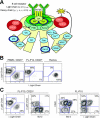
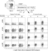
The B-cell receptor (BCR) transmits life and death signals throughout B-cell development, and altered BCR signaling may be required for survival of B-lymphoma cells. We used single-cell signaling profiles to compare follicular lymphoma (FL) B cells and nonmalignant host B cells within individual patient biopsies and identified BCR-mediated signaling events specific to lymphoma B cells. Expression of CD20, Bcl-2, and BCR light chain isotype (kappa or lambda) distinguished FL tumor B-cell and nontumor host B-cell subsets within FL patient biopsies. BCR-mediated signaling via phosphorylation of Btk, Syk, Erk1/2, and p38 occurred more rapidly in tumor B cells from FL samples than in infiltrating nontumor B cells, achieved greater levels of per-cell signaling, and sustained this level of signaling for hours longer than nontumor B cells. The timing and magnitude of BCR-mediated signaling in nontumor B cells within an FL sample instead resembled that observed in mature B cells from the peripheral blood of healthy subjects. BCR signaling pathways that are potentiated specifically in lymphoma cells should provide new targets for therapeutic attention.
Figures


Similar articles
-
B-cell signaling networks reveal a negative prognostic human lymphoma cell subset that emerges during tumor progression.Proc Natl Acad Sci U S A. 2010 Jul 20;107(29):12747-54. doi: 10.1073/pnas.1002057107. Epub 2010 Jun 11. Proc Natl Acad Sci U S A. 2010. PMID: 20543139 Free PMC article.
-
Mass Cytometry of Follicular Lymphoma Tumors Reveals Intrinsic Heterogeneity in Proteins Including HLA-DR and a Deficit in Nonmalignant Plasmablast and Germinal Center B-Cell Populations.Cytometry B Clin Cytom. 2017 Jan;92(1):79-87. doi: 10.1002/cyto.b.21498. Cytometry B Clin Cytom. 2017. PMID: 27933753 Free PMC article.
-
DC-SIGN-expressing macrophages trigger activation of mannosylated IgM B-cell receptor in follicular lymphoma.Blood. 2015 Oct 15;126(16):1911-20. doi: 10.1182/blood-2015-04-640912. Epub 2015 Aug 13. Blood. 2015. PMID: 26272216 Free PMC article.
-
Follicular lymphoma (FL): Immunological tolerance theory in FL.Hum Immunol. 2017 Feb;78(2):138-145. doi: 10.1016/j.humimm.2016.09.010. Epub 2016 Sep 30. Hum Immunol. 2017. PMID: 27693433 Review.
-
New concepts in follicular lymphoma biology: From BCL2 to epigenetic regulators and non-coding RNAs.Semin Oncol. 2018 Oct;45(5-6):291-302. doi: 10.1053/j.seminoncol.2018.07.005. Epub 2018 Oct 23. Semin Oncol. 2018. PMID: 30360879 Review.
Cited by
-
B cell activator PAX5 promotes lymphomagenesis through stimulation of B cell receptor signaling.J Clin Invest. 2007 Sep;117(9):2602-10. doi: 10.1172/JCI30842. J Clin Invest. 2007. PMID: 17717600 Free PMC article.
-
The Bruton tyrosine kinase inhibitor PCI-32765 ameliorates autoimmune arthritis by inhibition of multiple effector cells.Arthritis Res Ther. 2011 Jul 13;13(4):R115. doi: 10.1186/ar3400. Arthritis Res Ther. 2011. PMID: 21752263 Free PMC article.
-
A novel method for detection of phosphorylation in single cells by surface enhanced Raman scattering (SERS) using composite organic-inorganic nanoparticles (COINs).PLoS One. 2009;4(4):e5206. doi: 10.1371/journal.pone.0005206. Epub 2009 Apr 15. PLoS One. 2009. PMID: 19367337 Free PMC article.
-
Single Cell Profiling Distinguishes Leukemia-Selective Chemotypes.bioRxiv [Preprint]. 2024 May 3:2024.05.01.591362. doi: 10.1101/2024.05.01.591362. bioRxiv. 2024. PMID: 38826485 Free PMC article. Preprint.
-
Low level exposure to inorganic mercury interferes with B cell receptor signaling in transitional type 1 B cells.Toxicol Appl Pharmacol. 2017 Sep 1;330:22-29. doi: 10.1016/j.taap.2017.06.022. Epub 2017 Jun 28. Toxicol Appl Pharmacol. 2017. PMID: 28668464 Free PMC article.
References
-
- Fleming HE, Paige CJ. Pre-B cell receptor signaling mediates selective response to IL-7 at the pro-B to pre-B cell transition via an ERK/MAP kinase-dependent pathway. Immunity. 2001;15: 521-531. - PubMed
-
- Reth M, Wienands J. Initiation and processing of signals from the B cell antigen receptor. Annu Rev Immunol. 1997;15: 453-479. - PubMed
-
- Allman D, Srivastava B, Lindsley RC. Alternative routes to maturity: branch points and pathways for generating follicular and marginal zone B cells. Immunol Rev. 2004;197: 147-160. - PubMed
-
- Hartley SB, Cooke MP, Fulcher DA, et al. Elimination of self-reactive B lymphocytes proceeds in two stages: arrested development and cell death. Cell. 1993;72: 325-335. - PubMed
-
- Casellas R, Shih TA, Kleinewietfeld M, et al. Contribution of receptor editing to the antibody repertoire. Science. 2001;291: 1541-1544. - PubMed
Publication types
MeSH terms
Substances
Grants and funding
LinkOut - more resources
Full Text Sources
Other Literature Sources
Miscellaneous

