. 2006 Jul;3(7):e270.
doi: 10.1371/journal.pmed.0030270.

MPLW515L is a novel somatic activating mutation in myelofibrosis with myeloid metaplasia

Affiliations

MPLW515L is a novel somatic activating mutation in myelofibrosis with myeloid metaplasia

Yana Pikman et al. PLoS Med. 2006 Jul.

Abstract

Background: The JAK2V617F allele has recently been identified in patients with polycythemia vera (PV), essential thrombocytosis (ET), and myelofibrosis with myeloid metaplasia (MF). Subsequent analysis has shown that constitutive activation of the JAK-STAT signal transduction pathway is an important pathogenetic event in these patients, and that enzymatic inhibition of JAK2V617F may be of therapeutic benefit in this context. However, a significant proportion of patients with ET or MF are JAK2V617F-negative. We hypothesized that activation of the JAK-STAT pathway might also occur as a consequence of activating mutations in certain hematopoietic-specific cytokine receptors, including the erythropoietin receptor (EPOR), the thrombopoietin receptor (MPL), or the granulocyte-colony stimulating factor receptor (GCSFR).

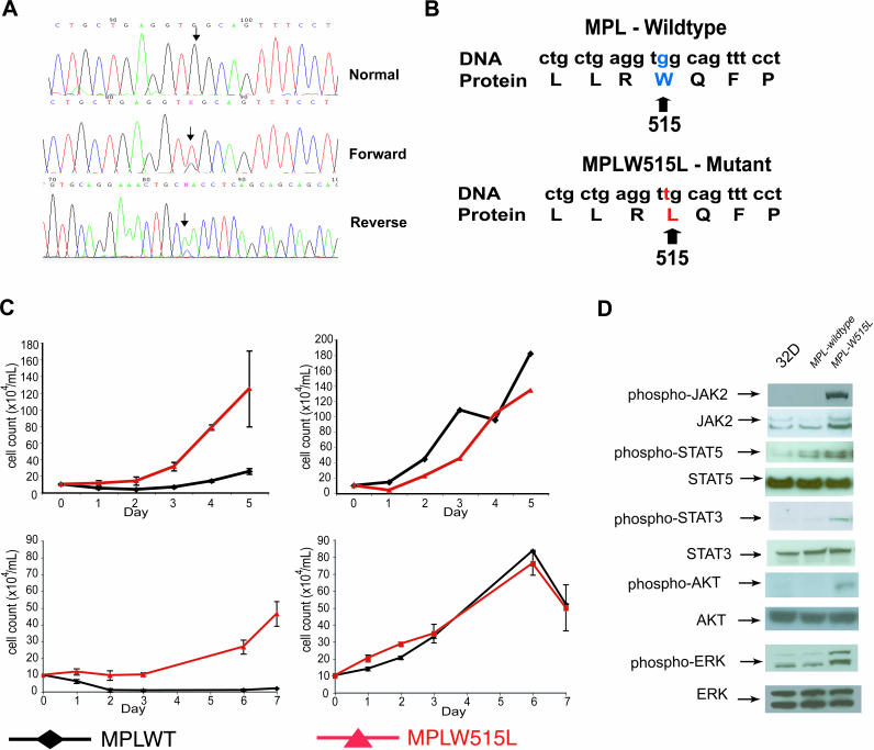
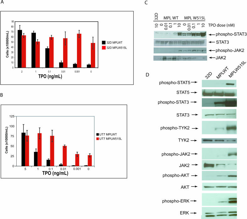
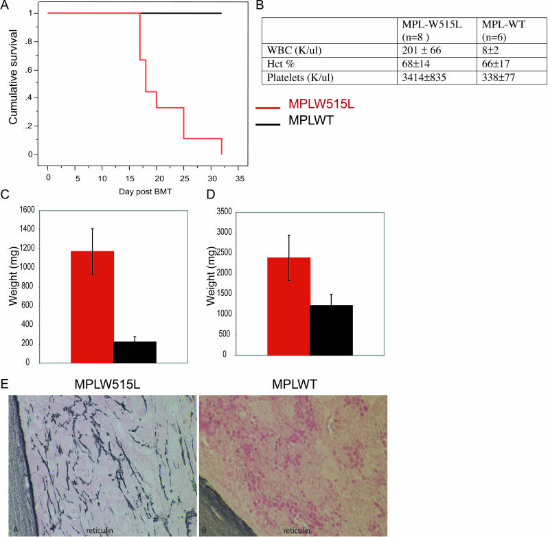
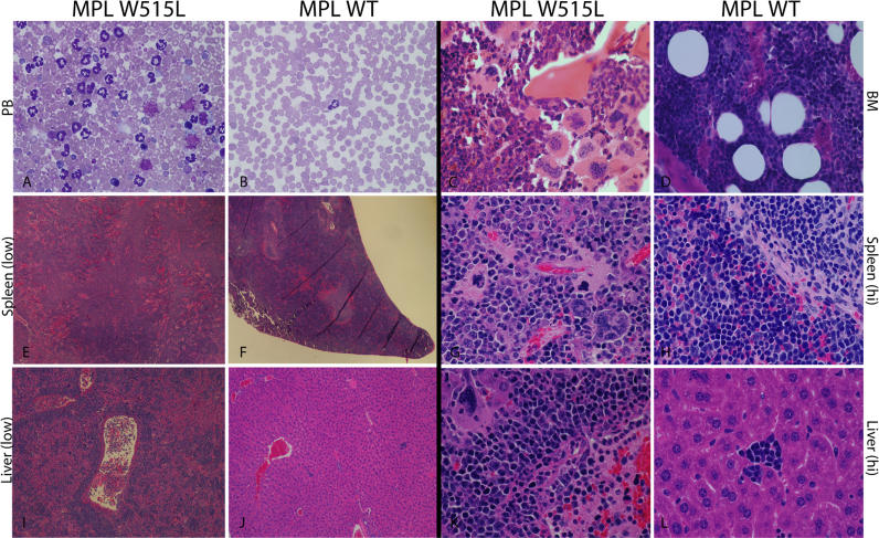
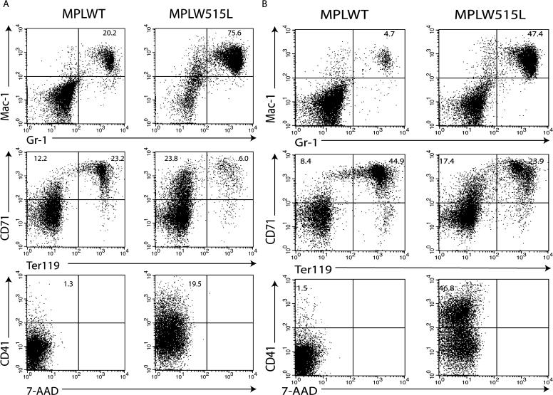
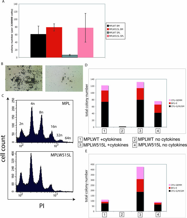
Methods and findings: DNA sequence analysis of the exons encoding the transmembrane and juxtamembrane domains of EPOR, MPL, and GCSFR, and comparison with germline DNA derived from buccal swabs, identified a somatic activating mutation in the transmembrane domain of MPL (W515L) in 9% (4/45) of JAKV617F-negative MF. Expression of MPLW515L in 32D, UT7, or Ba/F3 cells conferred cytokine-independent growth and thrombopoietin hypersensitivity, and resulted in constitutive phosphorylation of JAK2, STAT3, STAT5, AKT, and ERK. Furthermore, a small molecule JAK kinase inhibitor inhibited MPLW515L-mediated proliferation and JAK-STAT signaling in vitro. In a murine bone marrow transplant assay, expression of MPLW515L, but not wild-type MPL, resulted in a fully penetrant myeloproliferative disorder characterized by marked thrombocytosis (Plt count 1.9-4.0 x 10(12)/L), marked splenomegaly due to extramedullary hematopoiesis, and increased reticulin fibrosis.

Conclusions: Activation of JAK-STAT signaling via MPLW515L is an important pathogenetic event in patients with JAK2V617F-negative MF. The bone marrow transplant model of MPLW515L-mediated myeloproliferative disorders (MPD) exhibits certain features of human MF, including extramedullary hematopoiesis, splenomegaly, and megakaryocytic proliferation. Further analysis of positive and negative regulators of the JAK-STAT pathway is warranted in JAK2V617F-negative MPD.


Conflict of interest statement

Competing Interests: The authors have declared that no competing interests exist.

Figures

Figure 1
Figure 1. MPLW515L Mutation Is Found in JAK2V617F-Negative MF and Causes Cytokine-Independent Growth in 32D and UT7 Cells, and Constitutively Activates the JAK-STAT Signaling Pathway
(A) Forward (middle trace) and reverse (lower trace) sequence traces demonstrating a heterozygous guanine to thymine substitution (arrows) present in granulocyte DNA from a patient with MF. The mutation is not present in buccal DNA from the same patient (upper trace). (B) DNA sequence and protein translation for both the wild-type and mutant MPL alleles. The mutation results in a tryptophan-to-leucine substitution at codon 515. (C) Upper: 32D cells transduced with MPLW515L exhibit cytokine-independent growth compared with MPLWT (left). Cell lines grown in the presence of IL3 show equal rates of growth (right). Error bars denote the standard deviation for each sample measured in triplicate. Lower: UT7 cells transformed with MPLW515L exhibit cytokine-independent growth compared with MPLWT (left). Cell lines grown in the presence of TPO (5 ng/mL) show equal rates of growth (right). Error bars denote the standard deviation for each sample measured in triplicate. (D) 32D cells, 32D MPLWT cells, and 32D MPLW515L cells were deprived of cytokines and then analyzed by Western blots, demonstrating phosphorylation of JAK2, STAT5, STAT3, AKT, and ERK in MPLW515L compared with MPLWT.
Figure 2
Figure 2. MPLW515L-Expressing Cells Show Hyper-Responsiveness to TPO
(A) 32D MPLWT cells and 32D MPLW515L cells were cultured in triplicate at an initial concentration of 1 × 10 5 cells/mL, in RPMI/10% FBS containing TPO 2 ng/mL, 1 ng/mL, 0.1 ng/mL, 0.01 ng/mL, 0.001 ng/mL, or in the absence of TPO for four days, and then cell numbers were assessed. Error bars denote the standard deviation for each sample measured in triplicate. (B) UT7 MPLWT cells and UT7 MPLW515L cells were cultured in triplicate, at an initial concentration of 1 × 10 5 cells/mL, in IMDM/10% FBS containing TPO 5 ng/mL, 1 ng/mL, 0.1 ng/mL, 0.01 ng/mL, 0.001ng/mL, or in the absence of TPO for six days, and then cell numbers were assessed. Error bars denote the standard deviation for each sample measured in triplicate. (C) 32D MPLWT or 32D MPLW515L cells were deprived of cytokine overnight and then stimulated with TPO 5 ng/mL, 1 ng/mL, 0.1 ng/mL, 0.01 ng/mL, or without TPO for seven minutes and then analyzed by Western blot for phosphorylation of JAK2 and STAT3, demonstrating increased JAK2/STAT3 phosphorylation in response to TPO in 32D MPLW515L cells as compared with 32D-MPLWT cells. (D) Phosphorylation of JAK2, TYK2, STAT3, STAT5, AKT, and ERK in response to seven-minute stimulation with TPO (50ng/mL). 32D MPLWT or 32 MPLW515L cells were deprived of cytokine overnight and then stimulated with TPO 50 ng/mL for seven minutes and then analyzed by Western blot for phosphorylation of JAK2, TYK2, STAT3, STAT5, AKT, and ERK, demonstrating increased phosphorylation of these proteins in response to TPO in 32D MPLW515L cells as compared with 32D MPLWT cells.
Figure 3
Figure 3. Bone Marrow Transplant with MPLW515L-Transduced Bone Marrow Causes a Rapid Myeloproliferative Disease
(A) Kaplan-Meier survival plot of Balb/C mice transduced with MPLW515L ( n = 9) and MPLWT ( n = 6) showing death of all MPLW515L mice between 17 and 32 days post-BMT compared with MPLWT ( n = 6), which were sacrificed for endpoint analysis without evidence of disease. (B) Complete blood counts show leukocytosis and thrombocytosis in MPLW515L BMT model compared with MPLWT. There is no difference in hematocrits. Standard deviation is indicated. (C) Spleen weights of MPLW515L and MPLWT mice shows splenomegaly in MPLW515L mice but not in MPLWT mice with average spleen weight equal to 1,171 mg. (D) Liver weights of MPLW515L and MPLWT mice shows hepatomegaly in MPLW515L mice but not in MPLWT mice, with average liver weight equal to 2,390 mg (compared with 1,222 mg in MPLWT mice). (E) Bone marrow shows significantly increased bone marrow fibrosis by reticulin staining at 17 days post-BMT in MPLW515L mice, but not in MPLWT-expressing mice.
Figure 4
Figure 4. MPLW515L and MPLWT Bone Marrow Transplant Model Histopathology
Histology of MPLW515L-transduced and MPLWT-transduced Balb/C mice showing images of peripheral blood (A and B) and histopathology in representative sections of bone marrow (C and D), spleen (E–H), and liver (I–L). Peripheral blood smear (B) (600×, Wright-Giemsa) of a representative MPLWT animal displays an unremarkable white blood cell and platelet count. In contrast, peripheral blood smear (A) (600×, Wright-Giemsa) of a representative MPLW515L mutant animal reveals marked thrombocytosis and leukocytosis comprising a predominant population of maturing myeloid cells as well as frequent nucleated erythroid forms. Bone marrow images from MPLWT animals display preserved marrow architecture with maturing trilineage hematopoiesis (D) (600×, hematoxylin and eosin [H&E]). Comparatively, bone marrow sections from MPLW515L mutant animals demonstrate marrow elements comprising a prominent population of maturing myeloid cells with increased numbers of megakaryocytes including atypical and dysplastic forms occurring in frequent clusters (C) (600×, H&E) and showing emperipolesis of neutrophils in megakaryocyte cytoplasm. Spleen sections from MPLW515L mice display complete effacement of normal splenic architecture (E) (40×, H&E) with a marked expansion of red pulp that is composed of an admixture of maturing myeloid and erythroid elements and numerous numbers of atypical megakaryocytes (G) (600×, H&E) compared with MPLWT spleens (F and H) (40× and 600×, H&E), which display a relative preservation of normal spleen architecture and the presence of only maturing erythroid forms in the red pulp. Liver images from MPLW515L mice illustrate evidence of extensive extramedullary hematopoiesis in a perivascular and sinusoidal distribution (I) (100×, H&E) composed predominantly of a population of maturing erythroid elements with frequent large atypical megakaryocytes and occasional admixed myeloid forms (K) (600×, H&E). In comparison, only small, focal areas of nucleated erythroid cells were observed in livers from MPLWT animals (J and L) (100× and 600×, H&E).
Figure 5
Figure 5. Flow Cytometry Analysis of BM and Spleen in Mice Transduced with MPLW515L and MPLWT
(A) Flow-cytometry analysis of bone marrow cells shows a 4-fold increase in Mac1+/Gr1+ cells and a shift to a more immature erythroid population. There is a 15-fold increase in CD41+ cells in bone marrow expressing MPLW515L compared with MPLWT. (B) Flow-cytometry analysis of spleen cells shows a 10-fold increase in Mac1+/Gr1+ cells in MPLW515L. There is also a shift to a more immature erythroid population in MPLW515L with a greater percentage of CD71+/Ter119- cells. CD41+ cells are increased 30-fold in MPLW515L spleen cells compared with MPLWT spleen cells.
Figure 6
Figure 6. MPLW515L Increases the Number of Megakaryocyte and Myeloid Colonies in Spleen, without Affecting Megakaryocyte Ploidy, and Causes Cytokine-Independent Myeloid Colony Growth
(A) Megakaryocyte colony–forming assay in the presence of TPO, IL3, IL11, and IL6 demonstrates similar numbers of megakaryocyte colonies obtained from MPLW515L-expressing bone marrow and an increase in the number of megakaryocyte colonies from spleen cells compared with MPLWT. (B) Acetylcholinesterase staining of megakaryocyte colonies derived from bone marrow demonstrates much larger colony size in MPLW515L as compared with MPLWT megakaryocyte colonies. (C) Megakaryocyte ploidy analysis shows the same distribution for MPLW515L-expressing cells and MPLWT-expressing cells. (D and E) Total myeloid colony formation from bone marrow cells (D) and spleen cells (E) demonstrates cytokine-independent colony formation in MPLW515L bone marrow and spleen. There is no difference in colony distribution between MPLWT- and MPLW515L-expressing cells. Colony counts reflect only positively identifiable colonies, with thorough megakaryocyte colony analysis done in MegaCult assay ( Figure 6A), and thus are excluded from methylcellulose colony analysis. Colony numbers represent a total of three representative mice per group, in duplicate.
Figure 7
Figure 7. 32D MPLW515L Cells Are Sensitive to JAK Inhibitor I
(A) Dose-dependent inhibition of growth of 32D MPLW515L and 32D MPLWT cells but not 32D FIP1L1-PDGFRA cells with increasing doses of JAK Inhibitor I. (B) Reduction in STAT3 phosphorylation in 32D MPLW515L cells but not 32D FIP1L1-PDGFRA cells with increasing doses of JAK Inhibitor I. Cells were incubated with varying drug concentrations for four hours and then collected for Western blot analysis.

Similar articles

Cited by

References

    1. Dameshek W. Some speculations on the myeloproliferative syndromes. Blood. 1951;6:372–375. - PubMed
    1. Levine RL, Wadleigh M, Cools J, Ebert BL, Wernig G, et al. Activating mutation in the tyrosine kinase JAK2 in polycythemia vera, essential thrombocythemia, and myeloid metaplasia with myelofibrosis. Cancer Cell. 2005;7:387–397. - PubMed
    1. Jones AV, Kreil S, Zoi K, Waghorn K, Curtis C, et al. Widespread occurrence of the JAK2 V617F mutation in chronic myeloproliferative disorders. Blood. 2005;106:2162–2168. - PubMed
    1. James C, Ugo V, Le Couedic JP, StaERK J, Delhommeau F, et al. A unique clonal JAK2 mutation leading to constitutive signalling causes polycythaemia vera. Nature. 2005;434:1144–1148. - PubMed
    1. Baxter EJ, Scott LM, Campbell PJ, East C, Fourouclas N, et al. Acquired mutation of the tyrosine kinase JAK2 in human myeloproliferative disorders. Lancet. 2005;365:1054–1061. - PubMed

Publication types

MeSH terms

Substances