. 2006 Jul 21;313(5785):324-8.
doi: 10.1126/science.1129462. Epub 2006 Jun 22.

Alpha-synuclein blocks ER-Golgi traffic and Rab1 rescues neuron loss in Parkinson's models

Affiliations

Alpha-synuclein blocks ER-Golgi traffic and Rab1 rescues neuron loss in Parkinson's models

Antony A Cooper et al. Science. .

Abstract

Alpha-synuclein (alphaSyn) misfolding is associated with several devastating neurodegenerative disorders, including Parkinson's disease (PD). In yeast cells and in neurons alphaSyn accumulation is cytotoxic, but little is known about its normal function or pathobiology. The earliest defect following alphaSyn expression in yeast was a block in endoplasmic reticulum (ER)-to-Golgi vesicular trafficking. In a genomewide screen, the largest class of toxicity modifiers were proteins functioning at this same step, including the Rab guanosine triphosphatase Ypt1p, which associated with cytoplasmic alphaSyn inclusions. Elevated expression of Rab1, the mammalian YPT1 homolog, protected against alphaSyn-induced dopaminergic neuron loss in animal models of PD. Thus, synucleinopathies may result from disruptions in basic cellular functions that interface with the unique biology of particular neurons to make them especially vulnerable.


Figures
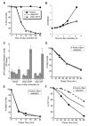
Fig. 1
Fig. 1
Expression of αSyn causes cell death and ER stress and impairs ERAD. (A) Survivorship curve during αSyn induction. After induction of αSyn-WT, αSyn-A53T expression, or control cells (Vector), cells with an optical density at 600 nm (OD600nm) of 1 were harvested and treated as described (24). Colony-forming units were determined and converted to relative percentages. (B) Growth curve during αSyn induction. After induction, the OD600nm for each sample was measured at the indicated times. (C) Cells induced for expression of αSyn-WT, αSyn-A53T, or control cells (Vector) were harvested at the times indicated; the level of UPR activation was then determined and plotted as relative units of ER stress. The degradation rate of Deg1-βGal (D), Sec61-2p (E), and CPY* (F), after 6 hours of either αSyn-WT or αSyn-A53T expression, was determined by pulse-chase immunoprecipitation as described (24) and compared to that of control cells (Vector).
Fig. 2
Fig. 2
αSyn accumulation causes a severe block in vesicular trafficking in the early secretory pathway. The trafficking of CPY (A and B) and ALP (C and D) was monitored in cells expressing αSyn-WT or αSyn-A53T at the times indicated by pulse-chase immunoprecipitation and compared to that of control cells (Vector). (B) Graphic representation of the amount of CPY remaining in the ER [p1/(p1 + p2 + mCPY)]. (D) Graphic representation of the amount of ALP remaining in the ER [p1/(p1 + p2 + mALP)]. For CPY, p1 is the ER form, p2 is the Golgi form, and m is the mature vacuolar form. For ALP, p is the ER form and m is the mature vacuolar form.
Fig. 3
Fig. 3
Plasmid overexpression screen identifies ER-Golgi trafficking genes as modifiers of αSyn toxicity. (A) Representative plates from αSyn modifier screen (24). αSyn-expressing cells were transformed individually with each of 3000 random ORFs under the control of a galactose-inducible promoter. Transformants were grown on synthetic media containing either glucose (control, αSyn ‘‘off’’) or galactose (to induce expression of αSyn and candidate ORFs, αSyn ‘‘on’’). Examples of strong- and moderate-toxicity suppressors are shown as black and blue circles, respectively. Examples of enhancers of αSyn-induced toxicity are shown as red circles, and a false-positive that did not reproduce upon further analysis is shown as a yellow circle. (B) Spotting assay shows that overexpression of ER-Golgi trafficking genes YPT1, YKT6, BRE5, UBP3, and ERV29 suppress αSyn-induced toxicity, whereas GYP8 and PMR1 overexpression enhances toxicity. (C) Suppression of toxicity is specific to the transport step facilitated by YPT1, because overexpression of other Rab GTPases has no effect on growth.
Fig. 4
Fig. 4
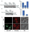
αSyn-induced cyto-toxicity and vesicular trafficking defects are modified by ER-Golgi trafficking components. The trafficking of CPY in cells expressing αSyn-WTand harboring either galactose-inducible GYP8 (A and B), galactose-inducible YPT1 (A and B), or SLY1-20 (C and D) was monitored by radio-labeling after 7 hours (C and D) or 8 hours (A and B) of induction and compared to trafficking in control cells (Vector). (B and D) Graphic representation of the amount of CPY remaining in the ER [p1/(p1 + p2 + mCPY)]. (E) Cells expressing αSyn-WT–GFP (green fluorescence protein) and HA (hemagglutin)–Ypt1p were examined by fluorescence and indirect immunofluorescence microscopy after 6 hours of αSyn induction.
Fig. 5
Fig. 5
Expression of Rab1 rescues DA neuron loss in animal models of PD. (A to C) Studies in Drosophila. (A and B) Aged fly brains immuno-stained for TH to highlight DA neurons; selective loss of TH immunostaining in the DM1 cluster is fully prevented by Rab1. Genotypes TH-GAL4 UAS-αSyn in trans to + or UAS-Rab1. (C) Quantitation of TH-positive neurons in DM1 cluster. Wild-type (αSyn) or mutant (A53T) αSyn causes loss of TH that is prevented by Rab1. Genotypes: TH-GAL4/+ (wildtype), TH-GAL4 UAS-αSyn or TH-GAL4 UAS-A53T in trans to + or UAS-Rab1 (two independent lines). (*) Values significantly different from αSyn or A53T, P < 0.00001, Student’s t test. (D) DA toxicity in C. elegans, reflected by degeneration of cephalic sensilla (CEP) neuronal processes in Pdat-1::GFP + Pdat-1::αSyn at the 7-day stage. The arrow highlights the single CEP process remaining. (E) Intact DA neurons in Pdat-1::GFP + Pdat-1::α Syn + Pdat-1::Rab1 at the 7-day stage. (F) Quantitation of Rab1 rescue of αSyn-induced neuron loss in three independent transgenic lines. (**) Values significantly different from αSyn, P < 0.05, Student’s t test. Scale bars: 20 μm. (G) Studies in rat midbrain primary neurons. Rab1 protects primary DA neurons from toxicity induced by αSyn-A53T. Primary midbrain cultures were transduced with lentivirus encoding αSyn-A53T alone, or αSyn-A53T plus β-Gal (LacZ), or αSyn-A53T plus Rab1. Control cells were cultured in the absence of lentivirus. Selective DA cell death was evaluated immunocytochemically. Viability is expressed as the percentage of MAP2-positive neurons that were also TH-positive (three independent experiments, at least 100 cells counted per experiment for each condition). The data are presented as the mean ± SD, n = 3 experiments; ***P < 0.01, analysis of variance with Newman-Keuls post-test.

Similar articles

Cited by

  • The unfolded protein response in neurodegenerative diseases: a neuropathological perspective.
    Scheper W, Hoozemans JJ. Scheper W, et al. Acta Neuropathol. 2015 Sep;130(3):315-31. doi: 10.1007/s00401-015-1462-8. Epub 2015 Jul 26. Acta Neuropathol. 2015. PMID: 26210990 Free PMC article. Review.
  • Golgi fragmentation is Rab and SNARE dependent in cellular models of Parkinson's disease.
    Rendón WO, Martínez-Alonso E, Tomás M, Martínez-Martínez N, Martínez-Menárguez JA. Rendón WO, et al. Histochem Cell Biol. 2013 May;139(5):671-84. doi: 10.1007/s00418-012-1059-4. Epub 2012 Dec 2. Histochem Cell Biol. 2013. PMID: 23212845
  • Advances in the genetics of Parkinson disease.
    Trinh J, Farrer M. Trinh J, et al. Nat Rev Neurol. 2013 Aug;9(8):445-54. doi: 10.1038/nrneurol.2013.132. Epub 2013 Jul 16. Nat Rev Neurol. 2013. PMID: 23857047 Review.
  • Strong association between glucocerebrosidase mutations and Parkinson's disease in Sweden.
    Ran C, Brodin L, Forsgren L, Westerlund M, Ramezani M, Gellhaar S, Xiang F, Fardell C, Nissbrandt H, Söderkvist P, Puschmann A, Ygland E, Olson L, Willows T, Johansson A, Sydow O, Wirdefeldt K, Galter D, Svenningsson P, Belin AC. Ran C, et al. Neurobiol Aging. 2016 Sep;45:212.e5-212.e11. doi: 10.1016/j.neurobiolaging.2016.04.022. Epub 2016 May 3. Neurobiol Aging. 2016. PMID: 27255555 Free PMC article.
  • Robust Sequence Determinants of α-Synuclein Toxicity in Yeast Implicate Membrane Binding.
    Newberry RW, Arhar T, Costello J, Hartoularos GC, Maxwell AM, Naing ZZC, Pittman M, Reddy NR, Schwarz DMC, Wassarman DR, Wu TS, Barrero D, Caggiano C, Catching A, Cavazos TB, Estes LS, Faust B, Fink EA, Goldman MA, Gomez YK, Gordon MG, Gunsalus LM, Hoppe N, Jaime-Garza M, Johnson MC, Jones MG, Kung AF, Lopez KE, Lumpe J, Martyn C, McCarthy EE, Miller-Vedam LE, Navarro EJ, Palar A, Pellegrino J, Saylor W, Stephens CA, Strickland J, Torosyan H, Wankowicz SA, Wong DR, Wong G, Redding S, Chow ED, DeGrado WF, Kampmann M. Newberry RW, et al. ACS Chem Biol. 2020 Aug 21;15(8):2137-2153. doi: 10.1021/acschembio.0c00339. Epub 2020 Aug 12. ACS Chem Biol. 2020. PMID: 32786289 Free PMC article.

References

    1. Vila M, Przedborski S. Nat Med. 2004;10(Suppl):S58. - PubMed
    1. Forman MS, Trojanowski JQ, Lee VM. Nat Med. 2004;10:1055. - PubMed
    1. Spillantini MG, Crowther RA, Jakes R, Hasegawa M, Goedert M. Proc Natl Acad Sci USA. 1998;95:6469. - PMC - PubMed
    1. Polymeropoulos MH, et al. Science. 1997;276:2045. - PubMed
    1. Kruger R, et al. Nat Genet. 1998;18:106. - PubMed

Publication types

MeSH terms