

Diffusion tensor imaging in amyotrophic lateral sclerosis: volumetric analysis of the corticospinal tract
- PMID: 16775271
- PMCID: PMC8133954
Diffusion tensor imaging in amyotrophic lateral sclerosis: volumetric analysis of the corticospinal tract
Abstract
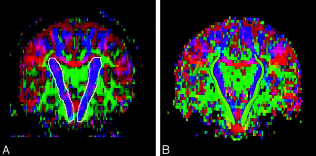
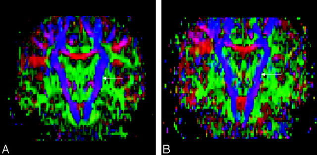
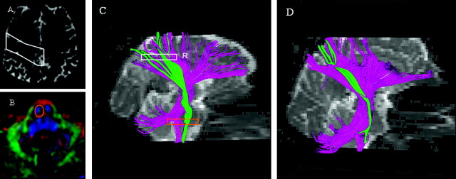
Background and purpose: Diffusion tensor imaging (DTI) allows direct visualization and volumetric analysis of the corticospinal tract (CST). The purpose of this study was to determine whether color maps and fiber tracking derived from DTI data are valuable in detecting and quantifying CST degeneration in patients with amyotrophic lateral sclerosis (ALS).
Methods: Sixteen patients with ALS with clinical signs of upper motor neuron (UMN) involvement and 17 healthy subjects were studied with the use of DTI. Disease severity was determined by means of the ALS Functional Rating Scale-Revised (ALSFRS-R) and an UMN involvement score. DTI was acquired with a 12-direction, single-shot, spin-echo echo-planar sequence. The CST from the lower pons to the corona radiata at the level of the corpus callosum on 4 contiguous coronal sections was manually segmented by using color maps generated from the DTI data. The left and right CST volumes were measured separately and normalized to the total intracranial volume. Normalized CST volumes were compared between patients with ALS and healthy subjects.
Results: The CST volumes of patients with ALS were significantly reduced (P < .01, unpaired t test) compared with healthy subjects, in both affected and nonaffected hemispheres. No significant correlation was found between CST volumes and any of the clinical parameters, including disease duration, ALSFRS-R, or UMN involvement score.
Conclusion: This study shows that volumetric analysis by using DTI-based color maps is valuable in detecting and monitoring structural degeneration of the CST. This will lead to objective and quantitative assessment of axonal degeneration in ALS.
Figures
Similar articles
-
Differential involvement of corticospinal tract (CST) fibers in UMN-predominant ALS patients with or without CST hyperintensity: A diffusion tensor tractography study.Neuroimage Clin. 2017 Feb 22;14:574-579. doi: 10.1016/j.nicl.2017.02.017. eCollection 2017. Neuroimage Clin. 2017. PMID: 28337412 Free PMC article.
-
Diffusion tensor imaging-based fractional anisotropy quantification in the corticospinal tract of patients with amyotrophic lateral sclerosis using a probabilistic mixture model.AJNR Am J Neuroradiol. 2007 Apr;28(4):724-30. AJNR Am J Neuroradiol. 2007. PMID: 17416829 Free PMC article.
-
A prospective harmonized multicenter DTI study of cerebral white matter degeneration in ALS.Neurology. 2020 Aug 25;95(8):e943-e952. doi: 10.1212/WNL.0000000000010235. Epub 2020 Jul 9. Neurology. 2020. PMID: 32646955 Free PMC article.
-
[Objective markers for upper motor neuron involvement in amyotrophic lateral sclerosis].Brain Nerve. 2007 Oct;59(10):1053-64. Brain Nerve. 2007. PMID: 17969345 Review. Japanese.
-
Amyotrophic lateral sclerosis and primary lateral sclerosis: The role of diffusion tensor imaging and other advanced MR-based techniques as objective upper motor neuron markers.Ann N Y Acad Sci. 2005 Dec;1064:61-77. doi: 10.1196/annals.1340.013. Ann N Y Acad Sci. 2005. PMID: 16394148 Review.
Cited by
-
Reliability of fiber tracking measurements in diffusion tensor imaging for longitudinal study.Neuroimage. 2010 Jan 15;49(2):1572-80. doi: 10.1016/j.neuroimage.2009.08.062. Epub 2009 Sep 8. Neuroimage. 2010. PMID: 19744567 Free PMC article.
-
Phosphorylated tau as a candidate biomarker for amyotrophic lateral sclerosis.JAMA Neurol. 2014 Apr;71(4):442-8. doi: 10.1001/jamaneurol.2013.6064. JAMA Neurol. 2014. PMID: 24492862 Free PMC article.
-
Quantitative diffusion tensor imaging in amyotrophic lateral sclerosis: revisited.Hum Brain Mapp. 2009 Nov;30(11):3657-75. doi: 10.1002/hbm.20794. Hum Brain Mapp. 2009. PMID: 19404990 Free PMC article.
-
Symptoms of degeneration of the pyramidal tracts in conventional magnetic resonance imaging and diffusion tensor imaging in a young woman with primary lateral sclerosis.J Postgrad Med. 2015 Jul-Sep;61(3):206-8. doi: 10.4103/0022-3859.150901. J Postgrad Med. 2015. PMID: 26119443 Free PMC article.
-
Diffusion tensor imaging-based research on human white matter anatomy.ScientificWorldJournal. 2012;2012:530432. doi: 10.1100/2012/530432. Epub 2012 Nov 25. ScientificWorldJournal. 2012. PMID: 23226983 Free PMC article.
References
-
- Rowland LP. Diagnosis of amyotrophic lateral sclerosis. J Neurol Sci 1998;160 Suppl 1:S6–24 - PubMed
-
- Chenevert TL, Brunberg JA, Pipe JG. Anisotropic diffusion in human white matter: demonstration with MR techniques in vivo. Radiology 1990;177:401–05 - PubMed
-
- Albayram S, Melhem ER, Mori S, et al. Holoprosencephaly in children: diffusion tensor MR imaging of white matter tracts of the brainstem–initial experience. Radiology 2002;223:645–51 - PubMed
-
- Melhem ER, Mori S, Mukundan G, et al. Diffusion tensor MR imaging of the brain and white matter tractography. AJR Am J Roentgenol 2002;178:3–16 - PubMed
MeSH terms
LinkOut - more resources
Full Text Sources
Medical
Miscellaneous