. 2006 Jul;116(7):1853-64.
doi: 10.1172/JCI27438. Epub 2006 Jun 8.

CaM kinase II selectively signals to histone deacetylase 4 during cardiomyocyte hypertrophy

Affiliations

CaM kinase II selectively signals to histone deacetylase 4 during cardiomyocyte hypertrophy

Johannes Backs et al. J Clin Invest. 2006 Jul.

Abstract

Class IIa histone deacetylases (HDACs) regulate a variety of cellular processes, including cardiac growth, bone development, and specification of skeletal muscle fiber type. Multiple serine/threonine kinases control the subcellular localization of these HDACs by phosphorylation of common serine residues, but whether certain class IIa HDACs respond selectively to specific kinases has not been determined. Here we show that calcium/calmodulin-dependent kinase II (CaMKII) signals specifically to HDAC4 by binding to a unique docking site that is absent in other class IIa HDACs. Phosphorylation of HDAC4 by CaMKII promotes nuclear export and prevents nuclear import of HDAC4, with consequent derepression of HDAC target genes. In cardiomyocytes, CaMKII phosphorylation of HDAC4 results in hypertrophic growth, which can be blocked by a signal-resistant HDAC4 mutant. These findings reveal a central role for HDAC4 in CaMKII signaling pathways and have implications for the control of gene expression by calcium signaling in a variety of cell types.


Figures

Figure 1
Figure 1. Selective response of HDAC4 to CaMKII.
(A and B) COS cells were transfected with GFP-HDAC4, GFP-HDAC5, FLAG-HDAC7, or GFP-MITR together with either an empty vector (pcDNA), constitutively active (c.a.) CaMKI, PKD1 (c.a.), CaMKIIδB-T287D, CaMKIIδC-T287D, or CaMKIIγA-T287D. CaMKI c.a. induced nuclear export of all HDACs and changed the predominant nuclear localization of MITR from punctate to homogenous. CaMKIIδB-T287D and CaMKIIγA-T287D selectively induced cytosolic accumulation of HDAC4 but did not affect the subcellular distribution of HDAC5, HDAC7, and MITR. (A) Representative images. Magnification, ×40. (B) Quantitative analysis. (C) Coimmunoprecipitation assays with COS cell lysates were analyzed with an antibody directed against endogenous (endog.) 14-3-3 protein. HDAC-input, HDAC4 and -5 present in the COS cell lysate before IP was performed; Kinase-input, CaMKI, PKD1, or CaMKIIδB-T287D present in the COS cell lysate before IP was performed.
Figure 2
Figure 2. Regulation of CaMKII subcellular localization.
(A) COS cells were transfected with CaMKIIδB-WT, CaMKIIδB-T287D, and CaMKIIδB-T287D/S332A. In contrast to the WT kinase, CaMKIIδB-T287D localized predominantly to the cytosol. Substitution of S332 rendered CaMKIIδB-T287D constitutively active and nuclear. (B) COS cells were cotransfected with CaMKIIδB-T287D/S332A and HDAC4 or HDAC5. Note that only HDAC4 was exported in response to the double CaMKIIδB mutant and colocalized with the kinase. (C and D) COS cells were first transfected with the indicated CaMKIIδB mutants and 12 hours later with HDAC4. Twelve hours after transfection with HDAC4, cells were treated for another 4 hours either with 1 nM leptomycin B (lower panel) or the vehicle ethanol (upper panel). Note that with leptomycin B, HDAC4 only accumulates in the nucleus in the presence of CaMKIIδB-T287D/S332A, which is active and nuclear, but not in the presence of CaMKIIδB-T287D or CaMKIIδB-T287D/K328,329N, which are active and cytosolic. (AC) Representative images. Magnification, ×40. (D) Quantitative analysis of experiment shown in C.
Figure 3
Figure 3. Detection of 14-3-3 binding sites of HDAC4 in response to CaMKI and CaMKII.
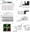
(A and B) Coimmunoprecipitation assays with COS cell lysates were analyzed with an antibody directed against endogenous 14-3-3 protein. The effects of CaMKI and CaMKII on various FLAG-HDAC4 mutants were tested. HDAC4-input, HDAC4 present in the COS cell lysate before IP was performed; CaMK-input, CaMKI or CaMKIIδB-T287D present in the COS cell lysate before IP was performed. (B) Fold-increase in 14-3-3 binding in response to CaMKII or CaMKI as compared with baseline. (C) The N terminal half of HDAC4 (amino acids 1–740) was fused to the GAL4 DNA-binding domain, and 14-3-3 was fused to the VP16 transcription activation domain. If GAL4-HDAC4 is not phosphorylated, it cannot recruit VP16–14-3-3 and cannot activate the GAL4-dependent luciferase reporter. (D) As indicated, different GAL4-HDAC constructs were used in this assay in the absence and presence of CaMKIIδB-T287D. COS cells were transfected with the indicated constructs. Increase in 14-3-3 binding is expressed as compared with control conditions without kinase. (E) CaMKI and CaMKII phosphorylation sites of HDAC4 are shown. ND, not detectable; NES, nuclear export signal.
Figure 4
Figure 4. Mapping the CaMKII-responsive region of HDAC4.
(A) Chimeric HDAC4/5 proteins (as indicated) were expressed in COS cells in the presence of CaMKIIδB-T287D. Subcellular localization was verified by immunocytochemistry. Amino acids 529–657 were revealed to be required for cytosolic accumulation of HDAC4 in response to CaMKIIδB-T287D. (B) An HDAC5 mutant, in which the CaMKII consensus sites were mutated to the corresponding sites in HDAC4, was expressed in COS cells alone or with CaMKI c.a. and CaMKIIδB-T287D. This mutant was responsive to CaMKI but not to CaMKIIδB-T287D. Magnification, ×40. (CF) Coimmunoprecipitation assays with COS cell lysates. (C) COS cells were cotransfected with FLAG-pcDNA, -HDAC4, or -HDAC5 and Myc-CaMKIIδB-T287D. Various stringency conditions of the immunoprecipitation buffer were tested as indicated. Only HDAC4 binds strongly to CaMKII. (D) Cotransfection of FLAG-HDAC4 with either WT or constitutively activated Myc–CaMKIIδB-T287D. Only the activated form of CaMKIIδB physically interacted with HDAC4. (E) IP of FLAG-HDAC4 deletion mutants coexpressed with Myc-CaMKIIδB-T287D to identify a domain of HDAC4 that binds to activated CaMKIIδB. (F) Based on the coimmunoprecipitation data, amino acids 585–608 of HDAC4 were identified to be required for physical interaction with CaMKIIδB-T287D and, therefore, define a CaMKII docking site.
Figure 5
Figure 5. R601 of HDAC4 is required for full responsiveness to CaMKIIδB-T287D.
(A and B) FLAG-HDAC4 mutants carrying point mutations in the CaMKII docking region were coexpressed with Myc-CaMKIIδB-T287D in COS cells. Coimmunoprecipitation revealed that substitution of R601 with either alanine or phenylalanine prevented a physical interaction with CaMKIIδB-T287D. (C and D) Myc-CaMKIIδB-T287D or CaMKI c.a. and either FLAG-HDAC4-WT, -R601A, or -R601F were coexpressed in COS cells, and HDAC4 localization was determined 1 day after transfection. HDAC4-R601A and -R601F were still responsive to CaMKI but not to CaMKIIδB-T287D and did not colocalize with the kinase. (C) Representative images. Magnification, ×40. (D) Quantitative analysis. (E) Mammalian 2-hybrid assay with GAL4-HDAC4 mutants and VP16–14-3-3. Substitution of R601 with phenylalanine and leucine prevented and with alanine and lysine markedly attenuated 14-3-3 binding. (F) In vitro kinase assay. Active His-CaMKIIδ induced phosphorylation of GST-HDAC4-WT (amino acids 419–670) but not of the GST-HDAC4-R601F (419–670) mutant. Ca2+-depletion by EGTA prevented HDAC4 phosphorylation by CaMKII. (G) GST pull-down assay with GST, GST-HDAC4-WT (419–670), GST-HDAC4-R601F (419–670) mutant, and active His-CaMKIIδ. (H) Coimmunoprecipitation revealed that HA-CaMKI docks to FLAG-HDAC4-WT and -R601F to the same degree, however, it docks to HDAC5 with greater affinity.
Figure 6
Figure 6. Cytosolic accumulation of HDAC4 in cardiomyocytes.
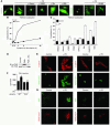
(AC) NRVMs were infected with adenoviral FLAG-HDAC4 or GFP-HDAC5. Subcellular distribution of HDAC4 and HDAC5 was verified following stimulation with PE (20 μM) for 4 hours. NRVMs were pretreated with the kinase inhibitors staurosporine (Stauro; 500 nM), KN93 (5 μM), KN62 (10 μM), AIPII-2 (500 nM), Bis (2.5 μM), Gö6976 (200 nM), or H89 (1 μM). (A) Representative images. (B) Quantitative analysis of time-dependent PE-induced cytosolic accumulation of HDAC4. (C) Effects of kinase inhibitors on PE-induced cytosolic accumulation of HDAC4. (D) NRVMs were treated with PMA with and without Bis. Immunoblotting was performed with antibodies against PKD (lower panel) and phospho-S744/S748 PKD (p-PKD; upper panel). (EG) NRVMs were infected with adenoviruses encoding FLAG-HDAC4-WT or FLAG-HDAC4-S246,467,632A (FLAG-HDAC4-S/A). One day after infection, cells were grown in serum-free media for 24 hours and then stimulated with PE (20 μM). Cells were fixed and stained with anti-sarcomeric α-actinin (red signal; 24 hours after PE) (E) or anti-ANP (perinuclear green signal; 12 hours after PE) (G). HDAC4-infected NRVMs were identified by anti-FLAG staining (green in E or red in G). (F) [3H]-leucine was added to NRVMs 2 hours after PE stimulation and [3H]-leucine incorporation was measured 24 hours later. *P < 0.05 vs. WT without PE and vs. S/A with PE. NS, not significant vs. S/A without PE. (A, E, and G) Representative images were captured at a magnification of ×40.
Figure 7
Figure 7. A model of CaMKII-dependent cytosolic accumulation of HDAC4.
α-Adrenergic stimulation activates CaMKII. Activated CaMKII interacts with HDAC4, which results in phosphorylation of HDAC4 and 14-3-3 protein–mediated nuclear export or a block of nuclear import. Cytosolic accumulation of HDAC4 derepresses MEF2 or other transcription factors, which associate with the nonphosphorylated form of HDAC4 when CaMKII is inactive.

Similar articles

Cited by

References

    1. Roth S.Y., Denu J.M., Allis C.D. Histone acetyltransferases. Annu. Rev. Biochem. 2001;70:81–120. - PubMed
    1. Grozinger C.M., Schreiber S.L. Deacetylase enzymes: biological functions and the use of small-molecule inhibitors. Chem. Biol. 2002;9:3–16. - PubMed
    1. Backs J., Olson E.N. Control of cardiac growth by histone acetylation/deacetylation. Circ. Res. 2006;98:15–24. - PubMed
    1. Verdin E., Dequiedt F., Kasler H.G. Class II histone deacetylases: versatile regulators. Trends Genet. 2003;19:286–293. - PubMed
    1. Grozinger C.M., Schreiber S.L. Regulation of histone deacetylase 4 and 5 and transcriptional activity by 14-3-3-dependent cellular localization. Proc. Natl. Acad. Sci. U. S. A. 2000;97:7835–7840. - PMC - PubMed

Publication types

MeSH terms

Substances