. 2006 Aug 1;296(1):219-30.
doi: 10.1016/j.ydbio.2006.04.459. Epub 2006 May 3.

A new Drosophila gene wh (wuho) with WD40 repeats is essential for spermatogenesis and has maximal expression in hub cells

Affiliations

A new Drosophila gene wh (wuho) with WD40 repeats is essential for spermatogenesis and has maximal expression in hub cells

Jianhong Wu et al. Dev Biol. .

Abstract

Through mutagenesis by P-element transposition, we identified a series of mutants with deletions in topoisomerase 3beta gene (top3beta) and an adjacent, previously uncharacterized gene CG15897, here named wuho (wh). Whereas top3beta truncation does not affect viability or fertility, wh null mutants display male sterile and female semi-sterile phenotypes. Furthermore, wh mutants can be fully rescued by wh transgenes, but not by top3beta transgenes, suggesting that the fertility phenotypes are caused by wh deletion. The alignment of WH protein sequence with other eukaryotic putative homologues shows they are evolutionarily conserved proteins with 5 WD40 repeats in the middle portion of the protein, and a bipartite nuclear localization signal at the carboxyl terminus. Yeast homologue with 5 WD40 repeats, Trm82, is the non-catalytic subunit of a tRNA methylase. Immunostaining shows that WH has the highest expression in hub cells, a niche for germline stem cells of testis. However, WH is not required for the maintenance of hub cells or the germline stem cells. In wh mutant males, spermatogenesis is arrested at the elongating stage of the developing spermatids, resulting in an absence of mature sperms in the seminal vesicles. The decreased fertility in wh mutant females is mostly due to defects in oogenesis. There are abnormal egg chambers present in the mutant females, in which the cystocytes fail to arrest their cell division at the fourth mitotic cycle, resulting in more than 16 cells in a single egg chamber. Additionally, these abnormal cystocytes do not undergo multiple rounds of endoreplication as the nurse cells do in a normal egg chamber. Therefore, the cytological analyses demonstrate that wh has a critical function in cellular differentiation for germline cells during gametogenesis.


Figures
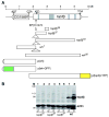
Fig. 1
Fig. 1
Deficiency mutants of wh and top3β. (A) Schematic diagram of the genomic structure of wh and top3β genes, their deficiency mutants and transgenic constructs. The P-element insertion mutant top3βEP(X)1432 was described previously (Wilson et al., 2000). At the P-element insertion site, top3β26, top3β37, top3β16 and wh7 have an insert of 170 bp, 2.3 kb, 3 kb and 632 bp, respectively. top3β26 has 400 bp deletion in the second exon. top3β37 has 712 bp deletion from the first exon through most of the second exon. top3β16 has 2798 bp deletion from the first exon through first 3 bp of the fourth intron. wh7 has 1426 bp deletion beyond the P-element insertion site, including 1041 bp of WH coding region. wh56 had 4016 bp deletion, including 368 bp of WH coding sequence, and the whole coding region of top3β gene. The transgenic construct diagrams show the start and end point of wh or top3β in the genomic DNA. Green and yellow box indicate the in-frame fusion of GFP and YFP, respectively. (B) top3β16, top3β26 and top3β37 are top3β null mutants. Adult males (lanes 1, 3, 5 and 7) and females (lanes 2, 4, 6 and 8) were homogenized with SDS sample buffer, subjected to SDS–PAGE, transferred to a nitrocellulose membrane and detected with rabbit anti-top3β (top panel), or rabbit anti-actin (bottom panel) serving as a loading control. M marks the lane for molecular mass standard, WT for precise excision wild-type control.
Fig. 2
Fig. 2
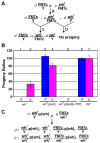
wh mutant males are sterile and can be rescued by wh transgene. (A) Males with wh7 mutation, generated from cross a, has no progeny (cross b). Thus, we cannot obtain wh7/wh7 homozygous female without the aid of a rescuing transgene. Stable stock of wh mutant can be maintained with cross c (or cross a). Similar phenotype was observed for wh56. (B) wh transgene can rescue the fertility of wh mutant, whereas the top3β transgene cannot. The male fertility was scored by mating the fly having the indicated genotype with wild-type females (columns 1, 3, 5 and 6), and female fertility was scored by mating the mutant females with wild-type males (columns 2, 4 and 7). wh mutant males are sterile, and mutant females have greatly reduced fertility (columns 1 and 2), when compared with wild-type controls (columns 6 and 7). wh transgene could rescue the fertility of both mutant males and females (compare columns 3 and 4 vs. columns 6 and 7). However, top3β transgene could not rescue the infertility of wh mutant males (column 5). (C) Genetic cross demonstrating that wh transgene can rescue wh mutant male to restore its fertility. From this cross, we could also generate wh homozygous females with or without a wh transgene, which were used in the experiments for monitoring effect of wh mutation on female fertility. Although the fly with wh7 mutant allele is shown here, similar cross was also used for wh56 mutant.
Fig. 3
Fig. 3
Both egg laying (embryo yield, columns 1, 3 and 5) and emerged larvae (embryo hatching, columns 2, 4 and 6) are reduced in wh female. The reduced fertility of wh mutant females is due to the decreased embryo yield and embryo hatching (columns 1 and 2) when compared with controls from either a transgene-rescued line (columns 3 and 4) or precise excision strain (columns 5 and 6).
Fig. 4
Fig. 4
wh7 and wh56 are wh null mutants. Adult males (lanes 1, 3, 5 and 7) and females (lanes 2, 4, 6 and 8) were homogenized with SDS sample buffer, subjected to SDS–PAGE, transferred to a nitrocellulose membrane and detected with rabbit anti-WH (A). Because the molecular mass of WH and actin is similar, a duplicate gel stained with Coomassie blue serves as a loading control (B). There is no WH expression in wh7 (lanes 1 and 2) or wh56 mutants (lanes 3 and 4), and p[wh] transgene can restore the WH expression in the null mutant background (lanes 5 and 6). WH expression in the precise excision strain is used as a control (lanes 7 and 8).
Fig. 5
Fig. 5
WH is an evolutionarily conserved WD40-repeat protein. (A) Protein sequence analysis (Stultz et al., 1997) of the deduced WH amino acid sequence showed WH had 5 WD40 repeats (underlined, I–V). A typical nuclear localization signal (NLS) was boxed at carboxyl terminus. The bipartite signal is composed of two basic sequence elements (dashed lines) separated by a spacer of ten residues (Dingwall et al., 1988). (B) Alignment of the WH deduced amino acid sequence and other putative homologues from human, mouse, rat, chicken, frog, worm (Caenorhabditis elegans), mosquito (Anopheles gambiae), fission yeast and baker’s yeast. Light shadow indicates the similarities and dark shadow indicates the identities. Five WD40 repeats (I–V) of WH are marked with black lines and NLS with a gray line.
Fig. 5
Fig. 5
WH is an evolutionarily conserved WD40-repeat protein. (A) Protein sequence analysis (Stultz et al., 1997) of the deduced WH amino acid sequence showed WH had 5 WD40 repeats (underlined, I–V). A typical nuclear localization signal (NLS) was boxed at carboxyl terminus. The bipartite signal is composed of two basic sequence elements (dashed lines) separated by a spacer of ten residues (Dingwall et al., 1988). (B) Alignment of the WH deduced amino acid sequence and other putative homologues from human, mouse, rat, chicken, frog, worm (Caenorhabditis elegans), mosquito (Anopheles gambiae), fission yeast and baker’s yeast. Light shadow indicates the similarities and dark shadow indicates the identities. Five WD40 repeats (I–V) of WH are marked with black lines and NLS with a gray line.
Fig. 6
Fig. 6
WH-GFP localization during oogenesis. Ovarioles were dissected from transgenic flies expressing WH-GFP fusion protein and stained with rabbit anti-GFP polyclonal antibody (A), mouse anti-adducin (B) and DAPI for DNA (C). Panel D is the merged image. The ovariole shown here has from left to right, germarium, and egg chambers at Stages 2–4. Arrow indicates the oocyte in Stage 4 egg chamber. Scale bar, 25 μm.
Fig. 7
Fig. 7
Oogenesis defects in wh mutant females. The same ovariole either from wh rescued transgenic line (A, C, E and G) or mutant line (B, D, F and H) is stained for DNA (blue, A and B), adducin (red, C and D) and vasa (green, E and F). The merged image is shown in panels G and H. Normal oogenesis is observed in wh transgenic females, whereas wh mutant ovariole has egg chambers with more than 16 germ cells. Notice that DNA staining in the mutant germ cells is significantly less than the nurse cells in wh rescued controls (compare A vs. B). Scale bar, 25 μm.
Fig. 8
Fig. 8
WH-GFP has the highest expression in hub cells. Testes were dissected from transgenic flies expressing WH-GFP fusion protein and stained with rabbit anti-GFP polyclonal antibody (A), mouse anti-Fasciclin III for marking hub cells (B) and DAPI for DNA (C). WH-GFP is colocalized with Fasciclin III (D, merged image). Scale bar, 25 μm.
Fig. 9
Fig. 9
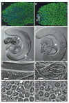
Arrest of spermatogenesis in wh mutant at the spermatids elongation stage. Confocal images were taken from fixed samples of testes (panels A and B; scale bar, 25 μm). Unfixed and squashed samples of testes were observed under a microscope with Nomarski differential interference contrast (panels C and D, with a scale bar of 50 μm) and phase-contrast (panels E–H; scale bar, 10 μm). (A and B) Testes were stained for Fasciclin III (red), vasa (green) and DNA (blue). No apparent defect was observed at the early stages of spermatogenesis when compared the mutant testis (B) with wh rescued testis as a wild-type control (A). Fasciclin III, a molecular marker of hub cell (Brower et al., 1981; Gonczy et al., 1992), is present in wh mutant testis (B), which indicates that the wh mutant testis contained the hub cells. Outlined are the presumed germline stem cells based on their morphology and location. (C) In the wh transgenic testis, elongated and bundled spermatids (arrow) are visible, and swollen seminal vesicles are filled with coiled mature sperms (arrowhead). (D) In wh mutant testis, no mature sperm bundles are visible, and seminal vesicles (arrowhead) are empty. (E and F) Spermatid bundles at the elongating stage (outlined). wh mutant spermatids are arrested at elongating step (F) as compared to the fully elongated spermatid bundles of wh transgenic males (E). (G and H) Developing spermatids at the early elongating stages. Arrowheads indicate the elongating mitochondria derivatives, which are morphologically distinct between the mutant and rescued control. Note the presence of vesicles (arrow) in wh mutant spermatids (H).

Similar articles

Cited by

References

    1. Alexandrov A, Martzen MR, Phizicky EM. Two proteins that form a complex are required for 7-methylguanosine modification of yeast tRNA. RNA. 2002;8:1253–1266. - PMC - PubMed
    1. Alexandrov A, Grayhack EJ, Phizicky EM. tRNA m7G methyl-transferase Trm8p/Trm82p: evidence linking activity to a growth phenotype and implicating Trm82p in maintaining levels of active Trm8p. RNA. 2005;11:821–830. - PMC - PubMed
    1. Brower DL, Smith RJ, Wilcox M. Differentiation within the gonads of Drosophila revealed by immunofluorescence. J Embryol Exp Morphol. 1981;63:233–242. - PubMed
    1. Chen D, McKearin DM. A discrete transcriptional silencer in the bam gene determines asymmetric division of the Drosophila germline stem cell. Development. 2003;130:1159–1170. - PubMed
    1. Dingwall C, Robbins J, Dilworth SM, Roberts B, Richardson WD. The nucleoplasmin nuclear location sequence is larger and more complex than that of SV-40 large T antigen. J Cell Biol. 1988;107:841–849. - PMC - PubMed

Publication types

MeSH terms