ADAM15 disintegrin is associated with aggressive prostate and breast cancer disease
- PMID: 16756724
- PMCID: PMC1600681
- DOI: 10.1593/neo.05682
ADAM15 disintegrin is associated with aggressive prostate and breast cancer disease
Abstract
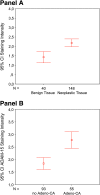
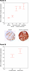
The aim of the current study was to evaluate the expression of ADAM15 disintegrin (ADAM15) in a broad spectrum of human tumors. The transcript for ADAM15 was found to be highly upregulated in a variety of tumor cDNA expression arrays. ADAM15 protein expression was examined in tissue microarrays (TMAs) consisting of 638 tissue cores. TMA analysis revealed that ADAM15 protein was significantly increased in multiple types of adenocarcinoma, specifically in prostate and breast cancer specimens. Statistical association was observed with disease progression within clinical parameters of predictive outcome for both prostate and breast cancers, pertaining to Gleason sum and angioinvasion, respectively. In this report, we also present data from a cDNA microarray of prostate cancer (PCa), where we compared transfected LNCaP cells that overexpress ADAM15 to vector control cells. In these experiments, we found that ADAM15 expression was associated with the induction of specific proteases and protease inhibitors, particularly tissue inhibitor of metalloproteinase 2, as validated in a separate PCa TMA. These results suggest that ADAM15 is generally overexpressed in adenocarcinoma and is highly associated with metastatic progression of prostate and breast cancers.
Figures


Similar articles
-
Overexpression of the A Disintegrin and Metalloproteinase ADAM15 is linked to a Small but Highly Aggressive Subset of Prostate Cancers.Neoplasia. 2017 Apr;19(4):279-287. doi: 10.1016/j.neo.2017.01.005. Epub 2017 Mar 8. Neoplasia. 2017. PMID: 28282546 Free PMC article.
-
The role of the disintegrin metalloproteinase ADAM15 in prostate cancer progression.J Cell Biochem. 2009 Apr 15;106(6):967-74. doi: 10.1002/jcb.22087. J Cell Biochem. 2009. PMID: 19229865 Review.
-
ADAM15 supports prostate cancer metastasis by modulating tumor cell-endothelial cell interaction.Cancer Res. 2008 Feb 15;68(4):1092-9. doi: 10.1158/0008-5472.CAN-07-2432. Cancer Res. 2008. PMID: 18281484
-
ADAM15 to α5β1 integrin switch in colon carcinoma cells: a late event in cancer progression associated with tumor dedifferentiation and poor prognosis.Int J Cancer. 2012 Jan 15;130(2):278-87. doi: 10.1002/ijc.25891. Epub 2011 Nov 9. Int J Cancer. 2012. PMID: 21190186
-
The role of tissue microarrays in prostate cancer biomarker discovery.Adv Anat Pathol. 2007 Nov;14(6):408-18. doi: 10.1097/PAP.0b013e318155709a. Adv Anat Pathol. 2007. PMID: 18049130 Review.
Cited by
-
Neoplasia: the second decade.Neoplasia. 2008 Dec;10(12):1314-24. doi: 10.1593/neo.81372. Neoplasia. 2008. PMID: 19048110 Free PMC article.
-
Characterization of oxygen-induced retinopathy in mice carrying an inactivating point mutation in the catalytic site of ADAM15.Invest Ophthalmol Vis Sci. 2014 Sep 23;55(10):6774-82. doi: 10.1167/iovs.14-14472. Invest Ophthalmol Vis Sci. 2014. PMID: 25249606 Free PMC article.
-
A transcriptome-wide association study identifies novel candidate susceptibility genes for prostate cancer risk.Int J Cancer. 2022 Jan 1;150(1):80-90. doi: 10.1002/ijc.33808. Epub 2021 Sep 25. Int J Cancer. 2022. PMID: 34520569 Free PMC article.
-
Analysis of ADAM12-Mediated Ephrin-A1 Cleavage and Its Biological Functions.Int J Mol Sci. 2021 Mar 1;22(5):2480. doi: 10.3390/ijms22052480. Int J Mol Sci. 2021. PMID: 33804570 Free PMC article.
-
HER2 and EGFR Overexpression Support Metastatic Progression of Prostate Cancer to Bone.Cancer Res. 2017 Jan 1;77(1):74-85. doi: 10.1158/0008-5472.CAN-16-1656. Epub 2016 Oct 28. Cancer Res. 2017. PMID: 27793843 Free PMC article.
References
-
- Bogenrieder T, Herlyn M. Axis of evil: molecular mechanisms of cancer metastasis. Oncogene. 2003;22(42):6524–6536. - PubMed
-
- Polette M, Nawrocki-Raby B, Gilles C, Clavel C, Birembaut P. Tumour invasion and matrix metalloproteinases. Crit Rev Oncol Hematol. 2004;49(3):179–186. - PubMed
-
- Wheelock MJ, Buck CA, Bechtol KB, Damsky CH. Soluble 80-kD fragment of cell-CAM 120/80 disrupts cell-cell adhesion. J Cell Biochem. 1987;34(3):187–202. - PubMed
-
- White JM. ADAMs: modulators of cell-cell and cell-matrix interactions. Curr Opin Cell Biol. 2003;15(5):598–606. - PubMed
-
- Peschon JJ, Slack JL, Reddy P, Stocking KL, Sunnarborg SW, Lee DC, Russell WE, Castner BJ, Johnson RS, Fitzner JN, et al. An essential role for ectodomain shedding in mammalian development. Science. 1998;282(5392):1281–1284. - PubMed
Publication types
MeSH terms
Substances
Grants and funding
LinkOut - more resources
Full Text Sources
Medical
