Dysregulated T cell expression of TIM3 in multiple sclerosis
- PMID: 16754722
- PMCID: PMC2118310
- DOI: 10.1084/jem.20060210
Dysregulated T cell expression of TIM3 in multiple sclerosis
Abstract
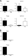
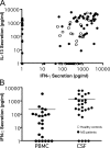
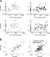
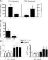
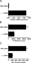
T cell immunoglobulin- and mucin domain-containing molecule (TIM)3 is a T helper cell (Th)1-associated cell surface molecule that regulates Th1 responses and promotes tolerance in mice, but its expression and function in human T cells is unknown. We generated 104 T cell clones from the cerebrospinal fluid (CSF) of six patients with multiple sclerosis (MS) (n = 72) and four control subjects (n = 32) and assessed their cytokine profiles and expression levels of TIM3 and related molecules. MS CSF clones secreted higher amounts of interferon (IFN)-gamma than did those from control subjects, but paradoxically expressed lower levels of TIM3 and T-bet. Interleukin 12-mediated polarization of CSF clones induced substantially higher amounts of IFN-gamma secretion but lower levels of TIM3 in MS clones relative to control clones, demonstrating that TIM3 expression is dysregulated in MS CSF clones. Reduced levels of TIM3 on MS CSF clones correlated with resistance to tolerance induced by costimulatory blockade. Finally, reduction of TIM3 on ex vivo CD4+ T cells using small interfering (si)RNA enhanced proliferation and IFN-gamma secretion, directly demonstrating that TIM3 expression on human T cells regulates proliferation and IFN-gamma secretion. Failure to up-regulate T cell expression of TIM3 in inflammatory sites may represent a novel, intrinsic defect that contributes to the pathogenesis of MS and other human autoimmune diseases.
Figures
Similar articles
-
T Cell Ig- and mucin-domain-containing molecule-3 (TIM-3) and TIM-1 molecules are differentially expressed on human Th1 and Th2 cells and in cerebrospinal fluid-derived mononuclear cells in multiple sclerosis.J Immunol. 2004 Jun 1;172(11):7169-76. doi: 10.4049/jimmunol.172.11.7169. J Immunol. 2004. PMID: 15153541
-
Blockade of T-cell immunoglobulin and mucin domain-containing molecule 3 aggravates T-helper cell 1 polarization in immune thrombocytopenia.Blood Coagul Fibrinolysis. 2019 Jun;30(4):133-139. doi: 10.1097/MBC.0000000000000805. Blood Coagul Fibrinolysis. 2019. PMID: 31090595
-
Phenotypical and functional characterization of T helper 17 cells in multiple sclerosis.Brain. 2009 Dec;132(Pt 12):3329-41. doi: 10.1093/brain/awp289. Brain. 2009. PMID: 19933767
-
The TIM gene family regulates autoimmune and allergic diseases.Trends Mol Med. 2005 Aug;11(8):362-9. doi: 10.1016/j.molmed.2005.06.008. Trends Mol Med. 2005. PMID: 16002337 Review.
-
TIM3 comes of age as an inhibitory receptor.Nat Rev Immunol. 2020 Mar;20(3):173-185. doi: 10.1038/s41577-019-0224-6. Epub 2019 Nov 1. Nat Rev Immunol. 2020. PMID: 31676858 Free PMC article. Review.
Cited by
-
A novel soluble form of Tim-3 associated with severe graft-versus-host disease.Biol Blood Marrow Transplant. 2013 Sep;19(9):1323-30. doi: 10.1016/j.bbmt.2013.06.011. Epub 2013 Jun 17. Biol Blood Marrow Transplant. 2013. PMID: 23791624 Free PMC article.
-
Interferon-β suppresses murine Th1 cell function in the absence of antigen-presenting cells.PLoS One. 2015 Apr 17;10(4):e0124802. doi: 10.1371/journal.pone.0124802. eCollection 2015. PLoS One. 2015. PMID: 25885435 Free PMC article.
-
LAG-3, TIM-3, and TIGIT: Distinct functions in immune regulation.Immunity. 2024 Feb 13;57(2):206-222. doi: 10.1016/j.immuni.2024.01.010. Immunity. 2024. PMID: 38354701 Review.
-
Cis association of galectin-9 with Tim-3 differentially regulates IL-12/IL-23 expressions in monocytes via TLR signaling.PLoS One. 2013 Aug 14;8(8):e72488. doi: 10.1371/journal.pone.0072488. eCollection 2013. PLoS One. 2013. PMID: 23967307 Free PMC article.
-
The costimulatory role of TIM molecules.Immunol Rev. 2009 May;229(1):259-70. doi: 10.1111/j.1600-065X.2009.00772.x. Immunol Rev. 2009. PMID: 19426227 Free PMC article. Review.
References
-
- Olsson, T. 1995. Cytokine-producing cells in experimental autoimmune encephalomyelitis and multiple sclerosis. Neurology. 45:S11–S15. - PubMed
-
- Agnello, D., C.S. Lankford, J. Bream, A. Morinobu, M. Gadina, J.J. O'Shea, and D.M. Frucht. 2003. Cytokines and transcription factors that regulate T helper cell differentiation: new players and new insights. J. Clin. Immunol. 23:147–161. - PubMed
-
- Szabo, S.J., B.M. Sullivan, S.L. Peng, and L.H. Glimcher. 2003. Molecular mechanisms regulating Th1 immune responses. Annu. Rev. Immunol. 21:713–758. - PubMed
-
- Monney, L., C.A. Sabatos, J.L. Gaglia, A. Ryu, H. Waldner, T. Chernova, S. Manning, E.A. Greenfield, A.J. Coyle, R.A. Sobel, et al. 2002. Th1-specific cell surface protein Tim-3 regulates macrophage activation and severity of an autoimmune disease. Nature. 415:536–541. - PubMed
Publication types
MeSH terms
Substances
Grants and funding
LinkOut - more resources
Full Text Sources
Other Literature Sources
Medical
Research Materials
Miscellaneous

