. 2006 Jun;26(12):4675-89.
doi: 10.1128/MCB.01830-05.

Nucleocytoplasmic shuttling modulates activity and ubiquitination-dependent turnover of SUMO-specific protease 2

Affiliations

Nucleocytoplasmic shuttling modulates activity and ubiquitination-dependent turnover of SUMO-specific protease 2

Yoko Itahana et al. Mol Cell Biol. 2006 Jun.

Abstract

Small ubiquitin-related modifier (SUMO) proteins are conjugated to numerous polypeptides in cells, and attachment of SUMO plays important roles in regulating the activity, stability, and subcellular localization of modified proteins. SUMO modification of proteins is a dynamic and reversible process. A family of SUMO-specific proteases catalyzes the deconjugation of SUMO-modified proteins. Members of the Sentrin (also known as SUMO)-specific protease (SENP) family have been characterized with unique subcellular localizations. However, little is known about the functional significance of or the regulatory mechanism derived from the specific localizations of the SENPs. Here we identify a bipartite nuclear localization signal (NLS) and a CRM1-dependent nuclear export signal (NES) in the SUMO protease SENP2. Both the NLS and the NES are located in the nonhomologous domains of SENP2 and are not conserved among other members of the SENP family. Using a series of SENP2 mutants and a heterokaryon assay, we demonstrate that SENP2 shuttles between the nucleus and the cytoplasm and that the shuttling is blocked by mutations in the NES or by treating cells with leptomycin B. We show that SENP2 can be polyubiquitinated in vivo and degraded through proteolysis. Restricting SENP2 in the nucleus by mutations in the NES impairs its polyubiquitination, whereas a cytoplasm-localized SENP2 made by introducing mutations in the NLS can be efficiently polyubiquitinated, suggesting that SENP2 is ubiquitinated in the cytoplasm. Finally, treating cells with MG132 leads to accumulation of polyubiquitinated SENP2, indicating that SENP2 is degraded through the 26S proteolysis pathway. Thus, the function of SENP2 is regulated by both nucleocytoplasmic shuttling and polyubiquitin-mediated degradation.


Figures

FIG. 1.
FIG. 1.
A sequence alignment of human SENP1 (hSENP1) and SENP2 potentially reveals NLS and NES in SENP2 in unconserved domains. (A) Amino acid sequence alignment of human SENP1 and SENP2 using the CLUSTAL W (version 1.81) multiple-sequence alignment program. The fully conserved residues and the conservation of strong groups are indicated by a black background and a light-gray background, respectively. The NLS and the NES sequences of SENP2 are indicated with underlines. The NLS of SENP1 is indicated with a box. Key amino acid residues involved in the catalytically active site are marked as asterisks. The conserved domain of SENP family proteins is shown with dotted lines above the sequence. (B) Diagram of human SENP2 with positions and amino acid sequences of the two predicted bipartite NLSs. The functionally important basic amino acids of each NLS are shown in boldface. Alanine substitutions to generate SENP2 NLS mutants are indicated. cNLS, C-terminally located putative NLS.
FIG. 2.
FIG. 2.
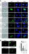
Identification of a bipartite NLS in SENP2. (A) Localization of wild-type SENP2 and SENP2 NLS mutants. U2OS cells were transfected with the indicated Myc-tagged SENP2 plasmids. Twenty-four hours after transfection, cells were fixed and immunostained with an anti-Myc antibody (α-Myc; 9E10.3), followed by staining with Cy2-conjugated secondary antibody. Immunofluorescent, phase-contrast, and DAPI-stained images are shown. (B) Quantification of SENP2 mutants in nuclear and cytoplasmic fractions. Low-magnification pictures of immunostained SENP2 are shown. The graphs show the percentage of cells in each subcellular fraction as determined by counting at least 200 cells in each sample.
FIG. 3.
FIG. 3.
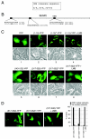
Identification of a CRM1-dependent NES in SENP2. (A) The consensus sequence for the CRM1-dependent nuclear export signal is shown; the conserved hydrophobic amino acid can be leucine (L), isoleucine (I), methionine (M), or phenylalanine (F). (B) Diagram of SENP2 with the positions and amino acid sequences of three NES-like sites indicated. Alanine substitutions for NES functional essential hydrophobic residues tested in the study are indicated. Superscript “m” indicates a mutant protein. (C) The SENP2 NES is a CRM1-dependent NES. Short peptides of the putative SENP2 NES were fused with a YFP vector, and localization of the fusion proteins was observed in transfected U2OS cells. Transfected cells were also treated with 10 nM LMB for 7 h to determine possible CRM1 dependency. Pictures were taken with living cells 24 h after transfection. Corresponding phase-contrast pictures are shown. (D) Quantification of cells. Low-magnification pictures of transfected cells are shown. The graph shows the percentages of cells with nuclear exclusion as determined by counting at least 200 cells.
FIG. 4.
FIG. 4.
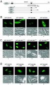
Function of NES in SENP2 export. (A) Schematic diagram of human SENP2 and its deletion mutants. Positions of the NLS and NES and of the N-terminal fusion of GFP are indicated. (B and C) Localization of GFP-SENP2 fusion proteins in transiently transfected U2OS cells. Plasmid DNA encoding each of the indicated GFP-SENP2 fusion proteins was transfected into U2OS cells, and the GFP signal was examined with a fluorescence microscope 24 h after transfection into live cells. Corresponding phase-contrast pictures are also shown. (D) The NES-mediated nuclear export of SENP2 is blocked by LMB. Plasmids expressing various GFP-SENP2 fusion proteins were transfected into U2OS cells. Twenty-four hours after transfection, the cells were treated with 10 nM LMB for 7 h and microscope pictures were taken with living cells.
FIG. 5.
FIG. 5.
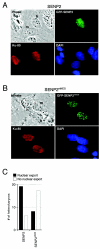
Interspecies heterokaryon assay shows that SENP2 is a shuttling protein. U2OS cells were transfected with plasmids expressing wild-type GFP-SENP2 (A) or the NES mutant GFP-SENP2mNES (B) fusion proteins. Twenty-four hours after transfection, the cells were washed and fused to wild-type MEF cells, and de novo protein synthesis was blocked by cycloheximide. Five hours after fusion, the cells were fixed and stained with an antibody to human Ku antigen, followed by staining with a rhodamine-conjugated secondary antibody. Representative heterokaryons were photographed. Nuclei of human (h) and mouse (m) origin in each heterokaryon are indicated. (C) Quantification of cells exported from the nucleus. Twenty-five randomly chosen heterokaryons from four independent heterokaryon assays described for panels A and B were examined, and the results were plotted.
FIG. 6.
FIG. 6.
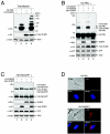
SUMO-modified PML, but not RanGAP1, is efficiently deconjugated by SENP2. (A to C) H1299 cells were transfected with the indicated plasmids, and cells were lysed 2 days after transfection with hot SDS (2%) lysis buffer. The cell lysate was resolved by SDS-PAGE and blotted with the indicated antibodies (α). GFP was used as a loading control for transfection efficiency. K, in thousands. (A and B) Unmodified and sumoylated His-tagged PML were detected with anti-His antibody. Unconjugated SUMO (free SUMO) was detected with anti-HA antibody. (C) Unmodified RanGAP1 and sumoylated His-tagged RanGAP1 were detected with an anti-His antibody. Unconjugated SUMO (free SUMO) was detected with anti-HA antibody. (D) Localization of PML and RanGAP1. U2OS cells were transfected with His-PML or His-RanGAP1. Twenty-four hours after transfection, cells were fixed and immunostained with an anti-His antibody, followed by staining with Cy2-conjugated or rhodamine-conjugated secondary antibody. Immunofluorescent, phase-contrast, and DAPI-stained images are shown.
FIG. 7.
FIG. 7.
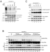
Disruption of the NLS, but not NES, impairs the protease function of SENP2. (A) H1299 cells were cotransfected with plasmids encoding HA-SUMO-3 and various Myc-SENP2 enzymes as indicated. The SENP2 constructs were transfected with wild-type SENP2 [WT]-SENPmNLS-SENPmNES-SENPmNLS/mNES at a ratio of 1:1.5:0.7:1.1 to obtain equal levels of SENP2 protein expression. Cells were lysed 2 days after transfection with a 2% hot SDS lysis buffer. Cell lysates were separated by SDS-PAGE, and the proteins were detected by immunoblotting with the indicated antibodies (α). GFP was used as a loading control for transfection efficiency. High-molecular-weight protein species detected with an anti-HA antibody indicate sumoylated proteins. Unconjugated SUMO-3 (free SUMO-3) is shown. (B) Desumoylation of PML by SENP2. H1299 cells were cotransfected with plasmids encoding His-PML and various Myc-SENP2 enzymes as indicated. The SENP2 constructs were transfected with wild-type Myc-SENP2-SENPmNLS-SENPmNES at a ratio of 1:1.2:0.7 to obtain similar levels of SENP2 protein expression. Cells were lysed 2 days after transfection with a 2% hot SDS lysis buffer. Cell lysates were subjected to SDS-PAGE and immunoblotted (IB) with the indicated antibodies. Unmodified and sumoylated His-PML was detected with anti-His antibody. Cat, a catalytically inactive SENP2 mutant. (C) Desumoylation of RanGAP1 by SENP2. H1299 cells were cotransfected with plasmids encoding His-RanGAP1 and the wild-type or mutant Myc-SENP2. The SENP2 constructs were transfected with wild-type Myc-SENP2-SENPmNLS-SENPmNES-SENP2Cat [Cat]) at a ratio of 1:1.5:0.7:1.5 to obtain equal levels of SENP2 protein expression. Cells were lysed 2 days after transfection with a 2% hot SDS lysis buffer, and the cell lysates were assayed as described for panel B.
FIG. 8.
FIG. 8.
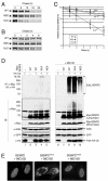
SENP2 is degraded through polyubiquitination-mediated proteolysis. (A) Half-life assay of SENP2. H1299 cells were transfected with plasmid DNA encoding wild-type Myc-tagged SENP2 (WT) or SENP2 mutants. Twenty-four hours after transfection, cells were pulse-labeled with [35S]methionine for 2 h and chased for the indicated lengths of time. Cell lysates were immunoprecipitated with anti-Myc antibody. The resulting SENP2 immunoprecipitates were separated by SDS-PAGE and visualized by autoradiography. (B) Half-life assay of SENP2 as described for panel A except with shorter time intervals, as indicated. The amount of labeled SENP2 protein at each time point was quantified on a PhosphorImager and normalized relative to the amount of radiolabeled SENP2 present in cells following the 0 h chase. Average results of four independent experiments were plotted in panel C. (D) In vivo ubiquitination assay of SENP2. H1299 cells were transfected with the indicated plasmid DNA. The GFP plasmid was also cotransfected as a control for transfection efficiency. The SENP2 constructs were transfected with wild-type Myc-SENP2-SENPmNLS-SENPmNES at a ratio of 1:1.5:0.7 to obtain equal levels of SENP2 protein expression. Two days after transfection, cells were treated with 20 μM MG132 for 6 h. Cells were lysed in 1% SDS lysis buffer and diluted 10 times with 0.1% NP-40-PBS. Diluted lysates were immunoprecipitated (IP) with anti-Myc antibody. The resulting SENP2 immunoprecipitates were separated by SDS-PAGE and immunoblotted (IB) with the indicated antibodies (α). K, in thousands; [Ub]n-SENP2, polyubiquitinated SENP2; expo, exposure. (E) Localization of wild-type SENP2 or its mutants after the treatment with MG132. U2OS cells were transfected with plasmid DNA encoding Myc-tagged SENP2. Twenty-four hours after transfection, cells were treated with 20 μM MG132 for 10 h, fixed, and immunostained with anti-Myc antibody, followed by staining with Cy2-conjugated secondary antibody.

Similar articles

Cited by

References

    1. Alarcon-Vargas, D., and Z. Ronai. 2002. SUMO in cancer—wrestlers wanted. Cancer Biol. Ther. 1:237-242. - PubMed
    1. Bailey, D., and P. O'Hare. 2004. Characterization of the localization and proteolytic activity of the SUMO-specific protease, SENP1. J. Biol. Chem. 279:692-703. - PubMed
    1. Bailey, D., and P. O'Hare. 2002. Herpes simplex virus 1 ICP0 co-localizes with a SUMO-specific protease. J. Gen. Virol. 83:2951-2964. - PubMed
    1. Bogerd, H. P., R. A. Fridell, R. E. Benson, J. Hua, and B. R. Cullen. 1996. Protein sequence requirements for function of the human T-cell leukemia virus type 1 Rex nuclear export signal delineated by a novel in vivo randomization-selection assay. Mol. Cell. Biol. 16:4207-4214. - PMC - PubMed
    1. Bohren, K. M., V. Nadkarni, J. H. Song, K. H. Gabbay, and D. Owerbach. 2004. A M55V polymorphism in a novel SUMO gene (SUMO-4) differentially activates heat shock transcription factors and is associated with susceptibility to type I diabetes mellitus. J. Biol. Chem. 279:27233-27238. - PubMed

Publication types

MeSH terms

LinkOut - more resources