. 2006 Jun;80(11):5423-34.
doi: 10.1128/JVI.02585-05.

Deletion of open reading frame UL26 from the human cytomegalovirus genome results in reduced viral growth, which involves impaired stability of viral particles

Affiliations

Deletion of open reading frame UL26 from the human cytomegalovirus genome results in reduced viral growth, which involves impaired stability of viral particles

Kerstin Lorz et al. J Virol. 2006 Jun.

Abstract

We previously showed that open reading frame (ORF) UL26 of human cytomegalovirus, a member of the US22 multigene family of betaherpesviruses, encodes a novel tegument protein, which is imported into cells in the course of viral infection. Moreover, we demonstrated that pUL26 contains a strong transcriptional activation domain and is capable of stimulating the major immediate-early (IE) enhancer-promoter. Since this suggested an important function of pUL26 during the initiation of the viral replicative cycle, we sought to ascertain the relevance of pUL26 by construction of a viral deletion mutant lacking the UL26 ORF using the bacterial artificial chromosome mutagenesis procedure. The resulting deletion virus was verified by PCR, enzyme restriction, and Southern blot analyses. After infection of human foreskin fibroblasts, the UL26 deletion mutant showed a small-plaque phenotype and replicated to significantly lower titers than wild-type or revertant virus. In particular, we noticed a striking decrease of infectious titers 7 days postinfection in a multistep growth experiment, whereas the release of viral DNA from infected cells was not impaired. A further investigation of this aspect revealed a significantly diminished stability of viral particles derived from the UL26 deletion mutant. Consistent with this, we observed that the tegument composition of the deletion mutant deviates from that of the wild-type virus. We therefore hypothesize that pUL26 plays a role not only in the onset of IE gene transcription but also in the assembly of the viral tegument layer in a stable and correct manner.


Figures

FIG. 1.
FIG. 1.
Structural analysis of HCMV BAC plasmids. (A and B) HindIII (A) and EcoRI (B) cleavage of wild-type (pHB15, lane 1), ΔUL26 (ΔUL26-2 and ΔUL26-5, lanes 2 and 3, respectively), and UL26 revertant (Rev-2 and Rev-5, lanes 4 and 5, respectively) BACs. The fragments unique to ΔUL26 are marked with dots. Southern blot analyses from BAC DNAs treated with HindIII and EcoRI using a biotinylated probe comprising the UL25-UL27 genomic region are shown in the bottom panels. Molecular size markers are indicated. (C) For verification of the correct recombination sites within the HCMV genome, PCR analyses of bacterial clones harboring the indicated BACs were performed using oligonucleotides specific for UL25 (left panel), UL26 (middle panel), and UL27 (right panel), respectively. The location of the primers in relation to the recombination cassette is shown in the scheme below. Lanes: 1, PCR with wild-type BAC pHB15; 2 and 3, PCRs with UL26 deletion BACs ΔUL26-2 and ΔUL26-5, respectively; 4 and 5, PCRs with revertant BACs Rev-2 and Rev-5, respectively. The scheme in D shows the HindIII and EcoRI restriction sites in relation to the relevant genomic segments of HCMV.
FIG. 2.
FIG. 2.
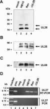
Analysis of protein and RNA expression of recombinant viruses. (A to C) HFF cells were mock infected (lane 1) or infected with HB15 (lane 2), Rev (lane 3), or ΔUL26 (lane 4) and harvested after confluent spreading of the CPE. Afterwards, the expression of pUL26 (A), pUL25 (B), and pUL69 (C) was examined by Western blot analyses using specific antibodies against the respective proteins. Molecular size markers are given at the left of each panel. (D) Reverse transcription-PCR analysis of UL27 RNA expression. (Upper panel) RNA of infected cells was reverse transcribed, followed by PCR amplification using UL27-specific primers. (Lower panel) PCR amplification was performed without prior reverse transcription of RNA. marker, molecular size marker; pos., HCMV DNA, used as a positive control for the PCR; neg., no DNA was added; mock, RNA from mock-infected cells; HB15, RNA from HB15-infected cells; Rev, RNA from Rev-infected cells; ΔUL26, RNA from ΔUL26-infected cells.
FIG. 3.
FIG. 3.
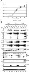
Accumulation of viral DNA and protein after infection with recombinant viruses. Infection was performed with viral inocula that were normalized for an equivalent uptake of viral DNA into cells. DNA and protein were harvested at 8, 24, 48, 72, and 96 h after infection. (A) HCMV-specific real-time PCR was performed to quantify viral genomes during the replicative cycle of HB15, Rev, or ΔUL26. Experiments were performed in triplicate; standard deviations are indicated. (B) Western blot analyses were performed to monitor the accumulation of viral proteins during the replicative cycle of HB15 (lanes 2 to 5), Rev (lanes 6 to 9), or ΔUL26 (lanes 10 to 13). The detected proteins are indicated at the right of each panel, nonspecific bands are indicated by asterisks, the sizes of molecular mass markers are given on the left. hpi, hours postinfection.
FIG. 4.
FIG. 4.
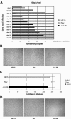
Analysis of plaque formation of recombinant viruses. (A) HFF cells were infected in duplicate with the recombinant viruses HB15 (gray bars), Rev (striped bars), and ΔUL26 (black bars) at 0.0005 infectious units/cell. The infection was followed by a conventional plaque overlay assay. After the appearance of plaques, the plaques for each virus were counted every day until confluence. Given are the mean plaque numbers for each duplicate infection. (B) Shown is a comparison of representative plaques in cultures infected with HB15, Rev, and ΔUL26 at day 15 postinfection. (C) IE1p72-expressing cells were infected in triplicate with HB15 (gray bar), Rev (white bar), or ΔUL26 (black bar) at 0.0005 infectious units/cell followed by a plaque overlay assay. The graph represents the number of plaques determined at day 12 after infection; standard deviations are indicated. (E) Shown is a comparison of representative plaques after infection of IE1p72-expressing cells with HB15, Rev, and ΔUL26 at day 12 postinfection.
FIG. 5.
FIG. 5.
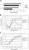
Growth kinetics of the recombinant viruses. For infection of HFF cells, the viral inoculum was standardized for equal IE1p72 expression 24 h postinfection. (A) HFFs were infected in parallel with HB15, Rev, or ΔUL26 at an MOI of either 0.1 or 0.01. Cell culture supernatants were harvested at 9 days postinfection, followed by quantification of infectious virus via IE1p72 fluorescence (see Materials and Methods). Error bars indicate standard deviations. (B) Multistep growth curves were performed using 0.01 infectious units/cell of the indicated recombinant viruses in triplicate. The supernatants were harvested at days 1, 3, 5, 7, 9, and 12 postinfection and frozen at −80°C. After thawing, the probes were again titrated via IE1p72 expression in triplicate. Standard deviations are indicated on the graph. (C) Quantification of viral genomes in the supernatant of infected HFF cells by real-time PCR. Aliquots of the supernatants obtained for the multistep growth curve shown in B were treated with proteinase K, incubated at 56°C for 1 h, and subsequently denatured at 95°C. Five microliters of each lysate was subjected to real-time PCR to quantify the genomic equivalents in the supernatants of the recombinant viruses. Evaluation was performed in triplicate from each of the three infections per virus at each time point. Standard deviations are indicated.
FIG. 6.
FIG. 6.
Reduced stability of the ΔUL26 virus. HFF cells were infected with 0.001 infectious units/cell of freshly thawed supernatant of the recombinant viruses HB15 (gray bars), Rev (striped bars), and ΔUL26 (black bars). After 1 h of incubation, the viral supernatants were removed, and fresh medium was added. The remaining virus stocks were stored at 20°C and used for infection of HFF cells for the following 5 days. Subsequently, the number of IE1p72-positive cells was determined for each virus at each time point 48 h after the infection was started. The remaining infectivity shown on the x axis was related to the infectivity of the supernatants at day 0, which was set as 100%. Determination of cell numbers was carried out in duplicate (both values for each virus are depicted as bars).
FIG. 7.
FIG. 7.
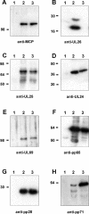
Tegument protein composition of wild-type (HB15) and mutant (ΔUL26) virions. Virus particles from the supernatant of HFF cells infected with HB15 or ΔUL26 were separated from cell fragments via low-speed centrifugation. Thereafter, the particles were purified by sedimentation in a glycerol-tartrate gradient, thus resulting in virion fractions, which were used for immunoblot analyses. Virion proteins were detected using monoclonal antibodies against MCP(UL86) (A), pUL25 (C), pUL24 (D), pp65(UL83) (F), and pp28(UL99) (G) or polyclonal antisera against pUL26 (B), ppUL69 (E), and pp71(UL82) (H). Lane 1 contains lysates of mock-infected cells, lane 2 contains lysates of HB15-infected cells, and lane 3 contains lysates of ΔUL26-infected cells (A to H).
FIG. 8.
FIG. 8.
Ultrastructural analysis of virus maturation and morphology. (a to d) HFF cells were infected with HB15 or ΔUL26 at an MOI of 0.5. Cells were fixed at 72 h postinfection and processed for electron microscopy. (a and b) Nuclei of HB15- and ΔUL26-infected cells, respectively. (c and d) Cytoplasm of HB15-and ΔUL26-infected cells. (e and f) Purified extracellular viral particles were visualized by negative staining with uranyl acetate. (e) HB15 virions. (f) ΔUL26 virion. All images were taken at ×46,460 magnification.

Similar articles

Cited by

References

    1. Adamo, J. E., J. Schroer, and T. Shenk. 2004. Human cytomegalovirus TRS1 protein is required for efficient assembly of DNA-containing capsids. J. Virol. 78:10221-10229. - PMC - PubMed
    1. Adler, H., M. Messerle, and U. H. Koszinowski. 2003. Cloning of herpesviral genomes as bacterial artificial chromosomes. Rev. Med. Virol. 13:111-121. - PubMed
    1. Almeida, J., D. Lang, and P. Talbot. 1978. Herpesvirus morphology: visualization of a structural subunit. Intervirology 10:318-320. - PubMed
    1. Andreoni, M., M. Faircloth, L. Vugler, and W. J. Britt. 1989. A rapid microneutralization assay for the measurement of neutralizing antibody reactive with human cytomegalovirus. J. Virol. Methods 23:157-167. - PubMed
    1. Arlt, H., D. Lang, S. Gebert, and T. Stamminger. 1994. Identification of binding sites for the 86-kilodalton IE2 protein of human cytomegalovirus within an IE2-responsive viral early promoter. J. Virol. 68:4117-4125. - PMC - PubMed

Publication types

LinkOut - more resources