. 2006 Jun;116(6):1514-24.
doi: 10.1172/JCI27564. Epub 2006 May 11.

IL-15 induces CD4 effector memory T cell production and tissue emigration in nonhuman primates

Affiliations

IL-15 induces CD4 effector memory T cell production and tissue emigration in nonhuman primates

Louis J Picker et al. J Clin Invest. 2006 Jun.

Abstract

HIV infection selectively targets CD4+ effector memory T (T EM) cells, resulting in dramatic depletion of CD4+ T cells in mucosal effector sites in early infection. Regeneration of the T EM cell compartment is slow and incomplete, even when viral replication is controlled by antiretroviral therapy (ART). Here, we demonstrate that IL-15 dramatically increases in vivo proliferation of rhesus macaque (RM) CD4+ and CD8+ T EM cells with little effect on the naive or central memory T (T CM) cell subsets, a response pattern that is quite distinct from that of either IL-2 or IL-7. T EM cells produced in response to IL-15 did not accumulate in blood. Rather, 5-bromo-2'-deoxyuridine (BrdU) labeling studies suggest that many of these cells rapidly disperse to extralymphoid effector sites, where they manifest (slow) decay kinetics indistinguishable from that of untreated controls. In RMs with uncontrolled SIV infection and highly activated immune systems, IL-15 did not significantly increase CD4+ T EM cell proliferation, but with virologic control and concomitant reduction in immune activation by ART, IL-15 responsiveness was again observed. These data suggest that therapeutic use of IL-15 in the setting of ART might facilitate specific restoration of the CD4 + T cell compartment that is the primary target of HIV with less risk of exhausting precursor T cell compartments or generating potentially deleterious regulatory subsets.


Figures
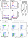
Figure 1
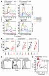
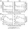
Figure 1. IL-15 induces memory, but not naive, CD4+ and CD8+ T cells to proliferate.
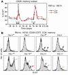
(A) The fraction of cells expressing the proliferation marker Ki-67 was determined in phenotypically defined naive versus memory, CD4+ and CD8+ T cells from the peripheral blood of 2 representative, normal RMs that were given sham injections first, followed by IL-15 injections 70 days later. (B) The change in the fraction of total CD4+ or CD8+, peripheral blood memory T cells expressing Ki-67 from baseline (defined as the average of the day –7 and day 0 values) to day 7 after the first dose is shown for all 6 RMs that were given the 2-dose IL-15 regimen. (C) The figure shows the correlated expression of CD28 and CCR7 on gated CD4+ and CD8+ memory T cells from the peripheral blood of a typical adult RM. Note that the double-negative TEM subset dominates the circulating CD8+ memory T cell population but constitutes only a very minor component of CD4+ memory T cells. CD4+ and CD8+ memory T cells in blood also differ in the presence of a significant CD28CCR7+ subset in the latter, but not the former, population. (D) The figure shows the typical profiles of CD28 versus CCR7 expression on lung airspace and small-intestinal mucosal CD4+ and CD8+ memory T cells of an adult RM. Note that the vast majority of these memory T cells display either a CD28+CCR7 transitional or a CD28CCR7 TEM phenotype.
Figure 2
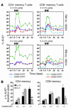
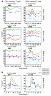
Figure 2. IL-15 selectively acts on TEM and transitional memory T cells.
(A) The CD4+ and CD8+ memory T cell subsets defined by CD28 and CCR7 expression (see Figure 1C) in peripheral blood were assessed for expression of Ki-67 before, during, and after IL-15 treatment (arrows). Note that CD4+ memory cells lack a significant CD28CCR7+ component, accounting for why only 3 populations are shown for this lineage. (B) The change in percentage Ki-67+ from baseline (the average of day –7 and day 0 values) to peak (either day 7 or day 10) is shown for the designated CD4+ (black) and CD8+ (red) memory subsets in 4 different RMs that received at least 3 doses of IL-15 (10 μg/kg) on days 0, 3, and 7. The IL-15–induced changes in percentage Ki-67 for each of the 3 comparable CD4+ versus CD8+ memory subsets (means provided in the figure) were not significantly different. (C) The figure shows the expression of CCR5 by the CD4+ memory subsets defined by CD28 and CCR7 in the peripheral blood of a representative adult RM. (D) The fraction of CCR5+, CD4+ peripheral blood memory T cells expressing Ki-67 is shown before, during, and after IL-15 treatment (arrows; 10 μg/kg) in the same 2 representative RMs depicted in A. As would be expected based on the staining pattern shown in C, IL-15 induced a substantial increase in proliferation within the CCR5-expressing, CD4+ memory population.
Figure 3
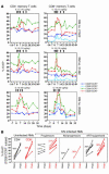
Figure 3. CD28 , CCR7 TEM cells are poorly responsive to IL-2.
(A) The CD4+ and CD8+ memory T cell subsets defined by CD28 and CCR7 expression in the peripheral blood in 2 representative RMs (of 4 total) were assessed for expression of Ki-67 before, during, and after 2 weeks of twice-weekly administration of IL-2. Note that IL-2 preferentially induced proliferation in the CD28+CCR7 component of both the CD4+ and the CD8+ memory populations. (B) The mean (± SEM) change in percentage Ki-67 from baseline (the average of day –7 and day 0 values) to peak response (day 7 or 10 after the first cytokine dose) is shown for the CD28/CCR7–defined peripheral blood memory T cell subsets in all 4 IL-2–treated RMs, and the 4 RMs that received at least 3 doses of IL-15 (the pre-peak doses on days 0, 3, and 7, matching the pre-peak IL-2 dosing schedule). While IL-2 demonstrated a superior (to CD4+) or similar (to CD8+) ability to induce proliferation among CD28+CCR7 transitional memory T cells, IL-15 was dramatically better than IL-2 in initiating proliferation within both the CD4+ and the CD8+, CD28CCR7 TEM subsets. *0.05 < P < 0.005; **P < 0.005.
Figure 4
Figure 4. IL-15 responsiveness is limited by the availability of response-capable cells, but not by development of an anti–IL-15 antibody response.
(A) The expression of Ki-67 within the CD28 subset of circulating CD4+ and CD8+ memory T cells is shown over time in a healthy, adult (IL-15–naive) RM receiving 2 courses of IL-15 therapy 70 days apart. Each black box represents 2 doses of IL-15 (each dose 10 μg/kg). Note that the second response was of a magnitude similar to or greater than that of the initial response. (B) The figure shows successive histograms of Ki-67 expression on gated CD28CCR7, CD4+ memory T cells from an RM (no. 18743) that received 10 μg/kg IL-15 on days 0, 3, 7, and 10 (see Figure 2 for overall quantitative data). The arrows (black, blue, red, and green) delineate successive waves of Ki-67 expression (see text). *IL-15 dose given on indicated days.
Figure 5
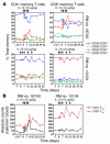
Figure 5. IL-15–responsive memory subsets do not accumulate in peripheral blood.
(A) The fraction of total CD4+ and CD8+ memory T cells in each of the CD28/CCR7–defined subsets in blood (see Figure 1A) is shown before, during, and after IL-15 treatment (the same IL-15–treated RMs shown in Figure 2). Note that despite vast differences in IL-15–induced proliferation between these subsets, their relative frequencies in blood (particularly the TCM-to-TEM ratio) either did not change or changed only transiently. (B) The absolute number of CD4+ and CD8+ CD28CCR7 TEM cells is shown in the same RMs. Again, profound IL-15–induced proliferation resulted in only transiently increased numbers of TEM cells in peripheral blood.
Figure 6
Figure 6. The progeny of IL-15–stimulated T cells are long-lived and rapidly migrate into an extralymphoid effector site.
The 6 RMs that received a 2-dose IL-15 regimen (10 μg/kg 3 days apart) were pulsed with 30 mg/kg BrdU on the 4 days after the second IL-15 dose, and then subsequently assessed for BrdU incorporation and decay in the CD28 (CD4+ and CD8+) memory subsets in peripheral blood (A) and the overall (CD4+ and CD8+) memory populations in BAL specimens (B). These IL-15–treated RMs were compared with a cohort of 12 (A) or 16 (B) untreated RMs that were subjected to the same 4-day BrdU labeling regimen. The figure shows the mean percentage BrdU+ cells (± SEM) in the populations indicated in the IL-15–treated (filled diamonds) versus control RMs (open diamonds), with the day of the last BrdU dose designated day 0. Note that for many data points, the SEM is sufficiently small so that the error bars are obscured by the symbols. The difference in BrdU+ cell frequencies between IL-15–treated and control RMs in the blood effector memory populations was significant at P < 0.0001 and P < 0.0004 at all time points for CD4+ and CD8+ T cells, respectively. In BAL, this difference was significant at P < 0.0004 at all time points for both CD4+ and CD8+ T cells.
Figure 7
Figure 7. IL-15 responsiveness is diminished in the highly activated immune systems of RMs with uncontrolled SIV infection but remains intact in a spontaneous controller.
(A) The figure shows the effects of 2 weeks of twice-weekly IL-15 therapy at the same dose used in previous experiments (10 μg/kg, RMs no. 21522 and no. 21462) and a dose 5 times higher (RM no. 21102) on the expression of Ki-67 by the peripheral blood CD4+ and CD8+ memory T cell subsets defined by CD28 and CCR7 expression in 3 representative RMs (of 6 total) with typical untreated, plateau-phase SIV infection (see Figure 9A for plasma viral loads). Note that circulating CD4+, CD28CCR7 TEM cells were absent in RM no. 21522 because of viral pathogenicity. (B) The same analysis is shown for an SIVmac239-infected RM (no. 21021) with spontaneous control of viral replication (plateau plasma viral loads < 5 × 10² copies/ml) treated with 10 μg/kg IL-15 at the same dosing schedule.
Figure 8
Figure 8. IL-15 responsiveness is restored with control of viral replication by ART.
(A) The figure shows the effects of IL-15 therapy on the expression of Ki-67 by the peripheral blood CD4+ and CD8+ memory T cell subsets defined by CD28 and CCR7 expression in 3 representative SIV-infected RMs (of 5 total) that were treated with ART and achieved a stable 3- to 5-log reduction in plasma viral load. Note that ART restored an IL-15–responsive CD4+, CD28CCR7 TEM cell population in all animals. (B) The maximal effect of IL-15 (baseline to peak) on CD4+ (black) and CD8+ (red) TEM (CD28CCR7) cell proliferation (percentage Ki-67) is shown for all study RMs given at least 3 doses of IL-15 (filled diamonds, 10 μg/kg; open diamonds, 50 μg/kg) on days 0, 3, and 7. The magnitude of the IL-15–induced proliferation in the CD4+ and CD8+ TEM cell populations of the uninfected and ART-treated, SIV-infected cohorts (means provided in the figure) was not significantly different; however, the IL-15–induced changes in TEM cell proliferation in both of these cohorts were significantly different from that observed in the SIV-infected RMs with uncontrolled (e.g., high-viral-load) infection (P < 0.02).
Figure 9
Figure 9. Short-term IL-15 therapy does not increase viral replication in either ART-treated or untreated RMs and influences proliferation of pathogen-specific T cells based on their TEM component.
(A and B) Plasma viral loads of untreated (A) or ART-treated (B), SIV-infected RMs are shown before, during, and after IL-15 therapy. (C) Cytokine flow cytometry was used to determine the pre-IL-15-treatment memory phenotype (CD28 versus CCR7) and follow the post-IL-15-treatment proliferative status (Ki-67 expression) of peripheral blood CD4+ and CD8+ memory T cells responsive to SIVgag or RhCMV in 3 SIV-infected animals previously shown to have IL-15–responsive total TEM cell populations (see Figure 8) — the 1 spontaneous controller (no. 21021) given 10 μg/kg, and 2 ART-treated RMs (nos. 21046 and 20955) given 50 μg/kg. Note that in all 3 RMs, CMV-specific CD4+ T cells included a large fraction of TEM cells and showed a marked in vivo proliferative response to IL-15; in contrast, SIVgag-specific CD4+ T cells included negligible TEM cells and little proliferative response to IL-15. Both SIVgag- and RhCMV-specific CD8+ T cells (in the 2 of 3 RMs that manifested such responses) included a large TEM component and manifested IL-15 responsiveness.

Similar articles

Cited by

References

    1. Douek D.C., Picker L.J., Koup R.A. T cell dynamics in HIV-1 infection. Annu. Rev. Immunol. 2003;21:265–304. - PubMed
    1. Veazey R., Lackner A. The mucosal immune system and HIV-1 infection. AIDS Rev. 2003;5:245–252. - PubMed
    1. Mehandru S., Tenner-Racz K., Racz P., Markowitz M. The gastrointestinal tract is critical to the pathogenesis of acute HIV-1 infection. . J. Allergy Clin. Immunol. 2005;116:419–422. - PubMed
    1. Sallusto F., Geginat J., Lanzavecchia A. Central memory and effector memory T cell subsets: function, generation, and maintenance. Annu. Rev. Immunol. 2004;22:745–763. - PubMed
    1. Brenchley J.M., et al. CD4+ T cell depletion during all stages of HIV disease occurs predominantly in the gastrointestinal tract. J. Exp. Med. 2004;200:749–759. - PMC - PubMed

Publication types

MeSH terms