. 2006 Jun 7;25(11):2275-86.
doi: 10.1038/sj.emboj.7601119. Epub 2006 May 11.

A dynein loading zone for retrograde endosome motility at microtubule plus-ends

Affiliations

A dynein loading zone for retrograde endosome motility at microtubule plus-ends

J H Lenz et al. EMBO J. .

Abstract

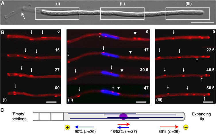
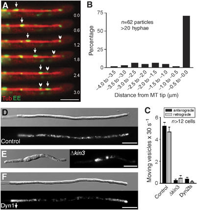
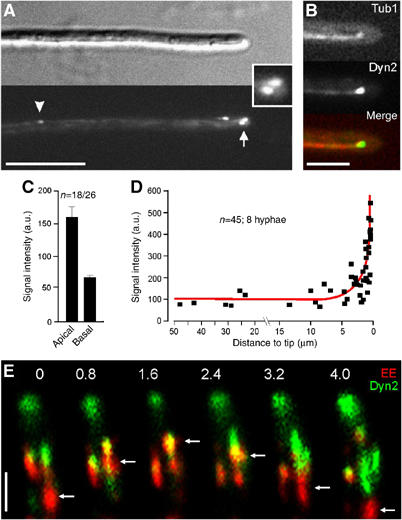
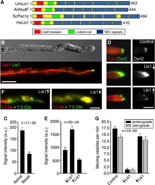
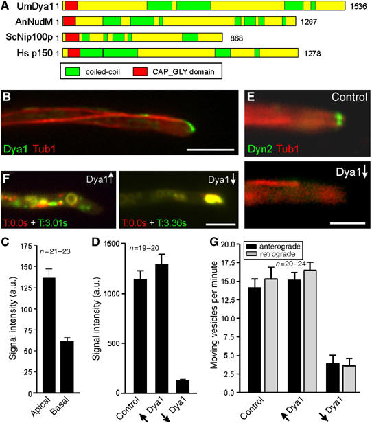
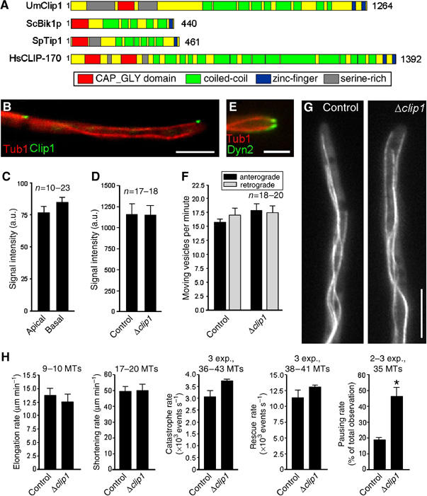
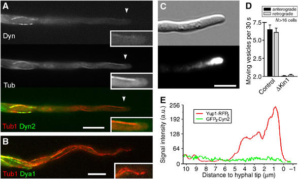
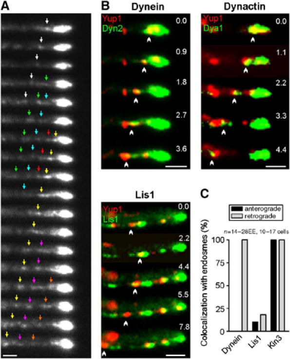
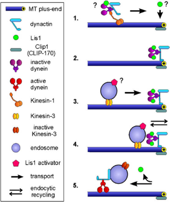
In the fungus Ustilago maydis, early endosomes move bidirectionally along microtubules (MTs) and facilitate growth by local membrane recycling at the tip of the infectious hypha. Here, we set out to elucidate the molecular mechanism of this process. We show that endosomes travel by Kinesin-3 activity into the hyphal apex, where they reverse direction and move backwards in a dynein-dependent manner. Our data demonstrate that dynein, dynactin and Lis1 accumulate at MT plus-ends within the hyphal tip, where they provide a reservoir of inactive motors for retrograde endosome transport. Consistently, endosome traffic is abolished after depletion of the dynein activator Lis1 and in Kinesin-1 null mutants, which was due to a defect in targeting of dynein and dynactin to the apical MT plus-ends. Furthermore, biologically active GFP-dynein travels on endosomes in retrograde and not in anterograde direction. Surprisingly, a CLIP170 homologue was neither needed for dynein localization nor for endosome transport. These results suggest an apical dynein loading zone in the hyphal tip, which ensure that endosomes reach the expanding growth region before they reverse direction.


Figures

Figure 1
Figure 1
Microtubule orientation in hyphae of U. maydis. (A) Overview of a hypha of U. maydis. Boxed regions (I–III) correspond to series given in 1B. Note that tip cells leave ‘empty' cell wall section behind (arrow) while they grow at their tip (right end). Bar: 10 μm. (B) Peb1-YFP (given in red) marks MT plus-ends that elongate towards the basal septum (I, arrows), as well as the hyphal tip (III, arrows), indicating that the plus-ends are orientated towards both poles of the hyphal cell. Around the nucleus (II; labeled by histone 4-CFP is given in blue), Peb1-signals move in both directions (arrow and arrowhead), indicating that MTs have an antipolar orientation. Elapsed time is given in seconds. Bars: 5 μm. (C) Quantitative analysis of growing MT ends demonstrates that the plus-ends (indicated by ‘plus') are focused to the tip (indicated by red arrows) and the subapical septum (indicated by blue arrows). An antipolar MT orientation in the middle of the cell suggests that minus-ends are located near the nucleus. Numbers indicate the percentage of Peb1-YFP signals that move in the direction indicated. Supplementary movie for Panel B is given on the EMBO web site.
Figure 2
Figure 2
Endosome organization and motility in the Kinesin-3 and dynein mutants. (A) EE that are labeled by a fusion protein of GFP and the membrane receptor Yup1 (arrowhead, arrow; Wedlich-Söldner et al, 2000) migrate bidirectioally along MTs, which are visualized by expression of RFP-α-tubulin. Elapsed time is given in seconds. Bar: 3 μm. (B) A quantitative analysis of the behavior of individual EE confirmed that most organelles reach the MT plus-ends before they reverse direction. (C) Quantitative analysis of the EE motility frequencies at 10 μm distance to the tip of control and mutant hyphae. Almost identical numbers of vesicles move towards the tip (anterograde) and the basal nuclear region (retrograde) of control hyphae. This motility is abolished in Kinesin-3 mutants (Δkin3) and in dynein mutants (Dyn2ts). (D) In control hyphae, Yup1-GFP labeled EE are motile and equally distributed along the length of the hyphae. Occasionally, transient endosome concentrations are found in the apex. Bar: 10 μm. (E) Δkin3 mutant hyphae are usually bipolar and much shorter (Schuchardt et al, 2005). The EE cluster near the nucleus, where the minus-ends of MTs are concentrated. Note that the MT orientation in Δkin3 hyphae was confirmed by analysis of Peb1-YFP motility (not shown). Bar: 10 μm. (F) In the conditional dynein mutants, depletion of dynein strengthens anterograde EE motility and leads to accumulations of EE at the MT plus-ends near the cell poles. Bar: 10 μm. Supplementary movie for Panels A and D–F is given on the EMBO web site.
Figure 3
Figure 3
Dynein localization and EE movement. (A) In U. maydis hyphae dynein, visualized by a fusion of 3xGFP and the endogenous Dyn2 forms motile comet-like structures (arrowheads). The strongest signals are always found in the hyphal apex (arrow, inset). Bar: 10 μm. (B) Colocalization of GFP3Dyn2 (Dyn, green in overlay) and RFP-α-tubulin labeled MTs (red) in control hyphae. Dynein localizes to the MT plus-ends that are reaching into the hyphal apex. Bar: 5 μm. (C) A quantitative analysis of the GFP3-Dyn2 signal intensity at MT plus-ends in the first 3 μm of the hyphal tip (apical) and in at least 12 μm distance from the tip (basal) reveals that the dynein accumulations at the hyphal tip is more than twice as strong as in basal parts of the cell. Note that the signal intensity has been normalized to a single GFP. (D) The average intensity of dynein signals at MT plus-ends was measured in relation to distance to the tip, demonstrating that the amount of dynein remains constant over most of the length of the hypha, but rapidly increases at the hyphal apex. (E) Colocalization of GFP3Dyn2 (Dyn, green in overlay series) and Yup-RFP2 (EE, red in overlay series) demonstrates that EE (arrows) migrate into the dynein signal before they reverse direction (colocalization results in yellow). Elapsed time is given in seconds. Bar: 2 μm. Supplementary movie for Panel E is given on the EMBO web site.
Figure 4
Figure 4
The role of a LIS1-homologue in dynein localization and endosome motility. (A) Lis1 from U. maydis (UmLis1, Accession Number: EAK84394) shares a similar domain structure with other LIS1-homologues, including those from A. nidulans (AnNudF, EAA57983), S. cerevisiae (ScPac1p, AAB00685) and human (HsLis1, NP 000421). (B) GFP-tagged endogenous Lis1 (green) localizes to plus-ends of RFP-α-tubulin containing MTs (red). Note that the tip contains the strongest signal. Bar: 5 μm. (C) Quantification of the GFP-Lis1 intensity at MT plus-ends in the apical 3 μm of the hyphal tip (apical) and in at least 12 μm distance from the tip (basal) demonstrates that Lis1 is concentrated at MT plus-ends in the tip. (D) The absence of Lis1 (Lis1↓) results in an increase of dynein at plus-ends within the hyphal tip. Overexpression of Lis1 (Lis1↑) reduces the apical dynein signal. Bar: 2 μm. (E) Quantitative analysis of GFP3Dyn2 intensity at MT plus-ends revealed that the absence of Lis1 (↓Lis1) significantly increases the amount of dynein at MT plus-ends. Consistently, Lis1 overexpression (↑Lis1) has the opposite effect. (F) Overlay of two images at different time points illustrates the motility of RFP2-tagged endosomes. Moving organelles appear in green or red, while stationary signals result in yellow color. Depletion of Lis1 led to an immobile accumulation of EE in the hyphal apex (strain FB2G3Dyn2_nLis1_YR2). Bar: 5 μm. Supplementary movies for Panel D and F are given on the EMBO web site. (G) A quantitative analysis of EE traffic reveals that overexpression of Lis1 (↑Lis1) affects EE motility only slightly, while nearly all motility is abolished in the absence of Lis1 (↓Lis1).
Figure 5
Figure 5
The role of a p150glued homologue in dynein localization and endosome motility. (A) The p150glued homologues from U. maydis (UmDya1, Accession Number: EAK84712) shares a similar domain structure with other p150glued homologues from A. nidulans (AnNudM, EAA58707), S. cerevisiae (ScNip100p, NP 015151) and human (Hsp150, NP 004073). (B) GFP3-tagged endogenous Dya1 (green) accumulates at the plus-ends of RFP-α-tubulin labeled MTs (red). The strongest signal is present in the hyphal apex. Bar: 5 μm. (C) Fluorescence intensity measurements of GFP3Dya1 at MT plus-ends in the first 3 μm of the hyphal tip (apical) and in basal parts of the cell (in >12 μm distance from the tip) reveals that dynactin was ∼2-times more concentrated at MT plus-ends in the hyphal tip than in subapical regions. (D) Quantification of the GFP3Dyn2 signals at MT plus-ends and in the apical cytoplasm shows that high levels of Dya1 (↑Dya1) has no significant effect on dynein localization (P=0.306), whereas dya1 repression (↓Dya1) leads to a dramatic decrease of the dynein signal. (E) No dynein is present at apical MT plus-ends after depletion of Dya1 (Dya1↓). Bar: 2.5 μm. (F) Overlay of two images at different time points illustrates the motility of GFP-tagged endosomes in strain FB2rDya1_YG. Moving organelles appear in green or red, while stationary signals result in yellow. Depletion of Dya1 leads to an immobile accumulation of EE in the hyphal apex. Bar: 5 μm. Supplementary movies for Panels E and F are given on the EMBO web site. (G) The expression of Dya1 (↑Dya1) has no effect on EE transport, whereas the absence of Dya1 (↓Dya1) abolishes almost all EE motility.
Figure 6
Figure 6
The role of a CLIP-170 homologue in dynein and endosome targeting. (A) The CLIP-170 homologues from U. maydis (UmClip1, Accession Number: EAK82811) shares a similar domain structure with other CLIP-170 members from S. cerevisiae (ScBik1p, NP 009901), Schizosaccharomyces pombe (SpTip1, P79065) and human (HsCLIP170, A43336). (B) GFP-tagged endogenous Clip1 (green) localizes to plus-ends of RFP-α-tubulin labeled MTs (red). Bar: 5 μm. (C) In contrast to dynein, dynactin and Lis1 intensities GFP-Clip1 fluorescence signals at MT plus-ends in the first 3 μm of the hyphae (apical) do not differ from those in basal parts of the cells (in >12 μm distance from tip). (D) In Δclip1 cells, the intensity of the dynein signal at MT plus-ends in the tip is not different from those of control cells. (E) In the clip1 deletion strain, GFP3-tagged dynein (green) still accumulates at plus-ends of RFP-labeled MTs (red). Bar: 2.5 μm. (F) Deletion of clip1 does not affect bidirectional EE motility. (G) In Δclip1 mutants, the organization of GFP-α-tubulin labeled MTs is not altered compared to control cells (strain FB2GT). Bar: 5 μm. Supplementary movies for Panels E and G are given on the EMBO web site. (H) Parameters of MT dynamic instability in control cells and Δclip1 mutant hyphae.
Figure 7
Figure 7
Kinesin-1-based targeting of dynein and dynactin to MT plus-ends in the hyphal tip. (A) Colocalization of GFP3Dyn2 (Dyn, green in overlay) and RFP-α-tubulin labeled MTs (Tub, red in overlay) in Δkin1 mutants reveals that dynein colocalizes with MTs in the subapical region. However, dynein does not reach the plus-ends in the hyphal tip (insets). Bar: 5 μm. (B) No GFP3Dya1 (green) appears at apical MT plus-ends (red) in Kinesin-1 null mutants, demonstrating that dynactin requires Kinesin-1 for long-distance plus-end targeting. Bar: 5 μm. (C) In Δkin1 hyphae EE cluster in the hyphal tip, indicating that deletion of Kinesin-1 strengthened plus-end directed transport, which is due to a lack of dynein in the hyphal tip. Bar: 5 μm. (D) Quantitative analysis of EE motility in the apical 10 μm of control and Kinesin-1 null mutant hyphae. Almost all motility is abolished in the absence of Kinesin-1. (E) Linescan analysis of intensities of GFP3-Dyn2 and Yup1-RFP2 in Δkin1 mutant hyphae (strain AB33ΔKin1_G3D2_YR2). In the absence of Kinesin-1, EE travel to the tip but are trapped there. Dynein is not enriched in the endosome cluster, which argues against a direct binding of the motor to anterogradely moving organelles. Supplementary movies for Panels A and C are given on the EMBO web site.
Figure 8
Figure 8
Dynein, dynactin, Lis1 on moving early endosomes. (A) In vivo observation of functional GFP3-Dyn2 at the hyphal tip demonstrates that dynein is continuously released from its apical accumulation. Individual signals marked by colored arrows. Interval between frames is 1 s. Bar: 1 μm. (B) Colocalization studies on strains expressing Yup1-RFP2 and GFP-tags fused to dyn2 (dynein), dya1 (dynactin) and lis1 (Lis1) demonstrate that the dynein/dynactin complex localizes to retrogradely moving organelles, whereas Lis1 only transiently appears on EE. Bars: 1 μm. (C) Quantitative analysis of EE that move for at least 3 μm. Kinesin-3 locates on all EE, whereas dynein was only found on retrogradely moving organelles. Supplementary movie for Panel B is given on the EMBO web site.
Figure 9
Figure 9
Dynein in minus-end directed traffic of early endosomes. (1) In the current model, Kinesin-1 takes dynein/dynactin to the MT plus-ends in the hyphal tip. Preliminary evidence indicates that a portion of Lis1 might hitchhike on the transported dynein/dynactin complex (not shown), but other mechanisms are also likely. (2) An inactive complex of dynein, dynactin and Lis1 accumulates at the plus-ends close to the growth region of the hypha. (3) Kinesin-3 transports EE along MTs to the inactive dynein/dynactin/Lis1 complex, thereby delivering an unknown activator of Lis1. (4) When EE reach the plus-ends at the hyphal apex they quickly exchange material with the growing tip. This might involve the uptake of material for transport towards the cell body as well as local membrane recycling processes. (5) Subsequently, the unknown activator triggers Lis1-dependent activation of the dynein/dynactin complex, which results in retrograde motility of the EE.

Similar articles

Cited by

References

    1. Berlin V, Styles CA, Fink GR (1990) BIK1, a protein required for microtubule function during mating and mitosis in Saccharomyces cerevisiae, colocalizes with tubulin. J Cell Biol 111: 2573–2586 - PMC - PubMed
    1. Bottin A, Kamper J, Kahmann R (1996) Isolation of a carbon source-regulated gene from Ustilago maydis. Mol Gen Genet 253: 342–352 - PubMed
    1. Brachmann A, Weinzierl G, Kamper J, Kahmann R (2001) Identification of genes in the bW/bE regulatory cascade in Ustilago maydis. Mol Microbiol 42: 1047–1063 - PubMed
    1. Brunner D, Nurse P (2000) CLIP170-like tip1p spatially organizes microtubular dynamics in fission yeast. Cell 102: 695–704 - PubMed
    1. Coquelle FM, Caspi M, Cordelieres FP, Dompierre JP, Dujardin DL, Koifman C, Martin P, Hoogenraad CC, Akhmanova A, Galjart N, De Mey JR, Reiner O (2002) LIS1, CLIP-170's key to the dynein/dynactin pathway. Mol Cell Biol 22: 3089–3102 - PMC - PubMed

Publication types

MeSH terms

LinkOut - more resources