. 2006 May;168(5):1531-41.
doi: 10.2353/ajpath.2006.050555.

p21Cip1 is required for the development of monocytes and their response to serum transfer-induced arthritis

Affiliations

p21Cip1 is required for the development of monocytes and their response to serum transfer-induced arthritis

John C Scatizzi et al. Am J Pathol. 2006 May.

Abstract

One of the central functions of cyclin-dependent kinase inhibitors, such as p21, p27, or p16, is to prevent entry into the cell cycle. However, the question remains as to whether they have other functions in the cell. We previously demonstrated that overexpression of p21 in fibroblasts isolated from patients with rheumatoid arthritis decreases the production of pro-inflammatory molecules. Overexpression of p21 has been also shown to reduce the development of experimental arthritis in mice and rats. To explore the role of endogenous p21 in the development of arthritis, we induced arthritis in p21(-/-) mice using the K/BxN serum transfer model of arthritis. Mice deficient in p21 were more resistant to serum transfer-induced arthritis (K/BxN) than wild-type (wt) control mice. Fewer macrophages were detected in p21(-/-) as compared to wt joints following transfer of K/BxN serum. Chemotaxis assays of bone marrow-derived macrophages from p21(-/-) and wt mice revealed no difference in migration. However, there was a substantial decrease in inflammatory monocytes circulating in peripheral blood and in monocyte precursors in bone marrow of p21(-/-) mice as compared to wt mice. Adoptive transfer of wt bone marrow-derived macrophages into p21(-/-) mice restored the sensitivity to serum transfer-induced arthritis. These data suggest a novel role for p21 in regulating the development and/or differentiation of monocytic populations that are crucial for the induction of inflammatory arthritis.


Figures
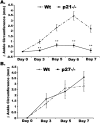
Figure 1
Figure 1
p21-deficient mice do not develop ankle inflammation associated with experimental arthritis. A: Quantitative analysis of the differential ankle circumference in p21−/− mice. 300 μl of pooled serum from K/BxN mice was injected intraperitoneally into wt and p21−/− mice. Ankle joints were examined for arthritis by measuring two perpendicular diameters of both joints (anteroposterior; mediolateral) by calipers. The change in ankle circumference at each time point is defined as the difference between the ankle circumference and the measurement at day 0. wt (n = 50) and p21−/− (n = 60) mice have been examined in multiple independent experiments. Shown are the composite data from those experiments (p21−/−: day 3, n = 29 mice; day 5, n = 20 mice; day 6, n = 21 mice; day 7, n = 17 mice; wt: day 3, n = 20 mice; day 5, n = 22 mice; day 6, n = 17 mice; day 7, n = 23 mice). The values represent the mean ± SE of ankles/time point, which were compared by Student’s t-test to wt mice under parallel conditions. **, P < 0.001 as compared to wt mice under parallel conditions. B: Quantitative analysis of the differential ankle circumference in p27−/− mice. wt and p27−/− mice were injected with K/BxN serum, and ankle joints were examined for arthritis as described above. Shown are the composite data from those experiments (p27−/−: day 3, n = 10 mice; day 5, n = 10 mice; day 7, n = 14 mice; wt: day 3, n = 10 mice; day 5, n = 10 mice; day 7, n = 16 mice). The values represent the mean ± SE of ankles/time point, which were compared by Student’s t-test.
Figure 2
Figure 2
Increased histological scores of ankle sections in wt mice compared to p21−/− mice. Ankles isolated from p21−/− and wt mice as described in Figure 1 were fixed, embedded in paraffin, sectioned, and stained with H&E or Safranin O and methyl green. Ankle sections were evaluated and scored by a pathologist blinded to the study as described in Materials and Methods. Shown are the composite data from two experiments (p21−/−: day 0, n = 14 ankles; days 3, 5, and 7, n = 8 ankles; days 4, 6, and 10, n = 16 ankles; wt: days 0, 3, and 5, n = 8 ankles; days 4, 6, and 10, n = 12 ankles; day 7, n = 6 ankles). The values represent the mean ± SE of ankles/time point, which were compared by Student’s t-test to wt mice under parallel conditions. *, P < 0.03 and **, P < 0.002 as compared to wt mice under parallel conditions.
Figure 3
Figure 3
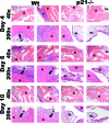
Increased inflammation and destruction in wt mice as compared to p21−/− mice following transfer of serum. At 4, 6, and 10 days following serum transfer, ankles as described in Figure 1 were harvested, sectioned, and stained with hematoxylin (blue) and eosin (pink). Shown are representative photomicrographs (40× or 200×) of the joint from wt and p21−/− mice. Arrows denotes invading pannus. SL, synovial lining; B, bone; P, pannus.
Figure 4
Figure 4
Fewer macrophages are recruited to p21−/− joints following injection of K/BxN serum. A: Quantitative fold increase of macrophage recruitment in ankle sections. Ankles from mice as described in Figure 1 were harvested, sectioned, and stained with antibodies that recognize the F4/80 antigen (brown staining) or isotype control (data not shown) and counterstained with hematoxylin. Macrophage counts were determined by a pathologist blinded to the study as described in Materials and Methods. Shown are the composite data from two experiments (p21−/−: day 0, n = 14 ankles; days 3, 5, and 7, n = 8 ankles; days 4, 6, and 10, n = 16 ankles; wt: days 0, 3, and 5, n = 8 ankles; days 4, 6, and 10, n = 12 ankles; day 7, n = 6 ankles). The values represent the mean ± SE of ankles/time point. The fold increase was defined as increase over untreated conditions. The data are representative of three independently performed experiments. *, P < 0.004; **, P < 0.0001 as compared to wt mice under parallel conditions. B: Photomicrographs (200×) of ankle sections from wt and p21−/− mice stained for F4/80 antigen. Ankles from mice as described in Figure 1 were harvested, sectioned, and stained with antibodies that recognize the F4/80 antigen (brown staining) or isotype control (data not shown) and counterstained with hematoxylin. SL, synovial lining; B, bone.
Figure 5
Figure 5
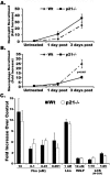
Reduced migration of monocytes to the inflamed peritoneum of p21−/− mice. p21−/− and wt mice were given intraperitoneal injections of 4% thioglycollate. Peritoneal cells were isolated 0 (n > 10), 1 (n > 8), and 3 (n > 12) days after injection and examined by flow cytometry as described in Materials and Methods. A: The fold increase of neutrophils recruited. Mice were treated as described above, neutrophils (CD45+Gr-1+) were identified using FACS analysis. B: The fold increase of macrophages recruited. Mice were treated as described above. Macrophages (CD45+F4/80+) were identified using FACS analysis. The data were a combination of four independently performed experiments. The values represent the mean ± SE of cells/time point, which were compared by Student’s t-test. The fold increase was defined as increase over untreated conditions. C: wt and p21−/− BMDMs respond similarly to chemotactic signals. Equal numbers of wt and p21−/− BMDMs were introduced to various chemoattractants to induce migration. Cell counts were expressed as -fold increase over background migration (no stimulus). Fkn, fractalkine/CX3CL1; Lkn, leukotactin/CCL15; fMLP, formyl-methionyl-leucyl-phenlyalanine; LCS, L-cell supernatant containing macrophage-colony stimulating factor.
Figure 6
Figure 6
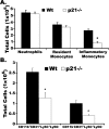
A: Immunophenotyping of peripheral blood reveals a deficiency in inflammatory monocytes in p21−/− mice. Peripheral blood was isolated by cardiac sticks from 5 to 8 week old wt (n = 23) and p21−/− (n = 16) mice. Cells were blocked for 10 minutes with Fc block and then stained with anti-CD45, anti-CD11b, anti-Gr-1, and anti-CD62L antibodies for 30 minutes. Following incubation with antibodies red blood cells were lysed and cells were fixed with BD Biosciences FACS lysing solution. Resident monocytes: CD45+CD11b++Gr-1CD62L; inflammatory monocytes: CD45+CD11b++Gr-1+CD62L++; neutrophils: CD45+CD11b++Gr-1++CD62L++. Values represent the mean ± SE, which were compared by Student’s t-test. *, P < 0.001 as compared to control under parallel conditions. B: Immunophenotyping of bone marrow reveals a deficiency in monocyte precursor populations in p21−/− mice. Bone marrow cells were isolated by tibia flush from 5 to 8 week old wt (n = 18) and p21−/− (n = 13) mice. Red blood cells were lysed, and the remaining cells were blocked for 10 minutes with Fc block and then stained with anti-CD45, anti-CD11b, anti-Gr-1, anti-CD31, anti-Ly6C, and anti-Ly-6G antibodies. Values represent the mean ± SE, which were compared by Student’s t-test. *, P < 0.002 as compared to wt under parallel conditions.
Figure 7
Figure 7
Adoptive transfer of wt BMDMs restores sensitivity to serum transfer in p21−/− mice. wt BMDMs were grown in culture as described in Materials and Methods and intravenously injected via tail vein into p21−/− mice at 1 day before serum transfer, 1 day after serum transfer, and 3 days after serum transfer. Ankle circumference was measured as described in Figure 1A. The values represent the mean ± SE of ankles/time point, which were compared by Student’s t-test to mice not receiving BMDMs under parallel conditions. (p21−/−: day 2, n = 12 mice; day 7, n = 20 mice; wt: day 2, n = 16 mice; day 7, n = 23 mice).

Similar articles

Cited by

References

    1. Sherr CJ. Cancer cell cycles. Science. 1996;274:1672–1677. - PubMed
    1. Gao CY, Zelenka PS. Cyclins, cyclin-dependent kinases and differentiation. BioEssays. 1997;19:307–315. - PubMed
    1. Kitaura H, Shinshi M, Uchikoshi Y, Ono T, Iguchi-Ariga SM, Ariga H. Reciprocal regulation via protein-protein interaction between c-Myc and p21(cip1/waf1/sdi1) in DNA replication and transcription. J Biol Chem. 2000;275:10477–10483. - PubMed
    1. Shim J, Lee H, Park J, Kim H, Choi EJ. A non-enzymatic p21 protein inhibitor of stress-activated protein kinases. Nature. 1996;381:804–806. - PubMed
    1. Coqueret O, Gascan H. Functional interaction of STAT3 transcription factor with the cell cycle inhibitor p21WAF1/CIP1/SDI1. J Biol Chem. 2000;275:18794–18800. - PubMed

Publication types

MeSH terms

Substances

LinkOut - more resources