Comparative Study
. 2006 Apr 19;26(16):4308-17.
doi: 10.1523/JNEUROSCI.0003-06.2006.

Activated microglia contribute to the maintenance of chronic pain after spinal cord injury

Affiliations
Comparative Study

Activated microglia contribute to the maintenance of chronic pain after spinal cord injury

Bryan C Hains et al. J Neurosci. .

Abstract

Traumatic spinal cord injury (SCI) results not only in motor impairment but also in chronic central pain, which can be refractory to conventional treatment approaches. It has been shown recently that in models of peripheral nerve injury, spinal cord microglia can become activated and contribute to development of pain. Considering their role in pain after peripheral injury, and because microglia are known to become activated after SCI, we tested the hypothesis that activated microglia contribute to chronic pain after SCI. In this study, adult male Sprague Dawley rats underwent T9 spinal cord contusion injury. Four weeks after injury, when lumbar dorsal horn multireceptive neurons became hyperresponsive and when behavioral nociceptive thresholds were decreased to both mechanical and thermal stimuli, intrathecal infusions of the microglial inhibitor minocycline were initiated. Electrophysiological experiments showed that minocycline rapidly attenuated hyperresponsiveness of lumbar dorsal horn neurons. Behavioral data showed that minocycline restored nociceptive thresholds, at which time spinal microglial cells assumed a quiescent morphological phenotype. Levels of phosphorylated-p38 were decreased in SCI animals receiving minocycline. Cessation of delivery of minocycline resulted in an immediate return of pain-related phenomena. These results suggest an important role for activated microglia in the maintenance of chronic central below-level pain after SCI and support the newly emerging role of non-neuronal immune cells as a contributing factor in post-SCI pain.


Figures

Figure 1.
Figure 1.
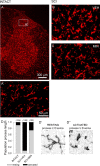
Immunolabeling for normal and reactive astroglia. Basal levels of GFAP signal were observed within the lumbar dorsal horn of intact animals (A). Higher magnification inset in A shows the typical resting astroglial morphology of long slender processes and small soma diameter. Distribution was uniform throughout white and gray matter. In SCI animals, on day 33, after 3 d of i.t. administration of vehicle (SCI+VEH), astroglia assumed an activated phenotype (B), whereby they exhibited a swollen appearance (B, inset). In SCI animals, on day 33, after 3 d of i.t. administration of the microglial inhibitor minocycline (SCI+MIN), signal remained unchanged compared with SCI+VEH (C). Astroglial morphological resembling an activated phenotype is shown (C, inset). The percentage of field analysis revealed significantly (*p < 0.05) elevated levels of astroglial activation in SCI+VEH and SCI+MIN groups, compared with intact animals (D). Error bars represent mean ± SD.
Figure 2.
Figure 2.
Immunolabeling for phosphorylated p-38 MAP kinase. Low levels of P-p38 signal were detected in the lumbar dorsal horn of intact animals (A). P-p38-positive cells exhibited a glial morphology with slender processes and were present primarily in gray matter. Thirty-three days after SCI and vehicle administration (SCI+VEH), P-p38 signal was increased, and P-p38-positive cells were more numerous (B). Positive cells exhibited morphological features of activated microglia: cell bodies were small, and several slender branched processes emerged from the soma. P-p38 (red) was colocalized to OX-42-positive cells (green) after SCI (C). In SCI animals, minocycline delivered i.t. for 3 d (SCI+MIN) resulted in a reduction in P-p38-positive cells (D). Morphological features of P-p38-positive cells were characteristic of quiescent microglia. Quantification revealed that after SCI, there was a significant (*p < 0.05) increase in the number of P-p38-positive cells when compared with intact animals (E). Compared with the SCI group, minocycline significantly (+p < 0.05) reduced the number of P-p38-positive cells (E). Error bars represent mean ± SD.
Figure 3.
Figure 3.
Immunolabeling for OX-42-positive microglia. OX-42 signal revealed moderate expression of resident microglia in both white and gray matter of the lumbar dorsal horn in an intact spinal cord (A). Microglia exhibited the resting type morphology: small compact somata bearing long, thin, ramified processes (A’). Thirty-three days after SCI, in vehicle-treated animals (SCI+VEH), microglia exhibited the activated phenotype: marked cellular hypertrophy and retraction of processes (B). Very few cells exhibiting resting morphological features were detected. In SCI animals, on day 33, after 3 d of i.t. administration of minocycline (SCI+MIN), microglia assumed the resting morphology (C). Quantification (D) of the proportion of resting (D’) and activated (D”) microglia confirmed that after SCI, there is a large shift from resting to activated microglia and that minocycline significantly reversed this shift.
Figure 4.
Figure 4.
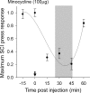
Thirty days after SCI, dorsal horn multireceptive units were sampled from the lumbar enlargement of SCI animals after acute topical administration of minocycline (arrow). The time course for maximum depression of evoked response to press stimulation of a receptive field located on the hindpaw after administration of minocycline revealed peak efficacy at 25–45 min for 100 μg. For subsequent electrophysiological experiments, units were sampled at time points of peak efficacy (shaded region).
Figure 5.
Figure 5.
Spontaneous background activity of lumbar dorsal horn multireceptive units was recorded in SCI animals 30 d after injury, after acute spinal administration of minocycline (A, arrow). Minocycline had no effect on ongoing activity over the course of 60 min. Expansion of waveform traces is shown for periods corresponding to time of minocycline administration (a1), peak effectiveness of drug (a2) (Fig. 4), and at the end of 60 min (a3). Quantification (B) of mean spontaneous firing revealed no significant differences at any point in response to minocycline (bin width, 4 s). Error bars represent mean ± SD. BK, Background.
Figure 6.
Figure 6.
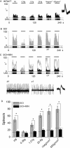
The effects of acutely administered minocycline on peripherally evoked activity in multireceptive units from SCI animals 30 d after injury. A representative unit from an intact animal displaying evoked responses to natural stimuli (PB, 0.39 g, 1.01 g, 20.8 g, 144 g/mm2, and 583 g/mm2 refer to phasic brush, von Frey filaments of increasing intensities, pressure, and pinch, applied for 20 s) is shown for comparison (A). The peristimulus time histogram shows that after SCI, evoked responses were increased to all peripheral stimuli (B). After SCI, evoked discharge rates were between 40 and 100 Hz. Phasic brush stimulation as well as compressive press and pinch stimuli resulted in high-frequency discharge. Von Frey filament stimulation resulted in graded increases in responsiveness of sampled units. At 30 min after administration, minocycline (SCI+MIN) resulted in decreased evoked responses to all peripheral stimuli (C). Predrug unit responses are overlaid on the SCI+MIN histogram. Example waveform of unit activity to press stimulation for SCI (b1) and SCI+MIN (c1) illustrate the effect of minocycline, which attenuated the post-SCI hyperresponsiveness. Minocycline significantly (*p < 0.05) reduced the evoked responses to all peripheral stimuli after SCI (D). Dashed lines indicated mean responses of intact animals. Error bars represent mean ± SD.
Figure 7.
Figure 7.
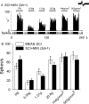
Evoked activity of a representative multireceptive unit 1 d after cessation of i.t.-delivered (3 d in duration) minocycline after SCI (SCI+MIN). After discontinuation of minocycline administration, evoked responses to peripheral stimulation (PB, 0.39 g, 1.01 g, 20.8 g, 144 g/mm2, and 583 g/mm2 refer to phasic brush, von Frey filaments of increasing intensities, pressure, and pinch, applied for 20 s) were elevated, indicating hyperresponsiveness (A). Quantification of responses to each stimulus revealed no significant differences between SCI and SCI animals after cessation of minocycline (B). Error bars represent mean ± SD.
Figure 8.
Figure 8.
Behavioral analysis of locomotor function and pain-related behaviors. Intact animals demonstrated expected levels of locomotor function, but 30 d after SCI, BBB scores revealed partial recovery of locomotor function for injured animals (A). In SCI animals, i.t. delivery of vehicle (SCI+VEH) or minocycline (SCI+MIN) had no significant effect during the period of administration (indicated by thick line) or for 2 d after cessation of administration, indicating no activation or depression of motor function that could compromise testing of nociceptive thresholds. After SCI, mechanical paw withdrawal thresholds had significantly (+p < 0.05) decreased in all groups when compared with intact animals (B). Minocycline resulted in an immediate increase in mechanical thresholds for the duration of administration. This effect was significant (*p < 0.05). Immediately after cessation of administration (day 33), mechanical thresholds returned to predrug levels that were equivalent to untreated SCI animals. Thermal paw withdrawal latencies (C) were significantly (+p < 0.05) lowered after SCI. Minocycline resulted in an immediate and significant (*p < 0.05) increase in paw withdrawal latencies. Minocycline sustained increased latencies for the duration of its administration. After cessation of delivery, latencies returned to predrug levels, which persisted for the duration of the experiment. Integral analysis (D) revealed that minocycline resulted in a significantly (*p < 0.05) more robust modulation of mechanical nociception compared with thermal nociception. Error bars represent mean ± SD. sec, Seconds.

Similar articles

Cited by

References

    1. Afrah AW, Fiska A, Gjerstad J, Gustafsson H, Tjolsen A, Olgart L, Stiller CO, Hole K, Brodin E (2002). Spinal substance P release in vivo during the induction of long-term potentiation in dorsal horn neurons. Pain 96:49–55. - PubMed
    1. Attal N, Guirimand F, Brasseur L, Gaude V, Chauvin M, Bouhassira D (2002). Effects of IV morphine in central pain: a randomized placebo-controlled study. Neurology 58:554–563. - PubMed
    1. Basso DM, Beattie MS, Bresnahan JC (1995). A sensitive and reliable locomotor rating scale for open field testing in rats. J Neurotrauma 12:1–21. - PubMed
    1. Cardenas DD, Warms CA, Turner JA, Marshall H, Brooke MM, Loeser JD (2002). Efficacy of amitriptyline for relief of pain in spinal cord injury: results of a randomized controlled trial. Pain 96:365–373. - PubMed
    1. Chaplan SR, Bach FW, Pogrel JW, Chung JM, Yaksh TL (1994). Quantitative assessment of tactile allodynia in the rat paw. J Neurosci Methods 53:55–63. - PubMed

Publication types