

Deletion of the Mycobacterium tuberculosis resuscitation-promoting factor Rv1009 gene results in delayed reactivation from chronic tuberculosis
- PMID: 16622237
- PMCID: PMC1459759
- DOI: 10.1128/IAI.74.5.2985-2995.2006
Deletion of the Mycobacterium tuberculosis resuscitation-promoting factor Rv1009 gene results in delayed reactivation from chronic tuberculosis
Abstract
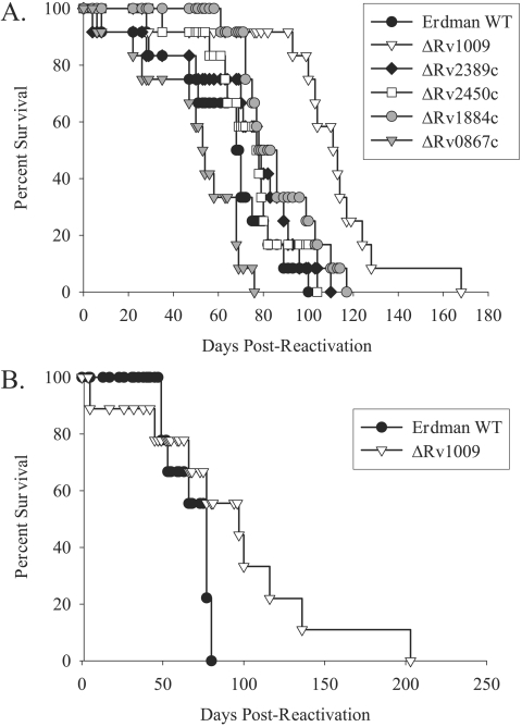
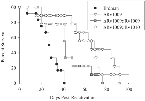
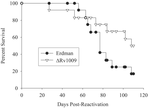
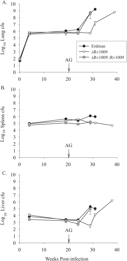
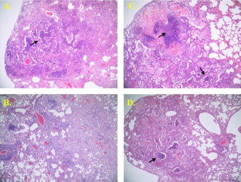
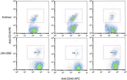
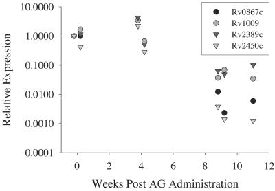
Approximately one-third of the human population is latently infected with Mycobacterium tuberculosis, comprising a critical reservoir for disease reactivation. Despite the importance of latency in maintaining M. tuberculosis in the human population, little is known about the mycobacterial factors that regulate persistence and reactivation. Previous in vitro studies have implicated a family of five related M. tuberculosis proteins, called resuscitation promoting factors (Rpfs), in regulating mycobacterial growth. We studied the in vivo role of M. tuberculosis rpf genes in an established mouse model of M. tuberculosis persistence and reactivation. After an aerosol infection with the M. tuberculosis Erdman wild type (Erdman) or single-deletion rpf mutants to establish chronic infections in mice, reactivation was induced by administration of the nitric oxide (NO) synthase inhibitor aminoguanidine. Of the five rpf deletion mutants tested, one (deltaRv1009) exhibited a delayed reactivation phenotype, manifested by delayed postreactivation growth kinetics and prolonged median survival times among infected animals. Immunophenotypic analysis suggested differences in pulmonary B-cell responses between Erdman- and deltaRv1009-infected mice at advanced stages of reactivation. Analysis of rpf gene expression in the lungs of Erdman-infected mice revealed that relative expression of four of the five rpf-like genes was diminished at late times following reactivation, when bacterial numbers had increased substantially, suggesting that rpf gene expression may be regulated in a growth phase-dependent manner. To our knowledge, deltaRv1009 is the first M. tuberculosis mutant to have a specific defect in reactivation without accompanying growth defects in vitro or during acute infection in vivo.
Figures
Similar articles
-
A Mycobacterium tuberculosis Rpf double-knockout strain exhibits profound defects in reactivation from chronic tuberculosis and innate immunity phenotypes.Infect Immun. 2008 Sep;76(9):4269-81. doi: 10.1128/IAI.01735-07. Epub 2008 Jun 30. Infect Immun. 2008. PMID: 18591237 Free PMC article.
-
The role of resuscitation promoting factors in pathogenesis and reactivation of Mycobacterium tuberculosis during intra-peritoneal infection in mice.BMC Infect Dis. 2007 Dec 17;7:146. doi: 10.1186/1471-2334-7-146. BMC Infect Dis. 2007. PMID: 18086300 Free PMC article.
-
Individual Mycobacterium tuberculosis resuscitation-promoting factor homologues are dispensable for growth in vitro and in vivo.Infect Immun. 2004 Jan;72(1):515-26. doi: 10.1128/IAI.72.1.515-526.2004. Infect Immun. 2004. PMID: 14688133 Free PMC article.
-
Rpf Proteins Are the Factors of Reactivation of the Dormant Forms of Actinobacteria.Biochemistry (Mosc). 2016 Dec;81(13):1719-1734. doi: 10.1134/S0006297916130095. Biochemistry (Mosc). 2016. PMID: 28260493 Review.
-
Resuscitation-promoting factors are important determinants of the pathophysiology in Mycobacterium tuberculosis infection.Crit Rev Microbiol. 2017 Sep;43(5):621-630. doi: 10.1080/1040841X.2017.1283485. Epub 2017 Feb 17. Crit Rev Microbiol. 2017. PMID: 28338360 Review.
Cited by
-
Mycobacterium tuberculosis gene expression at different stages of hypoxia-induced dormancy and upon resuscitation.J Microbiol. 2016 Aug;54(8):565-72. doi: 10.1007/s12275-016-6150-4. Epub 2016 Aug 2. J Microbiol. 2016. PMID: 27480637
-
Genome sequence of the Fleming strain of Micrococcus luteus, a simple free-living actinobacterium.J Bacteriol. 2010 Feb;192(3):841-60. doi: 10.1128/JB.01254-09. Epub 2009 Nov 30. J Bacteriol. 2010. PMID: 19948807 Free PMC article.
-
Serine/threonine protein kinase Stk is required for virulence, stress response, and penicillin tolerance in Streptococcus pyogenes.Infect Immun. 2011 Oct;79(10):4201-9. doi: 10.1128/IAI.05360-11. Epub 2011 Jul 25. Infect Immun. 2011. PMID: 21788381 Free PMC article.
-
A eukaryotic-like Ser/Thr kinase signals bacteria to exit dormancy in response to peptidoglycan fragments.Cell. 2008 Oct 31;135(3):486-96. doi: 10.1016/j.cell.2008.08.039. Cell. 2008. PMID: 18984160 Free PMC article.
-
Resuscitation-promoting factors are cell wall-lytic enzymes with important roles in the germination and growth of Streptomyces coelicolor.J Bacteriol. 2015 Mar;197(5):848-60. doi: 10.1128/JB.02464-14. Epub 2014 Dec 15. J Bacteriol. 2015. PMID: 25512314 Free PMC article.
References
-
- Barczak, A. K., P. Domenech, H. I. Boshoff, M. B. Reed, C. Manca, G. Kaplan, and C. E. Barry III. 2005. In vivo phenotypic dominance in mouse mixed infections with Mycobacterium tuberculosis clinical isolates. J. Infect. Dis. 192:600-606. - PubMed
-
- Beste, D. J., J. Peters, T. Hooper, C. Avignone-Rossa, M. E. Bushell, and J. McFadden. 2005. Compiling a molecular inventory for Mycobacterium bovis BCG at two growth rates: evidence for growth rate-mediated regulation of ribosome biosynthesis and lipid metabolism. J. Bacteriol. 187:1677-1684. - PMC - PubMed
-
- Bosio, C. M., D. Gardner, and K. L. Elkins. 2000. Infection of B cell-deficient mice with CDC 1551, a clinical isolate of Mycobacterium tuberculosis: delay in dissemination and development of lung pathology. J. Immunol. 164:6417-6425. - PubMed
-
- Cohen-Gonsaud, M., P. Barthe, C. Bagneris, B. Henderson, J. Ward, C. Roumestand, and N. H. Keep. 2005. The structure of a resuscitation-promoting factor domain from Mycobacterium tuberculosis shows homology to lysozymes. Nat. Struct. Biol. 12:270-273. - PubMed
Publication types
MeSH terms
Substances
Grants and funding
LinkOut - more resources
Full Text Sources
Other Literature Sources
Medical
Molecular Biology Databases
