. 2006 Apr;4(4):e115.
doi: 10.1371/journal.pbio.0040115. Epub 2006 Apr 4.

Purified Wnt5a protein activates or inhibits beta-catenin-TCF signaling depending on receptor context

Affiliations

Purified Wnt5a protein activates or inhibits beta-catenin-TCF signaling depending on receptor context

Amanda J Mikels et al. PLoS Biol. 2006 Apr.

Abstract

The Wnts comprise a large class of secreted proteins that control essential developmental processes such as embryonic patterning, cell growth, migration, and differentiation. In the most well-understood "canonical" Wnt signaling pathway, Wnt binding to Frizzled receptors induces beta-catenin protein stabilization and entry into the nucleus, where it complexes with T-cell factor/lymphoid enhancer factor transcription factors to affect the transcription of target genes. In addition to the canonical pathway, evidence for several other Wnt signaling pathways has accumulated, in particular for Wnt5a, which has therefore been classified as a noncanonical Wnt family member. To study the alternative mechanisms by which Wnt proteins signal, we purified the Wnt5a protein to homogeneity. We find that purified Wnt5a inhibits Wnt3a protein-induced canonical Wnt signaling in a dose-dependent manner, not by influencing beta-catenin levels but by downregulating beta-catenin-induced reporter gene expression. The Wnt5a signal is mediated by the orphan tyrosine kinase Ror2, is pertussis toxin insensitive, and does not influence cellular calcium levels. We show that in addition to its inhibitory function, Wnt5a can also activate beta-catenin signaling in the presence of the appropriate Frizzled receptor, Frizzled 4. Thus, this study shows for the first time that a single Wnt ligand can initiate discrete signaling pathways through the activation of two distinct receptors. Based on these and additional observations, we propose a model wherein receptor context dictates Wnt signaling output. In this model, signaling by different Wnt family members is not intrinsically regulated by the Wnt proteins themselves but by receptor availability.


Figures

Figure 1
Figure 1. Purified Wnt5a Protein Inhibits Canonical Wnt Reporter Activation
(A) Coomassie blue–stained gel showing the Wnt5a protein purification scheme. (B) The 24-h Wnt3a protein treatment activates the STF luciferase reporter in a dose-responsive manner. (C) Concomitant Wnt5a protein treatment inhibits Wnt3a-induced STF reporter activation. (D) Time-course analysis of Wnt5a-mediated inhibition of Wnt3a. At approximately 40 h post-transfection, cells were treated with Wnt3a protein for 0, 2, 4, 6, or 8 h alone or in conjunction with Wnt5a at t = 0, t = 1 h, or t = 3 h. (Filled diamonds indicate Wnt3a treatment alone; open diamonds indicate Wnt3a treatment at t = 0 plus Wnt5a protein treatment at t = 0; filled triangles indicate Wnt3a treatment at t = 0 plus Wnt5a protein treatment at t = 1; open circles indicate Wnt3a treatment at t = 0 plus Wnt5a protein treatment at t = 3.) (E) Wnt5a pretreatment does not enhance its inhibitory activity. At 24 h post-transfection, cells were pretreated with vehicle or Wnt5a protein (200 ng/ml) for 8 h. Cells were then treated with vehicle, Wnt3a alone, or Wnt3a concomitantly with Wnt5a for an additional 24 h, and luciferase assay was performed. (Filled squares indicate cells that were pretreated with vehicle for 8 h prior to Wnt addition; open squares indicate cells pretreated with Wnt5a protein for 8 h prior to Wnt addition.) Unless stated in text, luciferase activity was measured 48 h after transfection, approximately 24 h after Wnt treatment, and results are shown as the average fold change ± SD in luciferase activity over transfected, untreated negative controls.
Figure 2
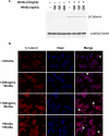
Figure 2. Wnt5a Does Not Affect β-Catenin Stabilization
(A) Wnt5a protein treatment does not affect cytosolic β-catenin protein levels. The 293 cells were treated for 3 h with the indicated concentrations of Wnt proteins and then hypotonically lysed for Western blot analysis of cytosolic fractions. (B) Wnt5a protein treatment does not alter β-catenin cellular localization. Cells grown on coverslips were treated with the indicated concentrations of Wnt proteins for 6 h and then fixed and stained as described in the text. Membrane localized β-catenin is indicated by arrowheads; membrane and cytosolic staining is indicated by arrows.
Figure 3
Figure 3. Wnt5a-Mediated Inhibition of Canonical Signaling Is Not Associated with Wnt-Stimulated Ca 2+ Flux
(A) Wnt5a-mediated inhibition is PTX insensitive. At 24 h post-transfection, 293 cells were pretreated with vehicle or 100 ng/ml PTX for 24 h. Cells were then treated with indicated Wnt proteins concomitantly with vehicle or PTX for an additional 24 h, and luciferase assay was performed. (B) PTX is active in 293 cells as PTX pretreatment of cells inhibits LPA-induced Erk1,2 activation. The 293 cells were pretreated with 100 ng/ml PTX for 24 h and then treated with 10 μM LPA or vehicle for 10 min. Western blot analysis was then performed on total cell lysates using anti–phospho-Erk1,2 antibody. Membrane was stripped and reprobed for total Erk1,2 as a loading control. (C) Wnt5a protein does not directly stimulate intracellular Ca 2+ flux. The 293 and 293Fz4 cells were loaded with Fura-2-dextran and then monitored for changes in intracellular Ca 2+ concentration as determined by the change in 340/380 excitation wavelength ratio following Wnt5a (500 ng/ml) and subsequent ionomycin treatment. Data represent the average 340/380 excitation wavelength ratio of three or more independent cells within the same field ± SD. Wnt5a has little effect on the ratio over the 15-min time course, whereas ionomycin treatment rapidly and robustly induces Ca 2+ flux in these cells. Prolonged treatment with Wnt5a (up to 1 h) did not result in changes in intracellular Ca 2+ levels (unpublished data).
Figure 4
Figure 4. Wnt5a Protein Activates β-Catenin Signaling Depending on Receptor Context
(A) Wnt5a treatment leads to β-catenin stabilization specifically in cells expressing mFz4. The 293 and 293Fz4 cells were treated with Wnt proteins and then assayed for cytosolic β-catenin protein accumulation via Western blot analysis. (B) Wnt5a treatment activates the STF reporter when LRP5 is coexpressed. At 24 h post-transfection with STF reporter and LRP5 or empty vector, 293Fz4 cells were treated with increasing concentrations of Wnt5a protein for an additional 24 h, and luciferase assay was performed as described in Figure 1. (C) Wnt5a does not inhibit Wnt3a-mediated reporter activation in 293Fz4 cells. The 293 and 293Fz4 cells were treated with Wnts as indicated 24 h post-transfection with reporters, and luciferase assay was performed. (D) Loss of Wnt5a-mediated reporter inhibition is specific to mFz4 overexpression. The 293, 293Fz4, and 293Fz8 cells were treated with the indicated Wnts for 24 h, 24 h post-transfection, and then luciferase assay was performed. Wnt5a maintains inhibitory activity when mFz8, but not mFz4, is overexpressed.
Figure 5
Figure 5. An Alternative Wnt Receptor, mRor2, Mediates Wnt5a's Inhibitory Activity
(A) Wnt5a synergizes with mRor2 to inhibit Wnt3a-mediated reporter activation. The 293 cells stably expressing exogenous mRor2 (293Ror2) were treated with Wnts as indicated, 24 h post-transfection with reporters, and luciferase assay was performed as outlined in Figure 1. (B) Wnt5a binds directly to the Fz4 and mRor2 CRD domains. Wnt5a protein specifically binds to the purified mFz4 and mRor2 CRDs; only background binding to the Smoothened CRD negative control is observed. While it may appear that Wnt5a binds to a similar extent to the Ror2 CRDs and Smo CRD, the input levels of the Smo CRD-IgG exceeded the Ror2 CRD-IgG levels, accounting for the increased background binding Smo CRD-IgG. (C) The mRor2 CRD domain is required for mediating Wnt5a inhibitory activity. The 293 cells were transiently transfected with reporters along with empty vector, mRor2, or mRor2ΔCRD constructs, treated with Wnts as described in text 24 h post-transfection for an additional 24 h, and then luciferase assay was performed as outlined in Figure 1.
Figure 6
Figure 6. Expression of mRor2 Is Required for Wnt5a-Mediated Inhibition of Canonical Signaling
(A) In 293 cells, mRor2 overexpression inhibits Wnt5a-mediated activation of the STF reporter. The 293 and 293Fz4 cells were transiently transfected with the reporters and LRP5 with or without mRor2 and then treated with Wnt proteins for 24 h, 24 h post-transfection. Overexpression of wild-type mRor2 inhibits Wnt5a-mediated activation of the STF reporter as well as allowing Wnt5a to inhibit Wnt3a-mediated STF reporter activation in 293Fz4 cells. (B) mRor2 overexpression inhibits Wnt5a-mediated activation of the STF reporter and allows Wnt5a to block Wnt3a-induced STF reporter activation in mouse L cells. L and LFz4 cells were transiently transfected with the reporters and LRP5 with or without mRor2 and then treated with Wnt proteins for 24 h, 24 h post-transfection. (C) Quantitative real-time PCR analysis of Ror2 expression in 293 and L cells. Reverse transcription followed by 45 cycles of quantitative real-time amplification of 293 cell RNA reveals a robust Ror2 product that is over 700 times more abundant than that produced from RNA derived from mouse L cells. Hprt quantification serves as an internal normalization control and RNA derived from 293Ror2 and LRor2 cells serves as a positive control for the reaction. The products from the PCR analysis are displayed below the charted quantitative data. (D) The cytoplasmic domain of mRor2 is required for its inhibitory function. Expression of a membrane-tethered variant of mRor2 possessing the extracellular domain of mRor2 fused to a GPI linkage (mRor2-GPI) reduces Wnt5a's ability to inhibit Wnt3a induced canonical Wnt signaling as well as inhibits wild type mRor2′s ability to enhance Wnt5a-mediated inhibition. (E) Expression of a membrane-tethered variant of mRor2 (mRor2-TM) that contains the extracellular and transmembrane domains of mRor2, but lacks the cytoplasmic domain, does not enhance Wnt5a mediated inhibition of canonical Wnt signaling. Expression of mRor2-TM reduces Wnt5a's ability to inhibit Wnt3a-induced canonical Wnt signaling when wild-type mRor2 is overexpressed. Western blot analysis shows that wild-type mRor2 levels do not change when the truncated mRor2 variants are coexpressed.
Figure 7
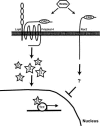
Figure 7. Model. Wnt5a Can Activate or Inhibit Canonical Wnt Signaling Depending on Receptor Context
In the presence of Fz4 and LRP5, Wnt5a can activate β-catenin/TCF signaling. However, when Ror2 is expressed, Wnt5a inhibits canonical Wnt signaling downstream of β-catenin stabilization, at the level of TCF-mediated transcription.

Comment in

Similar articles

Cited by

References

    1. Logan CY, Nusse R. The Wnt signaling pathway in development and disease. Annu Rev Cell Dev Biol. 2004;20:781–810. - PubMed
    1. Polakis P. Wnt signaling and cancer. Genes Dev. 2000;14:1837–1851. - PubMed
    1. Reya T, Clevers H. Wnt signalling in stem cells and cancer. Nature. 2005;434:843–850. - PubMed
    1. Shimizu H, Julius MA, Giarre M, Zheng Z, Brown AM, et al. Transformation by Wnt family proteins correlates with regulation of beta-catenin. Cell Growth Differ. 1997;8:1349–1358. - PubMed
    1. Wong GT, Gavin BJ, McMahon AP. Differential transformation of mammary epithelial cells by Wnt genes. Mol Cell Biol. 1994;14:6278–6286. - PMC - PubMed

Publication types

MeSH terms