. 2006 Apr 19;25(8):1590-602.
doi: 10.1038/sj.emboj.7601067. Epub 2006 Apr 6.

GGA function is required for maturation of neuroendocrine secretory granules

Affiliations

GGA function is required for maturation of neuroendocrine secretory granules

Or Kakhlon et al. EMBO J. .

Abstract

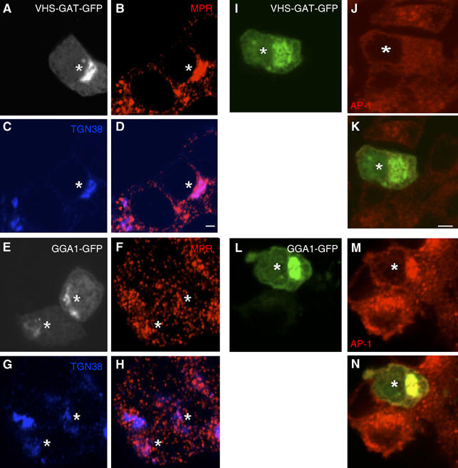
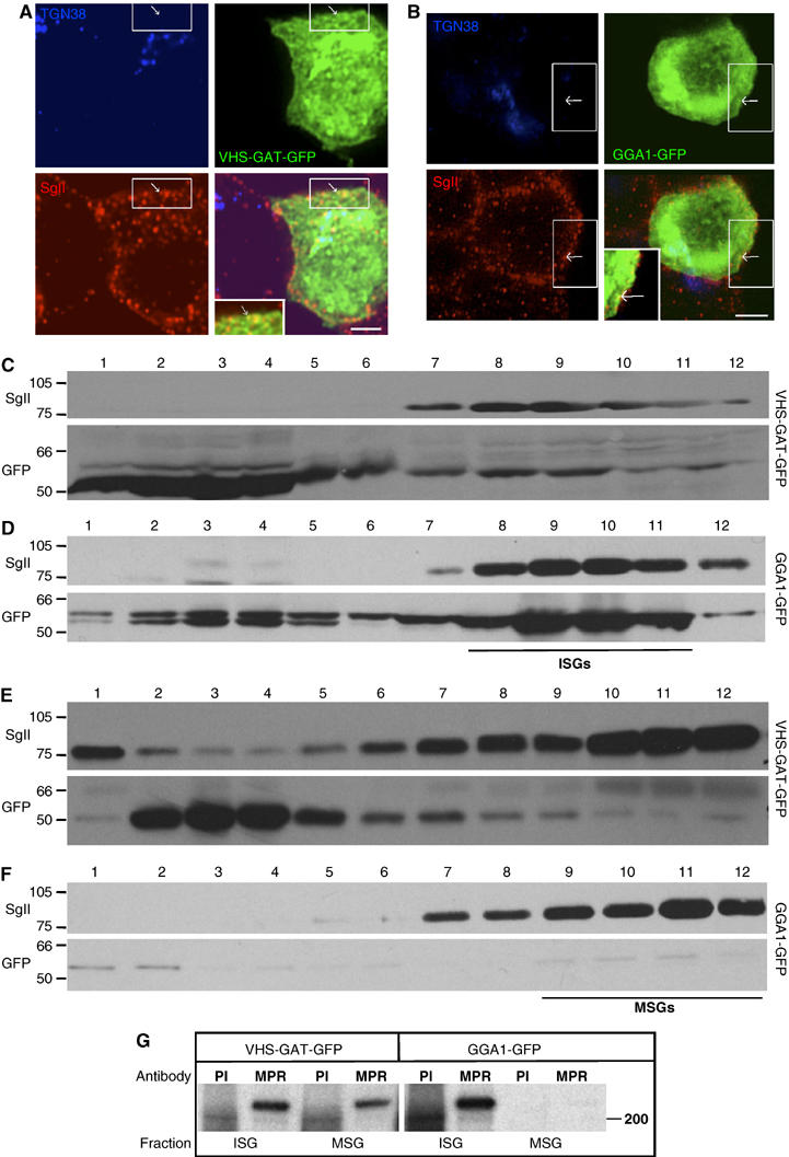
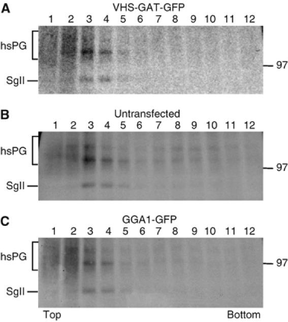
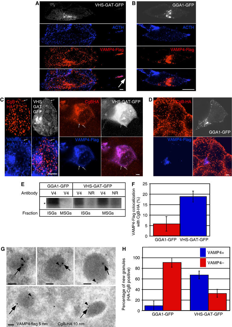
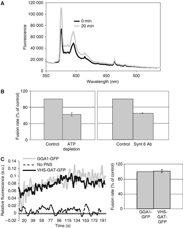
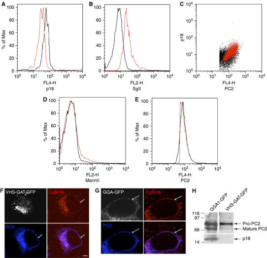
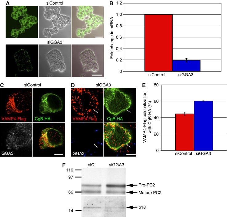
Secretory granule (SG) maturation has been proposed to involve formation of clathrin-coated vesicles (CCVs) from immature SGs (ISGs). We tested the effect of inhibiting CCV budding by using the clathrin adaptor GGA (Golgi-associated, gamma-ear-containing, ADP-ribosylation factor-binding protein) on SG maturation in neuroendocrine cells. Overexpression of a truncated, GFP-tagged GGA, VHS (Vps27, Hrs, Stam)-GAT (GGA and target of myb (TOM))-GFP led to retention of MPR, VAMP4, and syntaxin 6 in mature SGs (MSGs), suggesting that CCV budding from ISGs is inhibited by the SG-localizing VHS-GAT-GFP. Furthermore, VHS-GAT-GFP-overexpression disrupts prohormone convertase 2 (PC2) autocatalytic cleavage, processing of secretogranin II to its product p18, and the correlation between PC2 and p18 levels. All these effects were not observed if full-length GGA1-GFP was overexpressed. Neither GGA1-GFP nor VHS-GAT-GFP perturbed SG protein budding from the TGN, or homotypic fusion of ISGs. Reducing GGA3 levels by using short interfering (si)RNA also led to VAMP4 retention in SGs, and inhibition of PC2 activity. Our results suggest that inhibition of CCV budding from ISGs downregulates the sorting from the ISGs and perturbs the intragranular activity of PC2.


Figures

Figure 1
Figure 1
VHS-GAT-GFP but not GGA1-GFP accumulate MPR at a juxtanuclear region and redistribute AP1 to the cytosol. PC12 cells transfected with VHS-GAT-GFP (AD, IJ) or GGA1-GFP (EH, LN) were fixed 24 h post-transfection and labeled with (B, F) anti-MPR, anti-γ AP (J, M) Abs (red), or (C, G) anti-TGN38 (blue), and secondary Abs. Representative confocal images with the VHS-GAT-GFP (A, I) and GGA1-GFP (E, L) channels are shown. (D), (H), (K), and (N) are merged channels of (B) and (C, F and G, I and J, and L and M), respectively. MPR accumulation and AP1 redistribution can be observed in the VHS-GAT-GFP transfected cells (*). Bars=2 μm.
Figure 2
Figure 2
VHS-GAT-GFP is found on ISGs, and inhibits removal of MPR from ISGs. PC12 cells transfected with (A) VHS-GAT-GFP (green) or (B) GGA1-GFP (green) were fixed after 24 h and labeled with anti-TGN38 (blue) and anti-SgII (red) Abs and secondary Abs. Channel merges are shown in bottom right of each panel. Insets in the merged channels are the boxed region magnified. Arrows, GFP and SgII-positive, but TGN38-negative structures. Bar=2 μm. (CF) Fractions from EGs, which were loaded with (C, D) VG fractions 2–4, or (E, F) fractions 5–7 prepared from PNS of PC12 cells transfected with VHS-GAT-GFP (C, E) or GGA1-GFP (D, F). Proteins precipitated from the EG fractions were subjected to SDS–PAGE and immunoblotting using anti-SgII (upper panels) and anti-GFP (lower panels) Abs. Position of ISGs and MSGs on EGs is indicated. (G) ISG and MSG fractions prepared from [35S]-Met/Cys-labeled FACS-sorted cells expressing VHS-GAT-GFP or GGA1-GFP were subjected to IP with anti-MPR ab, or a preimmune control (PI).
Figure 3
Figure 3
TGN budding of ISGs is not affected by VHS-GAT-GFP and GGA1-GFP. PC12 cells were transfected with either VHS-GAT-GFP or GGA1-GFP and 24 h later FACS-sorted. Cells positive for VHS-GAT-GFP (A), unstransfected (B), or positive for GGA1-GFP (C) were reseeded, cultured overnight, pulsed for 5 min with [35S]-sulfate, and chased for 15 min to allow budding from the TGN. PNS was prepared from both cell populations and fractionated on a sucrose VG, and an aliquot of each fraction was subjected to SDS–PAGE and autoradiography. The peak of ISGs is found in fraction 3 and 4 under all conditions.
Figure 4
Figure 4
VHS-GAT-GFP but not GGA1-GFP inhibits the removal of VAMP4 from maturing ISGs. (A, B) AtT20 cells were cotransfected with either (A) VHS-GAT-GFP or (B) GGA1-GFP and VAMP4-Flag and fixed 24 h post-transfection. Anti-Flag (red) and anti-ACTH (blue) were visualized with secondary Abs by confocal microscopy. GFP channel is shown in white. Arrow, ACTH and VAMP4-Flag colocalizing at cell tip. Bars=5 μm. (C, D) PC12 cells were cotransfected with CgB-HA and VAMP4-Flag and either (C) VHS-GAT-GFP or (D) GGA1-GFP, and fixed 24 h post-transfection. Anti-HA (red) and anti-Flag (blue) Abs were visualized with secondary Abs by confocal microscopy. White channel, GFP. Arrows in (C), VAMP4-Flag positive granules. Bars=1 μm. (E) A PNS from VHS-GAT-GFP and GGA1-GFP FACS sorted, [35S]-Met/Cys-labeled cells was used to prepare ISG and MSG fractions. Membranes sedimented from these fractions were used for VAMP4 IP using a VAMP4 (V4) or an NR Ab. (F) Quantitation of the percentage of VAMP4-Flag structures colocalized with CgB-HA outside of the TGN/juxtanuclear area in PC12 cells transfected as in (C) (blue bar) and (D) (red bar). Percentage of VAMP4-Flag colocalization with CgB-HA was calculated based on the means of five independent observations, using the LSM 510 software. Student's t-test shows that the extent of colocalization is higher in the VHS-GAT-GFP-expressing cells than in cells expressing GGA1-GFP (P⩽1%). (G) Representative immuno-EM images of new granules positive for CgB-HA (10 nm gold, arrow) and VAMP4-Flag (5 nm gold, arrowhead). Images from VHS-GAT-GFP FACS-sorted cells are shown. (H) Quantitation of VAMP4-Flag-positive and -negative new granules in the two FACS-sorted populations. Fischer's exact test analysis shows the proportion of VAMP4-Flag-positive new granules in VHS-GAT-GFP cells is significantly higher than in GGA1-GFP cells (P⩽1%), n=3. Bar=100 nm.
Figure 5
Figure 5
A PHA-based ISG–ISG fusion assay shows that ISG–ISG fusion rate is not affected by VHS-GAT-GFP or GGA1-GFP overexpression. (A) The emission spectrum of a mixture of PHA-labeled ISGs, PNS, ATP-regenerating system, and GTP in FB (standard fusion conditions) was measured at 0 min (black), and after 20 min incubation (gray) at 37°C. After incubation, the PHA monomer fluorescence intensity (peaks at 377 and 397 nm) has increased. (B) Rates of fusion measured by the initial slope of a time-based monitoring of fluorescence, as shown in (C). Left panel, PHA-labeled ISGs were preincubated for 5 min on ice with PNS under standard fusion conditions (control), or with 25 U hexokinase and 3.3 mM glucose (ATP depletion). Following the preincubation, fluorescence was monitored over time as described in Materials and methods, and initial slopes calculated. Right panel, PHA-labeled ISGs were preincubated 10 min on ice under standard fusion conditions (as in left panel), or with 27 μg/ml Sx6 Ab, before monitoring fluorescence (n=3). Both ATP depletion and Sx6 Ab treatments caused a significant reduction in the initial slopes of fluorescence over time, as confirmed by Student's t-tests (P⩽1%). (C) A fusion assay performed using PHA-labeled ISGs in fusion buffer containing GTP- and ATP-regenerating system (dashed line), or with a PNS from 107 FACS-sorted VHS-GAT-GFP-positive cells (black line), or 107 FACS-sorted GGA1-GFP positive cells (gray line). The fluorescence of PHA monomers (ex. 330, em. 377) was recorded continuously at 37°C. Data shown are normalized to infinite dilution of PHA obtained by adding 1% Triton X-100 to each condition after the signal had reached a plateau. Fusion rates (averages of three experiments) as in (B) are shown in the right panel (n=3).
Figure 6
Figure 6
Inhibition of SgII processing in VHS-GAT-GFP- but not in GGA1-GFP-expressing cells. PC12 cells cotransfected with PC2 and either VHS-GAT-GFP (black lines in A, B, D, E), or as a negative control GGA1-GFP (red lines in A, B, D, E). At 24 h post–transfection, the cells were fixed and labeled with (A) and (C) anti-PC2 and anti-p18, (B) anti-SgII and anti-p18, (D) anti-PC2 and anti-Mann II, and (E) anti-PC2, and analyzed by flowcytometry with secondary Abs. Cells positive for either VHS-GAT-GFP (black) or GGA1-GFP (red) and PC2 (or p18 in B) were gated and analyzed for the distribution of p18 (A), SgII (B), MannII (D), and PC2 (E). Student's t-tests show that the ratios (median p18 in PC2-positive cells)/(median p18 in PC2-negative cells) in (A) and (median SgII in p18-positive cells)/(median SgII in p18-negative cells) in (B) are significantly (A) smaller (P⩽5%) or (B) larger (P⩽5%) in VHS-GAT-GFP- than in GGA1-GFP-positive cells. No significant difference was observed between the VHS-GAT-GFP and GGA1-GFP populations in (D) and (E). (C) Dot plots testing the correlation between p18 and PC2 levels in VHS-GAT-GFP- (black) and GGA1-GFP- (red) gated cells. One-way ANOVA shows that a significant (P⩽2.5%) linear trend exists between p18 and PC2 in GGA1-GFP-positive cells, but not in the VHS-GAT-GFP-positive cells. (F, G) PC12 cells were cotransfected with either VHS-GAT-GFP (F), or GGA1-GFP (G) and PC2 and fixed 24 h post-transfection. HA (red) and PC2 (blue) were visualized by confocal microscopy with secondary Abs, GFP channel (white). (H) PC12 cells cotransfected with PC2 and either GGA1-GFP (left lane), or VHS-GAT-GFP (right lane) were, after FACS sorting for GFP- and PC2-positive cells, lysed and subjected to SDS–PAGE and immunoblotting with anti-PC2 and anti-p18 Abs.
Figure 7
Figure 7
Depletion of GGA3 in PC12 cells inhibits VAMP4-Flag removal from maturing SGs and reduces pro-PC2 maturation. (A) PC12 cells were treated either with siControl (upper panel) or siGGA3 (lower panel), fixed, and labeled with anti-GGA3 Ab and secondary Ab (green). Representative confocal images, phase-contrast images, and channel merges are shown. Bar=20 μm. (B) Real-time RT–PCR analysis of GGA3 mRNA in PC12 cells transfected with siControl or siRNA against GGA3 (siGGA3). (CE) PC12 cells were transfected with siControl (C) or siGGA3 (D), and 48 h later, retransfected with the siRNAs, and with CgB-HA and VAMP4-Flag constructs, and fixed 24 h later. Anti-GGA3 (white), anti-HA (green), and anti-Flag (red) Abs were visualized with secondary Abs by confocal microscopy. (E) Quantitation of the percentage of all VAMP4-Flag structures colocalizing with CgB-HA, n=3. Colocalization was quantified using the LSM software. Student's t-test shows that the extent of VAMP4-Flag colocalized with CgB-HA is significantly (P⩽5%) higher in siGGA3- than in siControl-treated cells. (F) PC12 cells were treated with siControl or siGGA3 and cotransfected with the PC2 plasmid. PC2-positive cells were FACS sorted from the siControl- and siGGA3-treated cell populations, and analyzed as in Figure 6F with PC2 and p18 Abs. The maturation of pro-PC2 to PC2 was inhibited in siGGA3-treated cells resulting in, on average, a 180% increase in the ratio (pro-PC2/mature PC2) in siGGA3- relative to siControl-treated cells. p18 levels were also reduced in siGGA3 cells by 30% on average.

Similar articles

Cited by

References

    1. Arvan P, Castle D (1998) Sorting and storage during secretory granule biogenesis: looking backward and looking forward. Biochem J 332: 593–610 - PMC - PubMed
    1. Austin C, Hinners I, Tooze SA (2000) Direct and GTP-dependent interaction of ADP-ribosylation Factor 1 with clathrin adaptor protein AP-1 on immature secretory granules. J Biol Chem 275: 21862–21869 - PubMed
    1. Barr F, Huttner WB (1996) A role for ADP-ribosylation factor1, but not COP I, in secretory vesicle biogenesis from the trans-Golgi network. FEBS Lett 384: 65–70 - PubMed
    1. Boman AL, Zhang C, Zhu X, Kahn RA (2000) A family of ADP-ribosylation factor effectors that can alter membrane transport through the trans-Golgi. Mol Biol Cell 11: 1241–1255 - PMC - PubMed
    1. Bonifacino JS (2004) The GGA proteins: adaptors on the move. Nat Rev Mol Cell Biol 5: 23–32 - PubMed

MeSH terms

LinkOut - more resources