. 2006 Apr 19;25(8):1603-10.
doi: 10.1038/sj.emboj.7601070. Epub 2006 Apr 6.

Mba1, a membrane-associated ribosome receptor in mitochondria

Affiliations

Mba1, a membrane-associated ribosome receptor in mitochondria

Martin Ott et al. EMBO J. .

Abstract

The genome of mitochondria encodes a small number of very hydrophobic polypeptides that are inserted into the inner membrane in a cotranslational reaction. The molecular process by which mitochondrial ribosomes are recruited to the membrane is poorly understood. Here, we show that the inner membrane protein Mba1 binds to the large subunit of mitochondrial ribosomes. It thereby cooperates with the C-terminal ribosome-binding domain of Oxa1, which is a central component of the insertion machinery of the inner membrane. In the absence of both Mba1 and the C-terminus of Oxa1, mitochondrial translation products fail to be properly inserted into the inner membrane and serve as substrates of the matrix chaperone Hsp70. We propose that Mba1 functions as a ribosome receptor that cooperates with Oxa1 in the positioning of the ribosome exit site to the insertion machinery of the inner membrane.


Figures
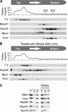
Figure 1
Figure 1
Mba1 cofractionates with mitochondrial ribosomes on sucrose gradients. (A) Wild-type mitochondria (1 mg) were lysed in Triton buffer and the extract was subfractionated by velocity centrifugation on a continuous sucrose gradient. After fractionation of the gradient, the protein concentration in each fraction was determined. The distribution of the following proteins in the fractions was assessed by Western blotting: F1β, the β-subunit of the ATPase; Mrps51 and Mrpl20, proteins of the small and large subunit of the mitochondrial ribosome, respectively; Mba1 and Oxa1. (B) Like A, with the exception that the mitochondrial extract was treated with 600 U/ml of bovine pancreatic RNase A for 30 min to disintegrate mitochondrial ribosomes. (C) Mitochondrial extracts were mock treated or incubated with 100 U/ml of RNase A to remove surface-exposed ribosomal RNA. Remaining ribosomal particles (P) were separated from the supernatant (S) by centrifugation through a high sucrose cushion. Unfractionated samples (T, total) are shown for control.
Figure 2
Figure 2
Ribosome binding of Mba1 is independent of Oxa1 and nascent chains. (A) Mitochondria (234 μg) isolated from Δoxa1 cells were incubated in translation buffer for 15 min. The sample was split and to one-half 2 mM puromycin was added. Following incubation for 5 min, the mitochondria were lysed and fractionated as described in Figure 1. The distribution of proteins was assessed by Western blotting. (B) Translation products were radiolabeled in isolated wild-type mitochondria (400 μg) for 15 min. The sample was split and one-half treated with puromycin (PM) as in (A). The reactions were divided into four aliquots. Mitochondria were directly applied to the gel (T, total) or lysed in 1% Triton X-100, 20 mM Tris, pH 7.4, containing 5 mM EDTA, 50 mM KCl (EDTA) or 5 mM Mg2+, 50 mM KCl (Mg2+) or 5 mM EDTA, 0.5 M KCl (0.5 M KCl EDTA). The extracts were cleared by centrifugation and applied onto a 1.2 M sucrose cushion. A ribosomal pellet fraction (P) was separated from residual proteins in the supernatant (S) by centrifugation for 60 min at 200 000 g. Proteins were resolved by SDS–PAGE and visualized by autoradiography. Fractions containing nascent chains, which appear as a smear due to their heterogenic size, are indicated by asterisks.
Figure 3
Figure 3
Mba1 binds mitochondrial ribosomes in vitro. (A) Recombinant MBP and MBP-Mba1 were purified and immobilized on an amylose resin. Mitochondrial translation products were radiolabeled in isolated mitochondria. Mitochondria were solubilized. The resulting extract was clarified by centrifugation and subjected to the immobilized MBP fusion proteins. Following extensive washing, bound proteins were eluted with sample buffer, resolved by SDS–PAGE and transferred to nitrocellulose. Transferred proteins were analyzed by staining with Ponceau S (upper panel) or by Western blotting with antibodies against proteins of the large (Mrpl36) and small (Mrps51) ribosomal subunit, Oxa1, aconitase (Aco1) and Tom70. The lower panel shows the radioactive translation products that were detected by autoradiography. Total lanes (T) show 10% of the mitochondrial extract that was applied to the resin. (B) Translation products were radiolabeled in mitochondria isolated from wild type (wt), a Δoxa1 mutant, a mutant containing a C-terminally truncated variant of Oxa1 (oxa1ΔC) and a rho0 strain. The binding of ribosomes and Oxa1 to the immobilized MBP-Mba1 fusion protein was assessed as in (A).
Figure 4
Figure 4
Deletion of Mba1 and the ribosome-binding domain of Oxa1 causes synergistic growth and insertion defects. (A) The strains indicated were grown on YPD medium to log phase. Serial 10-fold dilutions of the cultures were spotted on YP plates containing 2% glucose or 2% glycerol and plates were incubated at 30°C for 2 or 3 days, respectively. (B) The deletion of both Mba1 and the ribosome-binding domain of Oxa1 leads to the accumulation of unprocessed Cox2. Translation products were radiolabeled in mitochondria isolated from the strains indicated for 10 min at 25°C, separated by SDS–PAGE and visualized by autoradiography. (C) The deletion of Mba1 or of the ribosome-binding domain of Oxa1 causes a general insertion defect of mitochondrial translation products. Translation products were radiolabeled in mitochondria isolated from the strains indicated for 30 min at 30°C. The mitochondria were reisolated, washed and treated with 0.1 M Na2CO3 and 4.5 M urea. The suspension was adjusted to 1.6 M sucrose and layers of 1.4 and 0.25 M sucrose were placed on top of it. The gradient was centrifuged and fractionated. Proteins in the fractions were precipitated with 12% trichloroacetic acid and analyzed by SDS–PAGE and autoradiography. The radioactive signals of the translation products were quantified in each fraction and are depicted on the right.
Figure 5
Figure 5
In the absence of Mba1 and the ribosome-binding domain of Oxa1 mitochondrial translation products interact with mtHsp70. Translation products were radiolabeled for 20 min at 37°C in mitochondria from the strains indicated. The mitochondria were reisolated, treated with apyrase to deplete ATP and lysed in 0.1% Triton X-100, 150 mM NaCl, 4 mM EDTA, 20 mM Tris/HCl, pH 7.4, for 10 min at 4°C. The extract was cleared by centrifugation at 17 500 g for 10 min and used for immunoprecipitation with antibodies against mtHsp70 (α70) or preimmune serum (p.i.). Bound proteins were eluted with 1% SDS and analyzed by SDS–PAGE and autoradiography.
Figure 6
Figure 6
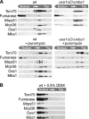
Mitochondrial ribosomes remain associated with the inner membrane in the absence of the ribosome-binding domain of Oxa1 and of Mba1. (A) Mitochondria from the strains indicated were incubated with or without puromycin and disintegrated by freeze thawing in 150 mM KCl, 5 mM EDTA, 20 mM Tris/HCl, pH 7.4. Then, the suspension was adjusted to 2 M sucrose and layers of 2 M sucrose, 1.5 M sucrose, 1 M sucrose in 60 mM KCl, 20 mM Tris/HCl, pH 7.4, were placed on top. After centrifugation at 215 600 g for 16 h at 2°C, the gradient was fractionated. Proteins in the fractions were precipitated by the addition of 12% trichloroacetic acid and analyzed by Western blotting with antibodies against the proteins indicated. (B) Isolated wild-type mitochondria were lysed in 0.5% dodecyl maltoside (DDM), 5 mM EDTA, 20 mM Tris/HCl, pH 7.4, for 15 min at 4°C and processed as in (A).
Figure 7
Figure 7
Model for membrane interaction of mitochondrial ribosomes. Mitochondrial ribosomes are tightly associated with the inner membrane. This interaction appears to rely on at least three mechanisms: (1) mitochondrial mRNAs are bound to the inner membrane by translational activators. (2) The membrane proteins Mba1 and Oxa1 are associated with the large subunit of the mitochondrial ribosome. The results shown in this study indicate that Mba1 and the C-terminal ribosome-binding domain of Oxa1 are critical to achieve close contact of the polypeptide exit tunnel of the ribosome and the insertion machinery of the inner membrane. (3) Additional factors, like Mdm38 or Cox11, contribute to the membrane binding of ribosomes independently of nascent chains, Mba1 or Oxa1.

Similar articles

Cited by

References

    1. Arlt H, Tauer R, Feldmann H, Neupert W, Langer T (1996) The YTA10–12 complex, an AAA protease with chaperone-like activity in the inner membrane of mitochondria. Cell 85: 875–885 - PubMed
    1. Bauer M, Behrens M, Esser K, Michaelis G, Pratje E (1994) PET1402, a nuclear gene required for proteolytic processing of cytochrome oxidase subunit 2 in yeast. Mol Gen Genet 245: 272–278 - PubMed
    1. Beckmann R, Bubeck D, Grassucci R, Penczek P, Verschoor A, Blobel G, Frank J (1997) Alignment of conduits for the nascent polypeptide chain in the ribosome–Sec61 complex. Science 278: 2123–2126 - PubMed
    1. Beckmann R, Spahn CM, Eswar N, Helmers J, Penczek PA, Sali A, Frank J, Blobel G (2001) Architecture of the protein-conducting channel associated with the translating 80S ribosome. Cell 107: 361–372 - PubMed
    1. Bonnefoy N, Chalvet F, Hamel P, Slominski PP, Dujardin G (1994) OXA1, a Saccharomyces cerevisiae nuclear gene whose sequence is conserved from prokaryotes to eukaryotes controls cytochrome oxidase biogenesis. J Mol Biol 239: 201–212 - PubMed

Publication types

MeSH terms