. 2006 Apr 17;203(4):1007-19.
doi: 10.1084/jem.20051342. Epub 2006 Apr 3.

Dystroglycan is selectively cleaved at the parenchymal basement membrane at sites of leukocyte extravasation in experimental autoimmune encephalomyelitis

Affiliations

Dystroglycan is selectively cleaved at the parenchymal basement membrane at sites of leukocyte extravasation in experimental autoimmune encephalomyelitis

Smriti Agrawal et al. J Exp Med. .

Abstract

The endothelial cell monolayer of cerebral vessels and its basement membrane (BM) are ensheathed by the astrocyte endfeet, the leptomeningeal cells, and their associated parenchymal BM, all of which contribute to establishment of the blood-brain barrier (BBB). As a consequence of this unique structure, leukocyte penetration of cerebral vessels is a multistep event. In mouse experimental autoimmune encephalomyelitis (EAE), a widely used central nervous system inflammatory model, leukocytes first penetrate the endothelial cell monolayer and underlying BM using integrin beta1-mediated processes, but mechanisms used to penetrate the second barrier defined by the parenchymal BM and glia limitans remain uninvestigated. We show here that macrophage-derived gelatinase (matrix metalloproteinase [MMP]-2 and MMP-9) activity is crucial for leukocyte penetration of the parenchymal BM. Dystroglycan, a transmembrane receptor that anchors astrocyte endfeet to the parenchymal BM via high affinity interactions with laminins 1 and 2, perlecan and agrin, is identified as a specific substrate of MMP-2 and MMP-9. Ablation of both MMP-2 and MMP-9 in double knockout mice confers resistance to EAE by inhibiting dystroglycan cleavage and preventing leukocyte infiltration. This is the first description of selective in situ proteolytic damage of a BBB-specific molecule at sites of leukocyte infiltration.


Figures
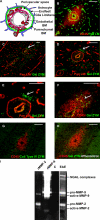
Figure 1.
Figure 1.
Gelatinase activity in EAE brains. Scheme of a post-capillary venule, showing cell layers and endothelial and parenchymal BMs separated by the perivascular space (A). EAE brain sections stained for pan-laminin and CD45 show leukocyte accumulation in the perivascular space and infiltration into the brain parenchyma (B). In situ zymography coupled with pan-laminin (C–E) or CD45 (F) immunofluorescence reveals protease activity in the CNS parenchyma subjacent to the parenchymal BM (arrows) at sites of leukocyte infiltration, only when gelatin (C–F) and not collagen type IV (G) is used as substrate. 1,10-phenanthroline abolishes gelatinase activity (H). (I) Gelatin gel zymography of healthy (C) and EAE brains shows proforms of MMP-9 and MMP-2 and the smaller active forms in EAE samples only. Higher mol wt bands are neutrophil gelatinase B–associated lipocalin (NGAL)–associated lipocalin complexes. rMMP-2 and rMMP-9 are standards. C–E are the same specimen at different magnifications; B, C–E, and F–H are serial sections. Data represent results from 10 mice. Bars, 40 μm.
Figure 2.
Figure 2.
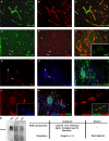
β-dystroglycan loss in inflamed vessels. Immunofluorescence for β-dystroglycan and pan-laminin reveals continuous β-dystroglycan staining bordering the parenchymal BM in noninflamed vessels (A–C) and loss of β-dystroglycan in inflamed vessels only (arrows in D and E). Triple staining for pan-laminin with β-dystroglycan and CD45 (G–I), or agrin and CD45 (J and K) reveals loss of β-dystroglycan at sites of leukocyte infiltration (G and I), despite continuous pan-laminin (H) and agrin (J and K) staining. Double β-dystroglycan and GFAP staining indicates presence of astrocyte endfeet surrounding inflamed vessels (arrows in F). Perlecan staining of the parenchymal BM is continuous around perivascular cuffs, with higher intensity staining in endothelial BMs (L). Western blot reveals ∼200 kD agrin core protein and the characteristic broad mol wt smear in stage 4 EAE and noninflamed brains (Cont.) and purified agrin (M). The table summarizes immunofluorescence and Western blot data. Images are from different specimens and represent results from 10 mice. Bars, 40 μm.
Figure 3.
Figure 3.
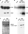
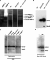
In vivo β-dystroglycan cleavage. Western blots for β- (A and B), α-dystroglycan (C), and secondary antibody control (D). A and C and WGA in B are glycoprotein-enriched CNS samples; Memb. is a CNS membrane fraction (B). All samples are run under reducing conditions. Intact β-dystroglycan at 43 kD is detected in stage 2 and 4 EAE and in noninflamed brains (Cont.) (A and B). An additional 30-kD β-dystroglycan fragment occurs only in EAE glycoprotein enriched and membrane fractions (A and B). Intact α-dystroglycan is observed at ∼120 kD in all samples (C). Lower bands in C are nonspecific cross-reactivity of the secondary antibody (D). The ∼80-kD band represents endogenous IgM μ chain, abundant in inflamed CNS, as shown in the negative control (D).
Figure 4.
Figure 4.
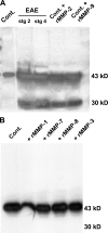
MMP-2 and MMP-9 cleave β-dystroglycan. Western blots for β-dystroglycan in glycoprotein-enriched samples from noninflamed brains (Cont.), stage 2 and 4 EAE brains, or Cont. samples treated with activated rMMP-2 or rMMP-9 result in the same 30-kD β-dystroglycan fragment as seen in EAE samples. Weak reaction of the secondary antibody with endogenous Ig as a result of serum influx into diseased brains is seen at ∼50 kD (A). Treatment of the same noninflamed sample with activated rMMP-1, rMMP-7, rMMP-8, or MMP-3 does not cleave β-dystroglycan (B).
Figure 5.
Figure 5.
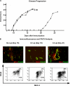
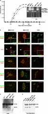
Macrophage depletion delays EAE onset. (A) Clodronate-liposome (Cl Lip) (▪)–treated mice are resistant to EAE up to day 21 after MOG immunization, whereas clinical symptoms appear by day 10 in PBS-liposome (♦)–treated and untreated mice (•). (B) FACS of CNS infiltrates reveals that CD45high/MAC3high macrophages represent 32% of the total CD45+ population at day 15 after immunization and reduction of this population to 13% in Cl Lip–treated mice at day 19. By day 21, the proportion of CD45high/MAC3high macrophages increases to 28% in Cl Lip–treated mice, coincident with EAE onset. Proportions of activated CD45low/MAC3high microglia do not vary between groups (7.5, 6.9, 7%). Immunofluorescence reveals MAC3 positive macrophages in CNS parenchyma of PBS-liposome–treated mice at day 15 after immunization that are absent in Cl Lip–treated mice until day 19, reappearing at day 21. Images are from different mice and represent results from six mice in each group.
Figure 6.
Figure 6.
T cells proliferate normally in clodronate-liposome–treated mice. Antigen-specific (MOG 35–55) (A) and anti-CD3–induced T cell proliferation (B) show no significant difference between clodronate- (▪ and black bars) or PBS-liposome (♦ and white bars)–treated mice at days 15 or 21 after MOG immunization. Data are means ± SEM from three experiments with n = 3.
Figure 7.
Figure 7.
Macrophages are crucial for gelatinase activity and β-dystroglycan cleavage. (A) Immunofluorescence for CD45 and β-dystroglycan in CNS sections of clodronate-treated mice at days 19 and 21 after MOG immunization and corresponding gelatin in situ zymographies (ZYM) coupled with staining for CD45 or pan-laminin reveal loss of β-dystroglycan staining and gelatinase activity at the parenchymal border (arrows) only in brains of clodronate-liposome–treated mice at day 21. (B) Gelatin zymography reveals absence of MMP-2 and MMP-9 in CNS samples from noninflamed (Cont.) and clodronate-liposome–treated mice at day 19 after immunization, but not in PBS-liposome–treated mice or clodronate-treated mice at day 21. (C) Corresponding Western blots for β-dystroglycan reveal the 30-kD fragment in PBS-liposome–treated mice and clodronate-liposome–treated mice at day 21 after immunization, but not at day 19. Images in A are either the same section or serial sections and represent results from eight mice in each group. Bars, 40 μm.
Figure 8.
Figure 8.
Macrophage-derived gelatinases cleave astrocyte and brain β-dystroglycan. (A) Gelatin zymography reveals pro- and active-MMP-2 and MMP-9 in macrophage-conditioned media (Mφ), but not in astrocyte lysates. (B) Western blots reveal intact β-dystroglycan in astrocyte lysates, which is cleaved to the 30-kD fragment by addition of macrophage-conditioned media (Mφ + astrocyte). (C) Cleavage of astrocyte- derived β-dystroglycan by macrophage-conditioned media is inhibited by MMP inhibitors (TIMP-1, TIMP-4), but not serine and cysteine protease inhibitors (AEBSF, E-64, Aprotinin). (D) Incubation of crude CNS extracts from healthy mice with macrophage-conditioned media is sufficient to cleave β-dystroglycan.
Figure 9.
Figure 9.
Combined MMP-2 and MMP-9 activity are crucial for EAE. (A) EAE progresses normally in MMP-2 KO (★) and MMP-9 KO (•), whereas MMP-2 and MMP-9 DKO (▪) mice are resistant to EAE up to 40 d after MOG immunization. The inset gel zymograph compares gelatinases in CNS extracts from EAE-induced WT, MMP-2 KO, MMP-9 KO, and DKO mice. (B) All sections were stained for pan-laminin (green) and either CD45, T cell receptor (TCRβ), macrophage/microglia (MAC3), DC (CD11c), or β-dystroglycan, revealing normal cellular infiltrates and associated loss of β-dystroglycan in MMP-2 KO and MMP-9 KO mice (arrows), but not in DKO mice. Images are from different specimens and represent results from eight mice for each of the single KO mice and five DKO mice. (C) Western blot confirms the presence of the 30-kD β-dystroglycan fragment in the CNS of WT, MMP-2 KO, and MMP-9 KO, but not DKO mice. (D) Incubation of brain extracts from healthy mice (non-EAE) with macrophage-conditioned media (Mφ) from MMP-2 KO, MMP-9 KO, or WT littermates, but not MMP-2/MMP-9 DKO, cleaves β-dystroglycan to the 30-kD fragment. Mφ alone did not contain intact or cleaved β-dystroglycan. Bars, 40 μm.

Similar articles

Cited by

References

    1. Wolf, K., R. Muller, S. Borgmann, E.B. Brocker, and P. Friedl. 2003. Amoeboid shape change and contact guidance: T-lymphocyte crawling through fibrillar collagen is independent of matrix remodeling by MMPs and other proteases. Blood. 102:3262–3269. - PubMed
    1. Yadav, R., K.Y. Larbi, R.E. Young, and S. Nourshargh. 2003. Migration of leukocytes through the vessel wall and beyond. Thromb. Haemost. 90:598–606. - PubMed
    1. Engelhardt, B. 1997. Lymphocyte trafficking through the central nervous system. In Adhesion Molecules and Chemokines in Lymphocyte Trafficking. A. Hamann, editor. Harwood Academic Publishers, Amsterdam. 173–200.
    1. Kawakami, N., U.V. Nagerl, F. Odoardi, T. Bonhoeffer, H. Wekerle, and A. Flugel. 2005. Live imaging of effector cell trafficking and autoantigen recognition within the unfolding autoimmune encephalomyelitis lesion. J. Exp. Med. 201:1805–1814. - PMC - PubMed
    1. Sixt, M., B. Engelhardt, F. Pausch, R. Hallmann, O. Wendler, and L.M. Sorokin. 2001. Endothelial cell laminin isoforms, laminin 8 and 10, play decisive roles in T-cell recruitment across the blood-brain-barrier in an EAE model. J. Cell Biol. 153:933–945. - PMC - PubMed

Publication types

MeSH terms