

doi: 10.1084/jem.20050783.
Epub 2006 Mar 13.
Regulatory T cells inhibit stable contacts between CD4+ T cells and dendritic cells in vivo
Affiliations
- PMID: 16533880
- PMCID: PMC2118249
- DOI: 10.1084/jem.20050783
Item in Clipboard
Regulatory T cells inhibit stable contacts between CD4+ T cells and dendritic cells in vivo
J Exp Med.
.
Abstract
Regulatory T (T reg) cells exert powerful down-modulatory effects on immune responses, but it is not known how they act in vivo. Using intravital two-photon laser scanning microscopy we determined that, in the absence of T reg cells, the locomotion of autoantigen-specific T cells inside lymph nodes is decreased, and the contacts between T cells and antigen-loaded dendritic cells (DCs) are of longer duration. Thus, T reg cells can exert an early effect on immune responses by attenuating the establishment of stable contacts during priming of naive T cells by DCs.
Figures
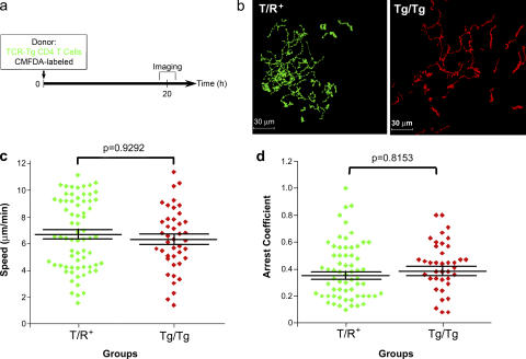
In the absence of immunization, T reg cells do not influence the movement of MBP-specific CD4+ T cells. (a) Experimental protocol. T/R+ or Tg/Tg mice, which have or lack endogenous T reg cells, respectively, received 107 CFSE-labeled MBP Ac1-11–specific CD4+ T cells by tail vein injection. 20 h later, PLNs were imaged. (b) Representative tracks of MBP-specific T cells in T/R+ and Tg/Tg recipients. (c) Mean speeds of MBP-specific CD4+ T cells in both types of recipient mice. (d) Arrest coefficient for CD4+ T cells in both types of recipient mice. Results are representative of three independent experiments.
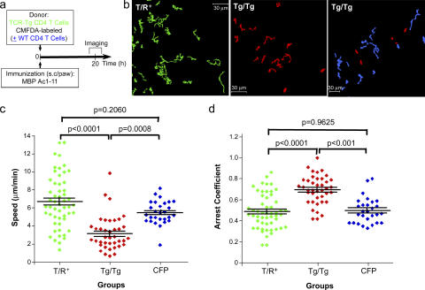
T reg cells release antigen-specific CD4+ T cells in the presence of antigen. (a) Experimental protocol for b–d. T/R+ or Tg/Tg mice, which have or do not have endogenous T reg cells, respectively, received 107 CFSE-labeled MBP Ac1-11–specific CD4+ T cells via the tail vein. As endogenous control, Tg/Tg animals received, by tail vein injection, 1–2 × 10⁷ CFSE-labeled MBP-specific T cells from Tg/Tg mice together with 107 CD4+CD25− T cells from syngeneic CFP-expressing mice. On the same day, mice received 50 μg MBP Ac1-11 peptide emulsified in IFA in the footpad. 20 h after immunization, draining PLNs were imaged. (b) Representative tracks of MBP-specific T cells (green) in T/R+ and Tg/Tg immunized recipients. (c) Mean speeds of MBP-specific CD4+ T cells in both types of recipient mice. (d) Arrest coefficient for CD4+ T cells in both types of recipient mice. Results are representative of three independent experiments. Representative tracks of WT CD4+CD25− T cells (blue) are shown in b. Arrest coefficients for WT CD4+ T cells (blue) are shown in d.
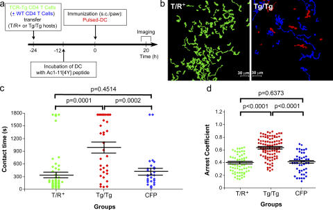
The presence of endogenous T reg cells results in shorter contact time between CD4+ T cells and peptide-pulsed DCs. (a) Experimental protocol. T/R+ or Tg/Tg mice, which have or lack endogenous T reg cells, respectively, received 107 CFSE-labeled MBP Ac1-11–specific CD4+ T cells together with 107 CD4+CD25− T cells from syngeneic CFP mice by tail vein injection and, 24 h later, received 5–10 × 105 CMTMR-labeled DCs pulsed with MBP Ac1-11[4Y] peptide in the footpad. 20 h after DC transfer, PLNs were imaged. (b) Representative tracks of MBP Ac1-11–specific CD4+ T cells in T/R+ and Tg/Tg DC-transferred recipients. Trackings of CFP-expressing WT CD4 T cells are represented in blue. (c) Contact time between MBP-specific or WT CD4 T cells and antigen-loaded DCs. (d) Arrest coefficient for transgenic (green or red) or WT (blue) CD4+ T cells in T/R+ and Tg/Tg DC-transferred recipients. Results in b–d are representative of three independent experiments.

Reconstitution of Tg/Tg animals with T reg cells diminishes the contact time between CD4+ T cells and peptide-pulsed DCs. (a) Experimental protocol. Tg/Tg mice received 107 CFSE-labeled MBP-specific CD4+ T cells via tail vein and, on the same day, 5–10 × 105 CD4+CD25+ T cells (T reg cells) or CD4+CD25− T cells (non–T reg cells) from a syngeneic WT animal in the footpad. 24 h later, CMTMR-labeled DCs pulsed with MBP Ac1-11[4Y] peptide were injected in the footpad, and PLNs were imaged 12 h after DC transfer. (b) Representative tracks of CD4 effector T cells in Tg/Tg animals reconstituted with T reg or non–T reg cells. (c) Contact time between CD4 effector T cells and DCs in Tg/Tg mice that received T reg or non– T reg cells. (d) Arrest coefficient of CD4+ T cells in Tg/Tg mice that received T reg or non–T reg cells. Results in b–d are representative of three independent experiments.
Comment in
-
In vivo sites and cellular mechanisms of T reg cell-mediated suppression.J Exp Med. 2006 Mar 20;203(3):489-92. doi: 10.1084/jem.20060214. Epub 2006 Mar 13. J Exp Med. 2006. PMID: 16533888 Free PMC article.
Similar articles
-
In vivo sites and cellular mechanisms of T reg cell-mediated suppression.J Exp Med. 2006 Mar 20;203(3):489-92. doi: 10.1084/jem.20060214. Epub 2006 Mar 13. J Exp Med. 2006. PMID: 16533888 Free PMC article.
-
Visualizing regulatory T cell control of autoimmune responses in nonobese diabetic mice.Nat Immunol. 2006 Jan;7(1):83-92. doi: 10.1038/ni1289. Epub 2005 Nov 27. Nat Immunol. 2006. PMID: 16311599 Free PMC article.
-
Stable T cell-dendritic cell interactions precede the development of both tolerance and immunity in vivo.Nat Immunol. 2005 Jul;6(7):707-14. doi: 10.1038/ni1210. Epub 2005 May 29. Nat Immunol. 2005. PMID: 15924144 Free PMC article.
-
Studying interactions between dendritic cells and T cells in vivo.Curr Opin Immunol. 2019 Jun;58:24-30. doi: 10.1016/j.coi.2019.02.002. Epub 2019 Mar 15. Curr Opin Immunol. 2019. PMID: 30884422 Free PMC article. Review.
-
Are dendritic cells central to regulatory T cell function?Immunol Lett. 2008 Aug 15;119(1-2):12-6. doi: 10.1016/j.imlet.2008.05.005. Epub 2008 Jun 13. Immunol Lett. 2008. PMID: 18585791 Review.
Cited by
-
Apoptotic cell administration enhances pancreatic islet engraftment by induction of regulatory T cells and tolerogenic dendritic cells.Cell Mol Immunol. 2013 Sep;10(5):393-402. doi: 10.1038/cmi.2013.16. Epub 2013 Jul 22. Cell Mol Immunol. 2013. PMID: 23872920 Free PMC article.
-
Near-Infrared Photoimmunotherapy Combined with CTLA4 Checkpoint Blockade in Syngeneic Mouse Cancer Models.Vaccines (Basel). 2020 Sep 14;8(3):528. doi: 10.3390/vaccines8030528. Vaccines (Basel). 2020. PMID: 32937841 Free PMC article.
-
Cytotoxic chemotherapy and CD4+ effector T cells: an emerging alliance for durable antitumor effects.Clin Dev Immunol. 2012;2012:890178. doi: 10.1155/2012/890178. Epub 2012 Feb 6. Clin Dev Immunol. 2012. PMID: 22400040 Free PMC article. Review.
-
Regulatory T cells exert checks and balances on self tolerance and autoimmunity.Nat Immunol. 2010 Jan;11(1):7-13. doi: 10.1038/ni.1818. Epub 2009 Dec 17. Nat Immunol. 2010. PMID: 20016504 Review.
-
CD4+CD25+Foxp3+ regulatory T cells induce alternative activation of human monocytes/macrophages.Proc Natl Acad Sci U S A. 2007 Dec 4;104(49):19446-51. doi: 10.1073/pnas.0706832104. Epub 2007 Nov 27. Proc Natl Acad Sci U S A. 2007. PMID: 18042719 Free PMC article.
References
-
- Bousso, P., and E. Robey. 2003. Dynamics of CD8+ T cell priming by dendritic cells in intact lymph nodes. Nat. Immunol. 4:579–585. - PubMed
-
- Mempel, T.R., S.E. Henrickson, and U.H. Von Andrian. 2004. T-cell priming by dendritic cells in lymph nodes occurs in three distinct phases. Nature. 427:154–159. - PubMed
-
- Stoll, S., J. Delon, T.M. Brotz, and R.N. Germain. 2002. Dynamic imaging of T cell-dendritic cell interactions in lymph nodes. Science. 296:1873–1876. - PubMed
-
- Sumen, C., T.R. Mempel, I.B. Mazo, and U.H. von Andrian. 2004. Intravital microscopy: visualizing immunity in context. Immunity. 21:315–329. - PubMed
Publication types
MeSH terms
Substances
Grants and funding
LinkOut - more resources
Full Text Sources
Other Literature Sources
Molecular Biology Databases
Research Materials
