. 2006 Mar;5(3):530-43.
doi: 10.1128/EC.5.3.530-543.2006.

Differential effects of heterochromatin protein 1 isoforms on mitotic chromosome distribution and growth in Dictyostelium discoideum

Affiliations

Differential effects of heterochromatin protein 1 isoforms on mitotic chromosome distribution and growth in Dictyostelium discoideum

Markus Kaller et al. Eukaryot Cell. 2006 Mar.

Abstract

Heterochromatin protein 1 (HP1) is a well-characterized heterochromatin component conserved from fission yeast to humans. We identified three HP1-like genes (hcpA, hcpB, and hcpC) in the Dictyostelium discoideum genome. Two of these (hcpA and hcpB) are expressed, and the proteins colocalized as green fluorescent protein (GFP) fusion proteins in one major cluster at the nuclear periphery that was also characterized by histone H3 lysine 9 dimethylation, a histone modification so far not described for Dictyostelium. The data strongly suggest that this cluster represents the centromeres. Both single-knockout strains displayed only subtle phenotypes, suggesting that both isoforms have largely overlapping functions. In contrast, disruption of both isoforms appeared to be lethal. Furthermore, overexpression of a C-terminally truncated form of HcpA resulted in phenotypically distinct growth defects that were characterized by a strong decrease in cell viability. Although genetic evidence implies functional redundancy, overexpression of GFP-HcpA, but not GFP-HcpB, caused growth defects that were accompanied by an increase in the frequency of atypic anaphase bridges. Our data indicate that Dictyostelium discoideum cells are sensitive to changes in HcpA and HcpB protein levels and that the two isoforms display different in vivo and in vitro affinities for each other. Since the RNA interference (RNAi) machinery is frequently involved in chromatin remodeling, we analyzed if knockouts of RNAi components influenced the localization of H3K9 dimethylation and HP1 isoforms in Dictyostelium. Interestingly, heterochromatin organization appeared to be independent of functional RNAi.


Figures
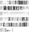
FIG. 1.
FIG. 1.
Alignment of HP1-like proteins. HcpA, HcpB, and HcpC were aligned with human HP1 orthologues HP1α, HP1β, and HP1γ. The highly conserved chromodomain and chromoshadow domain are underlined in black. Functionally important amino acid residues in the chromodomain are indicated as squares. Black shading indicates aromatic residues that form a pocket required for methyl-lysine binding; light gray shading indicates additional residues required for methyl-lysine recognition; dark gray shading indicates residues required for recognition of Ala 7 in the histone H3 N terminus (50). Functionally important amino acid residues in the chromoshadow domain are indicated as circles. Light gray circles indicate residues that form the CSD dimer interface (3). Residues required for recognition of the PxVxL motif in HP1-interacting proteins are shown in dark gray (central valine) and black (proline and leucine) circles (69). The nuclear localization signals within the hinge region are underlined in gray. Note that the previously described bipartite NLS within the hinge region of the human HP1 proteins (64) is missing in the Dictyostelium proteins but lies adjacent to the chromodomain. Abbreviations: Dd, Dictyostelium discoideum; Hs, Homo sapiens.
FIG. 2.
FIG. 2.
Localization of HP1 proteins. A) HcpA-GFP and HcpB-GFP localize to one major and several minor foci at the nuclear periphery. Since the plain of the major spot is shown, only one or two minor foci are seen. Although driven by identical promoters, the fusion proteins display significant differences in expression levels. Note the increased nucleoplasmic staining by HcpB-GFP. HcpA-GFP and HcpB-GFP both colocalize with histone H3K9me2 in the majority of subnuclear foci. Bar, 5 μm. B) Cotransformed HcpA-RFP and HcpB-GFP constructs demonstrate colocalization of HP1 isoforms. In this example, both proteins are seen in one major and one minor focus at the nuclear periphery. Bar, 5 μm. C) An isolated nucleus/centrosome complex from cells expressing HcpA-GFP is shown. The centrosome is marked with an asterisk. The three dark areas represent nuclear caps (nucleoli). The image on the right shows the labeling of HcpA-GFP with 5 nm gold-coupled anti-GFP antibodies at a higher magnification. The label is predominantly found in a strictly confined area of electron-dense material close to the centrosome. The nuclear envelope has been extracted by Triton X-100 during the isolation procedure. Bar, 200 nm.
FIG. 3.
FIG. 3.
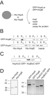
Homo- and heterodimer formation of HP1-like proteins in vitro. A) Scheme for pull-down analysis. Ni-Sepharose beads preloaded with either His-HcpA or His-HcpB were incubated with Dictyostelium discoideum cell lysates containing GFP-HcpA and GFP-HcpB either alone or in combination were treated as shown. B) GFP-HcpA and GFP-HcpB bind to both His-HcpA or His-HcpB but not to empty beads. I, input; S, supernatant; P, pellet (7- or 10-fold concentrated with respect to I and S). C) Neither HcpAΔC-GFP nor HcpBΔC-GFP can bind to His-HcpA. His-HcpA was immobilized on Ni-Sepharose beads and incubated with cell lysates containing either HcpAΔC-GFP or HcpBΔC-GFP. I, input; S, supernatant; P, pellet (10-fold concentrated with respect to I and S). D) Western blots showing proteins used in this study. Left, bacterially expressed His-HcpA and His-HcpB stained with an anti-His antibody. Right, cell lysates from the indicated Dictyostelium discoideum overexpression strains were probed with α-GFP antibody.
FIG. 4.
FIG. 4.
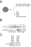
Homo- and heterodimer formation of HP1-like proteins in vivo. A) Scheme for pull-down analysis. Ni-Sepharose beads were incubated with total cell lysates from Dictyostelium discoideum cell lines cotransformed with His-HcpA and GFP-HcpA or GFP-HcpB. B) Both GFP-HcpA and GFP-HcpB are coeluted with His-HcpA from Ni-Sepharose beads. His-HcpA is under the control of an actin 15 promoter and expressed at very low levels in the cell. It is therefore hardly detectable in the input fraction but effectively enriched in the pellet fraction. GFP-HcpB runs as a double band in this gel. C) His-HcpA is detectable in nuclear extracts of cotransformants. Nuclear extracts were subjected to sodium dodecyl sulfate-polyacrylamide gel electrophoresis and Western blot analysis. His-HcpA was detected with α-His antibody. Abbreviations: I, input; S, supernatant; P, pellet. Pellet fractions were 20-fold concentrated with respect to input and supernatant.
FIG. 5.
FIG. 5.
Mitotic dynamics of Dictyostelium heterochromatin. A) Cells expressing HcpA-GFP; B) cells expressing HcpB-GFP. During interphase, heterochromatin localizes next to the nucleus-associated centrosome (see inset). During prophase, the heterochromatin cluster divides in up to six spots (see inset). During prometaphase, nucleoplasmic protein enters the cytoplasm, indicated by increased cytoplasmic fluorescence. The telophase panel for HcpB-GFP shows a telophase and an interphase cell to illustrate the loss of nuclear fluorescence during mitosis. Centrosomes and mitotic spindles are stained with an anti-DdCP224 antibody. a, interphase; b, prophase; c, prometaphase; d, metaphase; e, early anaphase; f, late anaphase; g, telophase. Bar, 5 μm.
FIG. 6.
FIG. 6.
Mitotic dynamics of HcpB-GFP and histone H3K9me2. Whereas H3K9me2 is stable during mitosis, the fluorescence intensity of HcpB-GFP at the putative centromeres and in the nucleoplasm significantly decreases. Images in vertical rows were captured with identical exposure times. a, interphase; b, prophase; c, early anaphase; d, telophase. Bar, 5 μm.
FIG. 7.
FIG. 7.
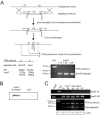
Disruption of endogenous genes of both Hcp isoforms. A) Scheme for hcpA gene targeting and subsequent Cre-mediated excision of the BSR cassette. Gene disruption and Cre recombination introduced restriction sites flanking the remaining loxP site at the endogenous locus. The recombinant hcpA allele (hcpAlp) is similar in size to the wild-type hcpA gene but can be distinguished from the wild-type allele by restriction digestions with the enzymes indicated. Restriction fragment analysis of PCR products derived from either wild-type (Ax2) or different recombinant clones was used to verify Cre-mediated recombination. Only the PCR products derived from the hcpAlp locus, but not from the wild-type hcpA locus, can be digested with BamHI. B) Expression of His-HcpA in hcpAlp cells. Nuclear extracts from hcpAlp-His-HcpA or Ax2 cells were probed with α-His antibody. C) Disruption of the hcpB gene. Clonal isolates of hcpAB mutant-His-HcpA cells transformed with the hcpB disruption construct were screened by PCR (see Materials and Methods) for either homologous recombination of the targeted construct (hcpB knockout [KO]) or an intact hcpB gene (hcpB wild type [WT]). Results for several independent knockout clones are shown in comparison to Ax2 wild-type cells and the single hcpB knockout cell line. The same clones were tested for presence of the Cre recombinant hcpAlp allele by PCR and restriction fragment analysis with NcoI as depicted for PCR product b in panel A. This PCR product that is longer than PCR product a (panel A) covers the 5′ noncoding region of the endogenous hcpA locus and allows us to distinguish it from the His-HcpA transgene, which only contains the coding sequence. Expression of His-HcpA was confirmed by Western blot analysis with an anti-His antibody (bottom panel).
FIG. 8.
FIG. 8.
Distinct phenotypes of cells expressing Hcp fusion proteins. A) Live-cell images of GFP-HcpA-, GFP-HcpB-, HcpAΔC-GFP-, and HcpBΔC-GFP-expressing cells. All images were taken with identical exposure times. Both truncated fusion proteins lacking the CSD still localize to the major heterochromatic cluster but do not form additional smaller foci. B) Expression levels of hcpA, hcpB, and thioredoxin in different background strains determined by semiquantitative RT-PCR. hcpA and hcpB transcript levels are significantly enriched in strains overexpressing GFP-HcpA or GFP-HcpB, respectively, but are not detectable in the knockout strains. Due to partial cross-sensitivity, primers used for hcpB can also bind to hcpA. Note the increase in signal intensity but slightly lower size of the PCR product in the GFP-HcpA overexpression strain. Numbers of PCR cycles for amplification were 32 (hcpA, hcpB) and 27 (thioredoxin). −RT, minus reverse transcription. C) Anaphase-bridge phenotype in the GFP-HcpA strain. A late mitotic cell, as identified by a long mitotic spindle, is shown. DAPI stain shows a DNA bridge between the main DNA masses. GFP-HcpA is found in small foci on the DNA bridge. The asterisk indicates the area of magnification in the upper right corner. Bar, 5 μm. D) Cell viability, as determined by plating efficiency, is significantly impaired by overexpression of HcpAΔC-GFP compared to the control transformant.
FIG. 9.
FIG. 9.
H3K9me2 localization in hcp and rrp knockout mutants. Knockout of either hcpA or hcpB has no effect on H3K9me2 levels or its subnuclear distribution. Similarly, knockouts of the RNA-dependent RNA polymerase genes rrpA, rrpB, or rrpC that are involved in RNA-mediated posttranscriptional gene silencing do not affect H3K9me2 levels and distribution. DAPI is in blue and H3K9me2 is in red. Bar, 5 μm.

Similar articles

Cited by

References

    1. Bannister, A. J., P. Zegerman, J. F. Partridge, E. A. Miska, J. O. Thomas, R. C. Allshire, and T. Kouzarides. 2001. Selective recognition of methylated lysine 9 on histone H3 by the HP1 chromo domain. Nature 410:120-124. - PubMed
    1. Bernard, P., J. F. Maure, J. F. Partridge, S. Genier, J. P. Javerzat, and R. C. Allshire. 2001. Requirement of heterochromatin for cohesion at centromeres. Science 294:2539-2542. - PubMed
    1. Brasher, S. V., B. O. Smith, R. H. Fogh, D. Nietlispach, A. Thiru, P. R. Nielsen, R. W. Broadhurst, L. J. Ball, N. V. Murzina, and E. D. Laue. 2000. The structure of mouse HP1 suggests a unique mode of single peptide recognition by the shadow chromo domain dimer. EMBO J. 19:1587-1597. - PMC - PubMed
    1. Cheutin, T., A. J. McNairn, T. Jenuwein, D. M. Gilbert, P. B. Singh, and T. Misteli. 2003. Maintenance of stable heterochromatin domains by dynamic HP1 binding. Science 299:721-725. - PubMed
    1. Couteau, F., F. Guerry, F. Muller, and F. Palladino. 2002. A heterochromatin protein 1 homologue in Caenorhabditis elegans acts in germline and vulval development. EMBO Rep. 3:235-241. - PMC - PubMed

Publication types

MeSH terms

LinkOut - more resources