. 2006 Apr;8(4):367-76.
doi: 10.1038/ncb1385. Epub 2006 Mar 5.

Coordination of microtubule and microfilament dynamics by Drosophila Rho1, Spire and Cappuccino

Affiliations

Coordination of microtubule and microfilament dynamics by Drosophila Rho1, Spire and Cappuccino

Alicia E Rosales-Nieves et al. Nat Cell Biol. 2006 Apr.

Abstract

The actin-nucleation factors Spire and Cappuccino (Capu) regulate the onset of ooplasmic streaming in Drosophila melanogaster. Although this streaming event is microtubule-based, actin assembly is required for its timing. It is not understood how the interaction of microtubules and microfilaments is mediated in this context. Here, we demonstrate that Capu and Spire have microtubule and microfilament crosslinking activity. The spire locus encodes several distinct protein isoforms (SpireA, SpireC and SpireD). SpireD was recently shown to nucleate actin, but the activity of the other isoforms has not been addressed. We find that SpireD does not have crosslinking activity, whereas SpireC is a potent crosslinker. We show that SpireD binds to Capu and inhibits F-actin/microtubule crosslinking, and activated Rho1 abolishes this inhibition, establishing a mechanistic basis for the regulation of Capu and Spire activity. We propose that Rho1, cappuccino and spire are elements of a conserved developmental cassette that is capable of directly mediating crosstalk between microtubules and microfilaments.


Figures
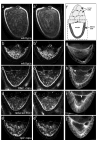
Figure 1
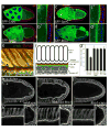
Figure 1. Rho1-capu, reduced Rho1, and capu-spire oocytes undergo premature ooplasmic streaming
(a-e’) Still confocal micrographs (a-e) and 5-frame confocal temporal projections of confocal time-lapse movies (a’-e’) of wildtype and mutant oocytes stained with trypan blue to visualize dynamic yolk granule movement. Granules appear as discrete spots of fluorescence in the still images. Linear patterns of light in the temporal projections indicate coordinated yolk granule movement generated by ooplasmic streaming. Anterior is up in all images. Wildtype stage 10 oocyte (a, a’) during cytoplasmic streaming. Note the spiral pattern formed by circular movement of yolk granules (a’).(b-F’) Unlike wildtype (b, b’), stage 7 oocytes from Rho1-capu(c, c’), reduced Rho1(d, d’), and spir-capu(e, e’) mutant mothers all undergo premature ooplasmic streaming, as indicated by the spiral patterns of fluorescence seen in the temporal projections (c’, d’, e’). (f) Schematic of a stage 10 egg chamber, consisting of the germline nurse cells (light gray) and oocyte (white), surrounded by somatic follicle cells (dark gray). Boxed area indicates region of egg chamber shown in panels a-j. Anterior is up.( g-j) Confocal micrographs of stage 7 oocytes from wildtype (g), Rho1-capu(h), reduced Rho1(i), and spir-capu(j) females stained with α-tubulin to visualize dynamic microtubules. Note subcortical arrays consistent with microtubule-dependent ooplasmic streaming (arrowheads in h-j). Scale bars: 50 μm.
Figure 2
Figure 2. Rho1, Capu and Spire expression is enriched at the oocyte cortex where stable microtubules are also localized
(a-d’) Rho1, Spire and Capu co-localize at the oocyte cortex. Stage 7 10 egg chambers from females containing transgenes expressing GFP-Capu (a), GFP-Spire isoform D (b), GFP-Spire isoform C (c), or GFP-Rho1 (d), and counterstained with phosphotyrosine (red; not used in d, d’) to outline the oocyte plasma membrane and DAPI (blue) to visualize the nuclei. Higher magnification views of the follicle cells and oocyte cortex are shown in a’-d’, respectively. We intermittently observe mosaicism of Rho1-GFP transgene expression in the somatic (follicle) cells (see Methods) as shown here to more clearly visualize Rho1 enrichment at the oocyte cortex, (e-f) Organization of the oocyte cortex, (e) High magnification view of the stage 10 wildtype oocyte cortex double labeled with α-tubulin (green) to visualize dynamic microtubules and Glu-tubulin (red) to visualize stabilized microtubules. Note separation between apical follicle cell membrane (arrowhead) and cortical band of Glu-microtubules (arrow), (f) A schematic diagram of the oocyte cortex showing the relative locations of the cytoskeletal components, (g) Quantification of the relative pixel intensity for Glu-microtubule staining at the anterior cortex (boxed region “A”) versus the posterior pole of the egg chambers (boxed region “P”) such as those shown in h-j. Error bars represent the standard deviation from the mean obtained from quantifying 20 egg chambers, (h-j’) Confocal micrographs of stage 10 oocytes stained with antibodies recognizing detyrosinated (Glu-) tubulin to visualize stabilized microtubules. Stage 10 egg chambers from wildtype (h), reduced Rho1(i), and Rho1-capu(j) females. Anterior is to the left, (h’-j’) Higher magnification view of the oocyte cortex in h-j, respectively. (h’’-j’’) Close up view of the posterior pole of the oocytes in h-j, respectively. Note the reduced level of Glu-microtubules at the posterior in wildtype (h’’) relative to the rest of the cortex. Levels of Glu-tubulin are not reduced at the posterior relative to the lateral cortex in Rho1 or Rho1-capu (i’’-j’’, respectively). Scale bars: (a-d) 50 μm; (a’-d’) 20 μm; (e) 10 μm; (h-j) 20 μm; (h’-j’’) 10 μm.
Figure 3
Figure 3. Protein-Protein interactions among Rho1, Capu, and Spire indicate a complex regulatory network
(a) Diagram of a canonical DRF protein and of the Capu protein. The Capu protein fragments used to map Capu/Spire/Rho1 protein-protein interactions are indicated, (b) Diagram of the Spire-A, -C, and -D protein isoforms. The Spire protein fragments used to map the Capu/Spire/Rho1 protein-protein interactions are indicated, (c) Rho1 binds directly to the N-terminus of Capu. 35S labeled in vitro translated (IVT) CapuN3 (fourth panel from top) binds preferentially to GST-RhoGTP (lane 4). This interaction is preserved in other Capu fragments that span CapuN3 (top 3 panels; lane 4). (d) All three Spire isoforms bind preferentially to RhoGTP.35S labeled IVT-SpireA (top panel), SpireD (middle panel) and SpireC (bottom panel) bind to GST-RhoGTP(lane 4).(e-f) Using the Spire protein pieces depicted in b, Rho1 binding to Spire-D and -C was mapped to the non-overlapping regions encompassed by SpireD3 (e) and SpireC3 (f). While we reproducibly observe low levels of Spire-D2 and -D4 binding to Rho1GTP, neither of these proteins affects Capu’s crosslinking or nucleation functions, (g) Immunoprecipitation from ovary lysate showing in vivo interaction between Rho1 and Capu. Capu is precipitated by an antibody to Rho1, but not when a non-relevant or no primary antibody is used, (h) Capu exhibits an intramolecular interaction. 35S labeled IVT-CapuC2 (input; lane 1) binds to GST-CapuN3 (lane 3). (i) The Capu FH2 domain binds directly to the WH2-containing SpireD3 domain. 35S labeled IVT-CapuFH2 (input; lane 1) binds preferentially to GST-SpireD (lane 3) and GST-SpireD3 (lane 6). (j) Capu binding to Spire is disrupted by GTP-bound Rho1. 35S labeled IVT-CapuFH2 (input; lane 1) binds to GST-SpireD (top panel, lane 3) and GST-SpireD3 (bottom panel, lane 3). The interaction between CapuFH2 and either SpireD or SpireD3 is disrupted by simultaneous addition of IVT-Rho1 in a dose-sensitive manner (lanes 4-6), and abolished when added after the interaction has stabilized (lane 7). (k) Capu FH2 domains dimerize. 35S labeled IVT-CapuFH2 (input; lane 1) binds to GST-CapuFH2 (lane 3). This interaction is observed with wildtype, as well as mutant (L768H and P597T), FH2 domains.
Figure 4
Figure 4. Capu and Spire affect actin dynamics
(a) Pyrene-actin polymerization assays were conducted with varying concentrations of CapuFH2 (indicated in μM). (b) Capu actin nucleation activity is not subject to auto-inhibition. CapuFH2 (1 μM) was pre-incubated with CapuN1 (1 μM) in G-buffer, or an equal volume of G-buffer alone, and added to pyrene actin polymerization assays. CapuN1 alone (1 μM) had no affect on actin dynamics, (c) The capu2F mutation does not affect actin nucleation. CapuFH2, CapuFH2[L768H], or CapuFH2[P597T], all at 1 μM, were added to polymerization assays. Note that the L768H mutation (corresponding to capuRK12) abolished nucleation activity whereas the P597T mutation (corresponding to capu2F) did not substantially affect nucleation. (d) CapuFHl-FH2 or was added to polymerization assays at varying concentrations (indicated in μM). (e) The affect of various concentrations of SpireD or SpireC (indicated in μM) on polymerization kinetics was assayed as above, (f) Capu and Spire do not affect each other’s actin nucleation activity. CapuFH1-FH2 (0.1 μM) or SpireD (0.8 μM) alone, or CapuFH1-FH2 pre-incubated with SpireD, was added to pyrene actin polymerization assays.
Figure 5
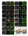
Figure 5. Rho1 regulates crosslinking of F-actin and microtubules by Capu and Spire
(a-n) Microtubule and microfilament bundling and crosslinking properties of Capu, Spire and Rho1. Stabilized microtubules (1:5 rhodamine-labeled:unlabeled; 1 μM; middle column) and F-actin (1 μM; left column) were incubated with Capu, Spire and Rho1 proteins, the mixture was diluted 1:4, plated on glass coverslips then visualized by confocal microscopy, (a) No protein added; (b) CapuN1; (c) CapuFH2; (d) CapuN1 and CapuFH2; (e) CapuFH22F (CapuFH2 containing P597T mutation); (f) CapuFH2RK12 (CapuFH2 containing L768H mutation); (g) SpireC isoform; (h) SpireD isoform; (i) CapuFH2 and SpireD; (j) CapuFH2 and Spire D1 protein piece; (k) CapuFH2 and Spire D3 protein piece; (l) CapuFH2 and Spire D with Rho1GTP; (m) CapuFH2 and Spire D with Rho1GDP; and (n) SpireC and SpireD. Final protein concentrations were: CapuFH2 (300 nM), CapuN (300 nM), CapuFH2-2F (300 nM), CapuFH2-LH (300 nM), SpireA (125 nM), SpireC (250 nM), SpireD (300 nM), SpireD1 (1 μM), SpireD3 (300 nM), and Rho1 (600 nM). Scale bar: 50 μm. Quantification of CapuFH2 wildtype and point mutant cross-linking by low speed co-sedimentation is shown in Supplementary Information Fig. S5b.(o-p) Model for the regulation of microtubule/microfilament crosslinking and ooplasmic streaming by Rho1, Capu, and Spire isoforms C and D. Schematic of a wildtype oocyte prior to the onset of ooplasmic streaming (o) and during ooplasmic streaming (p). Close-up views are shown in the insets. Microtubules are red and cortical microfilaments are green. Microtubule/microfilament crosslinking by SpireC and Capu is necessary to prevent the assembly of subcortical arrays of dynamic microtubules and the resulting streaming event (o). Active (GTP-bound) Rho1 promotes microtubule/microfilament crosslinking by sequestering SpireD, thereby preventing it from binding to SpireC and Capu. Upstream signaling events result in GTP hydrolysis by Rho1, allowing SpireD to bind to SpireC and Capu (p). This blocks microtubule/microfilament crosslinking, resulting in ooplasmic streaming.

Similar articles

Cited by

References

    1. Theurkauf WE. Premature microtubule-dependent cytoplasmic streaming in cappuccino and spire mutant oocytes. Science. 1994;265:2093–2096. - PubMed
    1. Manseau L, Schüpbach T. cappuccino and spire: two unique maternal-effect loci required for both the anteroposterior and dorsoventral patterns of the Drosophila embryo. Genes Dev. 1989;3:1437–1452. - PubMed
    1. Emmons S, et al. cappuccino, a Drosophila maternal effect gene required for polarity of the egg and embryo, is related to the vertebrate limb deformity locus. Genes Dev. 1995;9:2482–2494. - PubMed
    1. Wellington A, et al. Spire contains actin binding domains and is related to ascidian posterior end mark-5. Development. 1999;126:5267–74. - PubMed
    1. Cooley L, Theurkauf WE. Cytoskeletal functions during Drosophila oogenesis. Science. 1994;266:590–6. - PubMed

Publication types

LinkOut - more resources