. 2006 Jun 15;107(12):4790-7.
doi: 10.1182/blood-2005-07-3058. Epub 2006 Mar 2.

Interferons limit inflammatory responses by induction of tristetraprolin

Affiliations

Interferons limit inflammatory responses by induction of tristetraprolin

Ines Sauer et al. Blood. .

Abstract

Interferons (IFNs) are cytokines with pronounced proinflammatory properties. Here we provide evidence that IFNs also play a key role in decline of inflammation by inducing expression of tristetraprolin (Ttp). TTP is an RNA-binding protein that destabilizes several AU-rich element-containing mRNAs including TNFalpha. By promoting mRNA decay, TTP significantly contributes to cytokine homeostasis. Now we report that IFNs strongly stimulate expression of TTP if a costimulatory stress signal is provided. IFN-induced expression of Ttp depends on the IFN-activated transcription factor STAT1, and the costimulatory stress signal requires p38 MAPK. Within the Ttp promoter we have identified a functional gamma interferon-activated sequence that recruits STAT1. Consistently, STAT1 is required for full expression of Ttp in response to LPS that stimulates both p38 MAPK and, indirectly, interferon signaling. We demonstrate that in macrophages IFN-induced TTP protein limits LPS-stimulated expression of several proinflammatory genes, such as TNFalpha, IL-6, Ccl2, and Ccl3. Thus, our findings establish a link between interferon responses and TTP-mediated mRNA decay during inflammation, and propose a novel immunomodulatory role of IFNs.


Figures

Figure 1
Figure 1
p38 MAPK and STAT1 synergistically increase TTP mRNA expression. p38α(+/+) and p38α(−/−) MEFs were left untreated (w/o) or treated for 1 h with IFN-γ (g), anisomycin (a) or both (a/g) (A), or treated with IFN-β (b), anisomycin (a) or both (a/b) (B). STAT1(+/+) and STAT1(−/−) fibroblasts were stimulated as described for panel A and B, respectively (C and D). Total RNA was isolated and TTP mRNA levels were determined using qRT-PCR. To obtain TTP mRNA induction, values were normalized to those of untreated cells. Error bars indicate SD. *, p < 0.01 (a/g) or (a/b) versus (a) treatment in (+/+) cells; †, p < 0.01 (+/+) versus (−/−) MEFs by univariate linear regression models; n = 3 experiments.
Figure 2
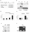
Figure 2
TTP promoter contains a functional GAS element that binds STAT1. (A) Schematic representation of the TTP promoter as cloned into the reporter construct pGL2-TTP, comprising the region from −2025 to +25. The TTP promoter contains a GAS element at the position −1976 to −1964. The reporter plasmid pGL2-TTP-GAS contains the TTP-GAS with flanking sequences (−2085 to −1891). The consensus for STAT1-binding GAS is shown (according to IUPAC nomenclature H stands for A, C, or T; S stands for G or C; D stands for G, A, or T). Mutated TTP-GAS (TTP-GASMut) was obtained by introducing 2 point mutations. (B) p38α(+/+) cells stably transfected with a reporter plasmid containing the luciferase gene under the control of a 2 kb fragment of the TTP promoter (pGL2-TTP) were left untreated (w/o) or treated for 1 h with IFN-γ (g), anisomycin (a) or both (a/g). Total RNA was isolated and luciferase expression was assayed by qRT-PCR. Error bars indicate SD. *, p < 0.01 (a/g) versus (a) treatment by univariate linear regression models; n = 3 experiments. (C) For nuclear run-on assay p38α(+/+) cells were left untreated (w/o) or treated for 25 min with IFN-γ (g), anisomycin (a) or both (a/g), nuclei were prepared and run-on reaction was performed. Nuclear RNA was isolated and hybridized to membranes containing cDNA of GAPDH (as a control) and TTP. (D) Bac 1.2F5 mouse macrophages were stimulated for 30 min with IFN-γ (g) or left untreated (−), and whole cell extracts were assayed for binding to a radioactively labeled TTP-GAS probe using EMSA. The results demonstrate IFN-γ-inducible binding of STAT1 to TTP-GAS but not to mutated TTP-GAS (TTP-GASMut). The presence of STAT1 in the complexes was confirmed by super shift using STAT1 antibodies (lanes marked with +S1). (E) A luciferase reporter containing TTP-GAS (pGL-TTPGAS) was transfected into STAT1(−/−) MEFs reconstituted with STAT1 cDNA (STAT1(+/+)) and for control into the parental STAT1(−/−) cells. After the transfection, cells were divided in two halves and 24 h later treated for 6 h with IFN-γ (g) or left untreated. Error bars indicate SD. †, p < 0.01 (+/+) versus (−/−) MEFs by univariate linear regression models; n = 3 experiments. (F) For ChIP, STAT1(+/+) MEFs were treated for 30 min with IFN-γ (g) or left untreated (−). Recruitment of STAT1 to the TTP promoter was assayed using STAT1 antibodies (+S1). A control ChIP was performed using rabbit pre-immune serum (C). Equal amount of material used in the ChIP experiments was confirmed by PCR-amplification of the input DNA (input).
Figure 3
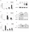
Figure 3
STAT1 is required for full expression of TTP in LPS-treated primary macrophages. (A) Macrophages derived from bone marrow (BMM) of STAT1WT and STAT1KO mice were left untreated or treated with IFN-γ (g), LPS (L) or both (L/g) and TTP mRNA induction was measured by qRT-PCR and normalized to samples from untreated cells. Error bars indicate SD, n = 3 experiments. (B) BMM isolated from STATWT mice were treated as explained for panel A, except the time of the treatment was 3 h instead of 1 h. TTP protein levels were analyzed by Western blotting of whole cell extracts using a TTP antibody. The blot was reprobed with a panERK antibody to control for equal protein loading. (C) BMM from STAT1WT and STAT1KO mice were treated for 1 and 4 h with LPS or left untreated, and mRNA induction was analyzed as described in panel A. Error bars indicate SD, n = 3 experiments. (D) Same cells as those used in panel C were treated for 2 and 4 h with LPS or left untreated. TTP protein was detected by Western blotting of whole cell extracts using a TTP antibody. Activation of IFN signaling by endogenous production of type I interferon in LPS-treated macrophages was demonstrated using antibody to tyrosine-phosphorylated STAT1 (pY701-S1). Equal protein loading was confirmed using a STAT1 antibody and a panERK antibody. (E) BMM from IFN-β knock out (IFNbKO) and wild type controls (IFNbWT) were stimulated with LPS for the times indicated. Total RNA was isolated and qRT PCR was performed. Error bars indicate SD, n = 3 experiments. (F) Whole cell extracts of the same cells as used in panel E were stimulated with LPS as indicated. TTP protein was detected by Western blotting using a TTP antibody.
Figure 4
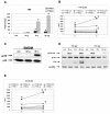
Figure 4
Interferon-stimulated TTP expression limits induction of TNFα and IL-6. (A) BMM derived from TTP wild type (TTPWT) and deficient (TTPKO) mice were left untreated or treated with IFN-γ (g), LPS (L) or both (L/g) for 3 h and TNFα mRNA induction was determined by qRT-PCR and normalized to untreated samples. *, p < 0.05 treated versus untreated cells by un ivariate linear regression models. (B) BMM from TTPWT and TTPKO were stimulated for the times indicated with IFN-γ (G), LPS or both (L/G) or left untreated (0). Supernatants were collected and analyzed for TNFα cytokine by ELISA. *, p < 0.01 4h versus 2h in L/G treated KO cells; †, p < 0.01 KO versus WT BMM by univariate linear regression models, n = 3 experiments. (C) TTPWT BMM were treated for 30 min (30′) with IFN-γ and LPS, and activation of p38 was demonstrated using an antibody against phosphorylated p38 MAPK (pp38). Equal protein loading was confirmed using a p38 antibody. (D) BMM from TTPWT and TTPKO mice were treated as explained for panel A. Absence of TTP protein in TTPKO cells was confirmed by Western blotting of whole cell extracts using a TTP antibody. Activation of IFN signaling by endogenous production of type I interferon in LPS-treated macrophages was demonstrated using an antibody to tyrosine-phosphorylated STAT1 (pY701-S1). Equal protein loading was confirmed using a STAT1 antibody and a panERK antibody. (E) BMM from TTPWT and TTPKO were stimulated for the times indicated with IFN-β (B), LPS or both (L/B) or left untreated (0). Supernatants were collected and IL-6 was measured by ELISA. *, p < 0.01 6h versus 4h in L/B treated KO cells; †, p < 0.01 KO versus WT BMM by univariate linear regression models, n = 3 experiments.
Figure 5
Figure 5
Interferon- and LPS-induced TTP expression has a suppressive effect on Ccl2 and Ccl3 mRNA production. BMM derived from TTP wild type (TTPWT) and deficient (TTPKO) mice were left untreated or treated with IFN-β (g), LPS (L) or both (L/g) for 3 h. Total RNA was extracted and the induction of Ccl2 (A), Ccl3 (B), Gbp2 (C) and Cxcl10 (D) genes was detected by real time RT-PCR using Taqman assays from Applied Biosystems. To obtain mRNA induction, qRT-PCR values were normalized to those of untreated cells. *, p < 0.05 treated versus untreated cells by univariate linear regression models.

Similar articles

Cited by

References

    1. Sen GC. Viruses and interferons. Annu Rev Microbiol. 2001;55:255–281. - PubMed
    1. Bogdan C. The function of type I interferons in antimicrobial immunity. Curr Opin Immunol. 2000;12:419–424. - PubMed
    1. Schroder K, Hertzog PJ, Ravasi T, Hume DA. Interferon-gamma: an overview of signals, mechanisms and functions. J Leukoc Biol. 2004;75:163–189. - PubMed
    1. Schindler C, Brutsaert S. Interferons as a paradigm for cytokine signal transduction. Cell Mol Life Sci. 1999;55:1509–1522. - PMC - PubMed
    1. Levy DE, Darnell JE., Jr. Stats: transcriptional control and biological impact. Nat Rev Mol Cell Biol. 2002;3:651–662. - PubMed

Publication types

MeSH terms