

doi: 10.1128/IAI.74.3.1933-1940.2006.
Binding of the streptococcal surface glycoproteins GspB and Hsa to human salivary proteins
Affiliations
- PMID: 16495569
- PMCID: PMC1418684
- DOI: 10.1128/IAI.74.3.1933-1940.2006
Item in Clipboard
Binding of the streptococcal surface glycoproteins GspB and Hsa to human salivary proteins
Infect Immun.
2006 Mar.
Abstract
GspB and Hsa are homologous surface glycoproteins of Streptococcus gordonii that bind sialic acid moieties on platelet membrane glycoprotein Ibalpha. Since this species is an important member of the oral flora, we examined the direct binding of these adhesins to human salivary proteins. Both GspB and Hsa bound low-molecular-weight salivary mucin MG2 and salivary agglutinin. Hsa also bound several other salivary proteins, including secretory immunoglobulin A. Screening of six oral streptococcal isolates revealed that at least two of the strains expressed GspB homologues. These results indicate that GspB-like adhesins may be important for oral bacterial colonization.
Figures
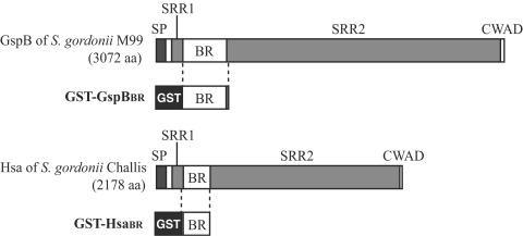
Structures of GspB of S. gordonii strain M99 and Hsa of S. gordonii strain Challis and diagrams of GST fusion proteins. The number of amino acids (aa) in GspB and Hsa is indicated in parentheses. GST, glutathione S-transferase; SP, signal peptide; SRR1, first serine-rich region; BR, basic region; SRR2, second serine-rich region; CWAD, cell wall-anchoring domain.
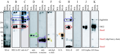
Binding of fusion proteins to human salivary proteins. Salivary proteins were separated by electrophoresis through 3 to 8% polyacrylamide gradient gels and then stained with silver (A) or subjected to Western blotting (B-F), lectin blotting (G, H), or far-Western blotting (I-K). Each lane contains 5 μl (for silver staining, antilactoferrin antibody, anti-α-amylase antibody, MAL-II, GST, GST-GspBBR, and GST-HsaBR), 3 μl (for anti-sLex monoclonal antibody), 2 μl (for anti-human IgA antibody), 0.5 μl (for HECA-452), or 0.1 μl (for Lens culinaris agglutinin) of the saliva samples. WS, whole saliva; SMSL, submandibular and sublingual saliva; PG-R, parotid saliva from the right duct. Salivary proteins identified the following proteins: high-molecular-weight mucin MG1 (MUC5B) (a), salivary agglutinin (gp340) (b), low-molecular-weight mucin MG2 (MUC7) (c), proline-rich glycoprotein (d), lactoferrin (e), α-amylase (f), and sIgA heavy chain (g). LCA, Lens culinaris agglutinin.
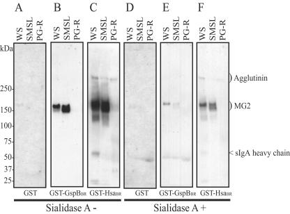
Effect of sialidase A treatment on fusion protein binding to human salivary proteins. Salivary proteins were separated by electrophoresis through 3 to 8% polyacrylamide gradient gels and transferred to nitrocellulose membranes. Membranes were incubated in DPBS in the absence (A to C) or presence (D to F) of sialidase A and then probed with GST (A, D), GST-GspBBR (B, E), or GST-HsaBR (C, F). Each lane contains 5 μl saliva. WS, whole saliva; SMSL, submandibular and sublingual saliva; PG-R, parotid saliva from the right duct.
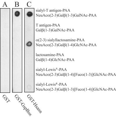
Binding of GST fusion proteins to different oligosaccharide structures. One microgram of each oligosaccharide-conjugated polyacrylamide was spotted onto three separate nitrocellulose membranes. Each membrane was incubated with GST (A), GST-GspBBR (B), or GST-HsaBR (C) followed by anti-GST serum and peroxidase-conjugated anti-rabbit IgG. NeuAc, N-acetylneuraminic acid; Gal, galactose; GalNAc, N-acetylgalactosamine; GlcNAc, N-acetylglucosamine; Fuc, fucose; PAA, polyacrylamide.
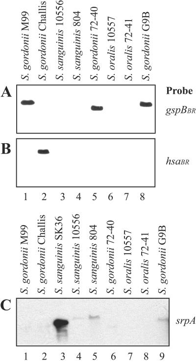
Detection of gspB homologues in six strains of oral streptococci by Southern hybridization. Genomic DNAs from M99, Challis, SK36, and the six oral streptococcal isolates were digested with HindIII, separated by agarose gel electrophoresis, transferred to nylon membranes, and hybridized with digoxigenin-labeled probes for gspB (A), hsa (B), or srpA (C).
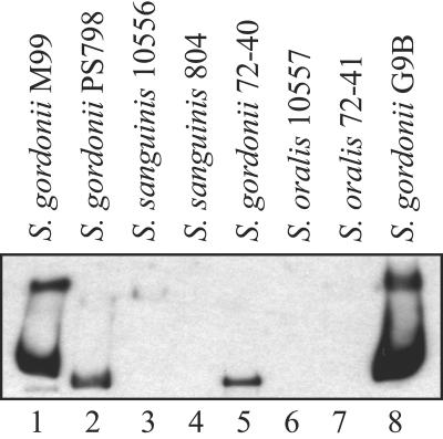
Surface expression of GspB homologues in the six oral strains of viridans group of streptococci. Cell wall proteins were separated by electrophoresis through a 3 to 8% polyacrylamide gradient gel and then analyzed by Western blotting using a polyclonal anti-GspB serum. All proteins shown in this figure migrated above the largest standard (250 kDa). Each lane contains cell wall proteins extracted from bacteria in 200 μl of a broth culture. PS798, secA2-complemented Challis.
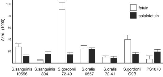
Binding of the six oral streptococcal strains to sialic acid moieties. Microtiter wells were coated with fetuin or asialofetuin (50 μg/well). Binding of washed bacteria to the immobilized glycoproteins was assessed as described previously (2), except that the bound bacteria were detected by staining with crystal violet, dissolving the stain in 5% acetic acid, and then reading the absorbance at 570 nm. Binding is expressed as the mean ± standard deviation (error bar) (n = 6).

Western blot analysis of expression of the GspB homologue by S. gordonii 72-40 (lane 1) and the mutant strain PS1070 (lane 2). (Top) Cell wall proteins were separated by electrophoresis through a 3 to 8% polyacrylamide gradient gel, transferred to nitrocellulose, and then probed with the anti-GspB serum. (Bottom) Proteins were precipitated from the spent culture medium using trichloroacetic acid, separated by electrophoresis on a 3 to 8% polyacrylamide gradient gel, transferred to nitrocellulose, and then probed with an anti-FLAG monoclonal antibody (Sigma).
Similar articles
-
Binding of the Streptococcus gordonii surface glycoproteins GspB and Hsa to specific carbohydrate structures on platelet membrane glycoprotein Ibalpha.Mol Microbiol. 2005 Oct;58(2):380-92. doi: 10.1111/j.1365-2958.2005.04830.x. Mol Microbiol. 2005. PMID: 16194227
-
The Streptococcus gordonii surface proteins GspB and Hsa mediate binding to sialylated carbohydrate epitopes on the platelet membrane glycoprotein Ibalpha.Infect Immun. 2004 Nov;72(11):6528-37. doi: 10.1128/IAI.72.11.6528-6537.2004. Infect Immun. 2004. PMID: 15501784 Free PMC article.
-
Functions of cell surface-anchored antigen I/II family and Hsa polypeptides in interactions of Streptococcus gordonii with host receptors.Infect Immun. 2005 Oct;73(10):6629-38. doi: 10.1128/IAI.73.10.6629-6638.2005. Infect Immun. 2005. PMID: 16177339 Free PMC article.
-
Streptococcal adhesion and colonization.Crit Rev Oral Biol Med. 1997;8(2):175-200. doi: 10.1177/10454411970080020601. Crit Rev Oral Biol Med. 1997. PMID: 9167092 Review.
-
Cell surface protein receptors in oral streptococci.FEMS Microbiol Lett. 1994 Aug 15;121(2):133-40. doi: 10.1111/j.1574-6968.1994.tb07089.x. FEMS Microbiol Lett. 1994. PMID: 7926661 Review.
Cited by
-
Can salivary activity predict periodontal breakdown in A. actinomycetemcomitans infected adolescents?Arch Oral Biol. 2013 Jun;58(6):611-20. doi: 10.1016/j.archoralbio.2012.10.009. Epub 2012 Dec 6. Arch Oral Biol. 2013. PMID: 23219180 Free PMC article.
-
Streptococcus gordonii Hsa environmentally constrains competitive binding by Streptococcus sanguinis to saliva-coated hydroxyapatite.J Bacteriol. 2007 Apr;189(8):3106-14. doi: 10.1128/JB.01535-06. Epub 2007 Feb 2. J Bacteriol. 2007. PMID: 17277052 Free PMC article.
-
Streptococcus mitis phage-encoded adhesins mediate attachment to {alpha}2-8-linked sialic acid residues on platelet membrane gangliosides.Infect Immun. 2009 Aug;77(8):3485-90. doi: 10.1128/IAI.01573-08. Epub 2009 Jun 8. Infect Immun. 2009. PMID: 19506011 Free PMC article.
-
Role of Neuraminidase-Producing Bacteria in Exposing Cryptic Carbohydrate Receptors for Streptococcus gordonii Adherence.Infect Immun. 2018 Jun 21;86(7):e00068-18. doi: 10.1128/IAI.00068-18. Print 2018 Jul. Infect Immun. 2018. PMID: 29661931 Free PMC article.
-
Implications of salivary protein binding to commensal and pathogenic bacteria.J Oral Biosci. 2013 Nov 1;55(4):169-174. doi: 10.1016/j.job.2013.06.004. J Oral Biosci. 2013. PMID: 24707190 Free PMC article.
References
-
- Becerra, L., R. V. Soares, L. S. Bruno, C. C. Siqueira, F. G. Oppenheim, G. D. Offner, and R. F. Troxler. 2003. Patterns of secretion of mucins and non-mucin glycoproteins in human submandibular/sublingual secretion. Arch. Oral Biol. 48:147-154. - PubMed
-
- Bensing, B. A., D. Takamatsu, and P. M. Sullam. 2005. Determinants of the streptococcal surface glycoprotein GspB that facilitate export by the accessory Sec system. Mol. Microbiol. 58:1468-1481. - PubMed
-
- Bensing, B. A., and P. M. Sullam. 2002. An accessory sec locus of Streptococcus gordonii is required for export of the surface protein GspB and for normal levels of binding to human platelets. Mol. Microbiol. 44:1081-1094. - PubMed
Publication types
MeSH terms
Substances
Grants and funding
LinkOut - more resources
Full Text Sources
