. 2006 Mar;80(5):2515-28.
doi: 10.1128/JVI.80.5.2515-2528.2006.

Nature of nonfunctional envelope proteins on the surface of human immunodeficiency virus type 1

Affiliations

Nature of nonfunctional envelope proteins on the surface of human immunodeficiency virus type 1

Penny L Moore et al. J Virol. 2006 Mar.

Abstract

Human immunodeficiency virus type 1 (HIV-1) neutralizing antibodies are thought be distinguished from nonneutralizing antibodies by their ability to recognize functional gp120/gp41 envelope glycoprotein (Env) trimers. The antibody responses induced by natural HIV-1 infection or by vaccine candidates tested to date consist largely of nonneutralizing antibodies. One might have expected a more vigorous neutralizing response, particularly against virus particles that bear functional trimers. The recent surprising observation that nonneutralizing antibodies can specifically capture HIV-1 may provide a clue relating to this paradox. Specifically, it was suggested that forms of Env, to which nonneutralizing antibodies can bind, exist on virus surfaces. Here, we present evidence that HIV-1 particles bear nonfunctional gp120/gp41 monomers and gp120-depleted gp41 stumps. Using a native electrophoresis band shift assay, we show that antibody-trimer binding predicts neutralization and that the nonfunctional forms of Env may account for virus capture by nonneutralizing antibodies. We hypothesize that these nonfunctional forms of Env on particle surfaces serve to divert the antibody response, helping the virus to evade neutralization.


Figures

FIG. 1.
FIG. 1.
Potential forms of Env on the HIV-1 membrane. gp120 is shown in red with the outer neutralizing face in light shading and the inner nonneutralizing face in darker shading. Carbohydrate moieties are depicted as “tree”-like structures. gp41 is comprised of N-terminal (yellow) and C-terminal (green) transmembrane domains, separated by a disulfide-constrained loop. The membrane-proximal gp41 region exposed on the trimer is depicted in dark green. A) Functional Env trimer; B) uncleaved gp160 precursor (depicted here as a trimer; however, it may also exist as other oligomeric forms); C) gp120 shedding; D) alternative trimer isoform exposing the nonneutralizing face of gp120; E) gp120/gp41 monomers.
FIG. 2.
FIG. 2.
SDS-PAGE and Western blot analysis of Env derived from VLPs. Monomeric JR-FL gp120 and VLPs bearing JR-FLgp160ΔCT SOS, WT, and uncleaved Envs (lanes 1 to 4, respectively) were analyzed by SDS-PAGE and Western blot in A) nonreducing or B) reducing conditions. Blots were probed with anti-gp120 MAbs PA1 and B12.
FIG. 3.
FIG. 3.
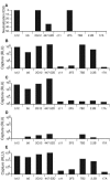
Comparison of MAb-VLP capture and neutralization. A) Neutralization (50% inhibitory concentration [IC50] [μg/ml]) by a panel of MAbs, as indicated, against JR-FL WT-VLPs. B to E) Capture of various JR-FL VLPs by the same MAbs. B) WT-VLPs; C) SOS-VLPs; D) UNC-VLPs; E) UNC-VLPs which also bear VSV-G. RLU, relative light units.
FIG. 4.
FIG. 4.
MAb epitope availability on various forms of Env. The MAbs that are able to recognize each form of Env depicted are given below in parentheses. For some forms of Env, MAb recognition is inferred from virus capture and previous studies (7, 37, 58). A) gp120/gp41 trimers (sCD4, b12, 2G12, 2F5, and Z13); B) gp120/gp41 monomers (sCD4, b12, b6, 447-52D, P7, c11, X5, HIVIG, 2F5, and Z13); C) monomeric gp120 (sCD4, b12, b6, 447-52D, X5, P7); D) trimeric and monomeric gp41 stumps (7B2, 2.2B, T3, and Z13); E) uncleaved gp160 oligomers (sCD4, b12, b6, 2G12, 447-52D, P7, 2F5, Z13 and T3).
FIG. 5.
FIG. 5.
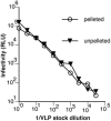
Effect of centrifugal pelleting on VLP infectivity. WT-VLPs were concentrated by high-speed centrifugation and resuspended in the same starting volume of tissue culture medium. The infectivity of pelleted (circles) compared to unpelleted (triangles) VLPs was then assessed using CF2.CD4Th.synCCR5 cells.
FIG. 6.
FIG. 6.
Effect of detergent type and concentration on the pattern of Env separation by BN-PAGE. A) Various detergents were used to solubilize WT-VLPs, as follows: 0.5% Tween 80 (lane 1); 0.5% Tween 20 (lane 2); 10 mM β-octylglucoside (lane 3); 0.25% each of Igepal and Triton X-100 (lane 4); 0.25% Igepal (lane 5); 0.25% Triton X-100 (lane 6); 0.25% sodium deoxycholate (lane 7); 25 mM CHAPS (cholamidopropyldimethylammoniopropanesulfonate) (lane 8). Monomeric gp120 was included as a control (lane 9). B) Progressively lower concentrations of Triton X-100 were used to lyse WT-VLPs in each lane, as indicated. Monomeric gp120 was included as a control in lane 1. Western blots were probed with the anti-gp120 cocktail (b12, 447-52D, and 2G12) (lanes 1 to 6) or an anti-gp41 cocktail (2F5 and 4E10) (lanes 7 to 10).
FIG. 7.
FIG. 7.
Effect of cross-linking on the proportion of functional and nonfunctional forms of Env. WT-VLPs (lanes 1, 2, 5, and 6 of part A; lanes 1 and 2 of part B) and SOS-VLPs (lanes 3, 4, 7, and 8 of part A; lanes 3 and 4 of part B) were incubated with (lanes 2, 4, 6, and 8) or without (lanes 1, 3, 5, and 7) 1 mM BS3 before BN-PAGE. Monomeric gp120 was included as a control (lane 9). Samples in lanes 5 to 8 were boiled in 1% SDS/50 mM dithiothreitol prior to loading. Western blots were probed with A) the anti-gp120 cocktail (b12, 447-52D, and 2G12) or B) the anti-gp41 cocktail (2F5 and 4E10).
FIG. 8.
FIG. 8.
Behavior of inactivated HIV preparations in BN-PAGE. Env from VLPs and virus preparations was compared. A) JR-FL gp160ΔCT WT-VLPs (lane 1); HIV-1ADA (lane 2); and HIV-1MN (lane 3) Western blots were probed with an anti-gp120 cocktail, consisting of b12, 447-52D, and 2G12. B) The same samples in panel A were probed with the anti-gp41 cocktail of 2F5 and 4E10. C) JR-FL WT-VLPs (lane 1) and peripheral blood mononuclear cell-produced JR-CSF (lane 2) were probed with the anti-gp120 cocktail.
FIG. 9.
FIG. 9.
Analysis of sCD4 binding to native Env proteins in BN-PAGE band shifts. SOS-VLPs were incubated either alone (lane 1) or with two-domain (lane 2) or four-domain (lane 3) sCD4 at a final concentration of 20 μg/ml. Samples were then separated by BN-PAGE. Western blots were probed with an anti-gp120 MAb cocktail containing 2G12, b12, and 447-52D. Ferritin 12-mer (220 kDa) and 24-mer (439 kDa) served as molecular size markers.
FIG. 10.
FIG. 10.
MAb binding to native Env in BN-PAGE band shifts. WT-VLPs (A and C), SOS-VLPs (B and D), inactivated MN virus (E), and UNC-VLPs (F) were incubated with Fabs at a final concentration of 20 μg/ml and then processed for BN-PAGE. Western blots in panels A, B, E, and F were probed with the anti-gp120 cocktail containing 2G12, b12, and 447-52D; blots in panels C and D were probed with the anti-gp41 cocktail containing 2F5 and 4E10. Labels: T, gp120/gp41 trimer; M, gp120/gp41 monomer; O, uncleaved oligomers (most likely dimers, trimers, and tetramers). The Fabs used (neutralization IC50 titers against JR-FL gp160ΔCT WT in μg/ml) are as follows: lane 1, no Fab; lane 2, b12 (0.1 μg/ml); lane 3, b6 (>40 μg/ml); lane 4, 2G12 (0.5 μg/ml); lane 5, 447-52D ( μg/ml); lane 6, P7 (>40 μg/ml); lane 7, X5 (>40 μg/ml); lane 8, HIVIG (>40 μg/ml); lane 9, 2F5 ( μg/ml); lane 10, Z13e1 ( μg/ml); lane 11, T2 (>40 μg/ml); lane 12, T3 (>40 μg/ml). In panels G and H four-domain sCD4 was titrated in band shifts (lanes 1 to 8 correspond to four-domain sCD4 concentrations of 0.003, 0.01, 0.03, 0.1, 0.3, 1, 3, and 10 μg/ml, respectively), and in a neutralization assay against WT-VLPs.
FIG. 11.
FIG. 11.
Separation of the MAb-VLP binding step from detergent treatment and sample preparation indicates that monomer is present on intact particles. We further investigated whether gp120/gp41 monomers are a relevant species on intact particles by separating MAb binding from sample preparation for BN-PAGE. Various Fabs were incubated with SOS-VLPs either before (lanes 2 to 7) or after (lanes 8 to 13) two washes with 1 ml PBS, after which VLPs were detergent treated and run on BN-PAGE as usual (lane 1, JR-FL gp120; lanes 2 and 8, no Fab; lanes 3 and 9, Fab b12; lanes 4 and 10, Fab b6; lanes 5 and 11, Fab 2G12; lanes 6 and 12, Fab Z13 WT; lanes 7 and 13, Fab LS4 directed to Ebola virus GP) for 1 h before being pelleted, washed two times to completely remove any unbound Fab (verified in parallel controls), and then the washed VLP sample was resuspended in PBS and prepared for BN-PAGE as shown.
FIG. 12.
FIG. 12.
Serologic and electron microscopic evidence that nonneutralizing MAb b6 binds to intact particles via a form of Env other than trimers. MAbs b12 (solid squares) and b6 (open squares) were titrated in ELISA against monomeric JR-FL gp120 (A) and in neutralization assays against JR-FL gp160ΔCT WT- (B). VLPs. Representative examples of electron micrographs of JR-FL gp160ΔCT WT-VLPs reacted with b12 (C; top panels) or b6 (D; bottom panels), at 10 μg/ml each, followed by protein G-conjugated 10-nm gold (one or two large black dots per virion were observed in these examples). Bar, 100 nm.
FIG. 13.
FIG. 13.
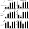
Competitive virus capture. The effect of incubating VLPs with MAbs on their ability to be captured by the same or different MAbs was investigated. WT- and VSV-G-VLPs (A, C, and E) or UNC- and VSV-G-VLPs (B, D, and F) were incubated with 10 μg/ml of MAb competitor (or none), as indicated, and then capture by MAbs b6 (A and B), b12 (C and D), and 2G12 (E and F) was determined.

Similar articles

Cited by

References

    1. Abacioglu, Y. H., T. R. Fouts, J. D. Laman, E. Claassen, S. H. Pincus, J. P. Moore, C. A. Roby, R. Kamin-Lewis, and G. K. Lewis. 1994. Epitope mapping and topology of baculovirus-expressed HIV-1 gp160 determined with a panel of murine monoclonal antibodies. AIDS Res. Hum. Retroviruses 10:371-381. - PubMed
    1. Abrahamyan, L. G., R. M. Markosyan, J. P. Moore, F. S. Cohen, and G. B. Melikyan. 2003. Human immunodeficiency virus type 1 Env with an intersubunit disulfide bond engages coreceptors but requires bond reduction after engagement to induce fusion. J. Virol. 77:5829-5836. - PMC - PubMed
    1. Abrahamyan, L. G., S. R. Mkrtchyan, J. Binley, M. Lu, G. B. Melikyan, and F. S. Cohen. 2005. The cytoplasmic tail slows the folding of human immunodeficiency virus type 1 Env from a late prebundle configuration into the six-helix bundle. J. Virol. 79:106-115. - PMC - PubMed
    1. Allaway, G. P., K. L. Davis-Bruno, G. A. Beaudry, E. B. Garcia, E. L. Wong, A. M. Ryder, K. W. Hasel, M. C. Gauduin, R. A. Koup, J. S. McDougal, et al. 1995. Expression and characterization of CD4-IgG2, a novel heterotetramer that neutralizes primary HIV type 1 isolates. AIDS Res. Hum. Retrovir. 11:533-539. - PubMed
    1. Bewley, C. A., J. M. Louis, R. Ghirlando, and G. M. Clore. 2002. Design of a novel peptide inhibitor of HIV fusion that disrupts the internal trimeric coiled-coil of gp41. J. Biol. Chem. 277:14238-14245. - PubMed

Publication types

MeSH terms

LinkOut - more resources