

Regulation of antibacterial defense in the small intestine by the nuclear bile acid receptor
- PMID: 16473946
- PMCID: PMC1450165
- DOI: 10.1073/pnas.0509592103
Regulation of antibacterial defense in the small intestine by the nuclear bile acid receptor
Abstract
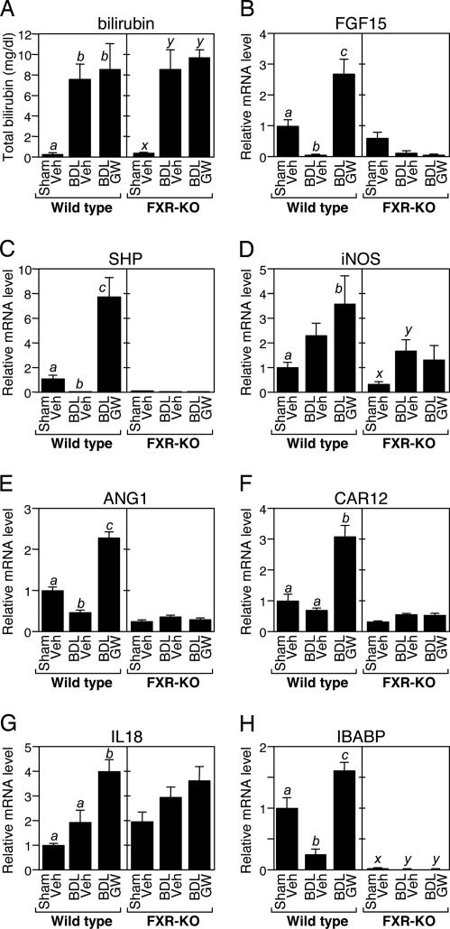
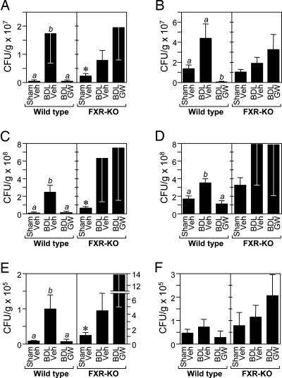
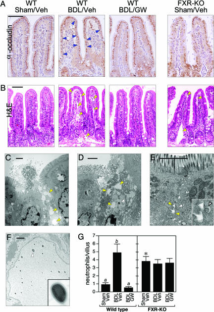
Obstruction of bile flow results in bacterial proliferation and mucosal injury in the small intestine that can lead to the translocation of bacteria across the epithelial barrier and systemic infection. These adverse effects of biliary obstruction can be inhibited by administration of bile acids. Here we show that the farnesoid X receptor (FXR), a nuclear receptor for bile acids, induces genes involved in enteroprotection and inhibits bacterial overgrowth and mucosal injury in ileum caused by bile duct ligation. Mice lacking FXR have increased ileal levels of bacteria and a compromised epithelial barrier. These findings reveal a central role for FXR in protecting the distal small intestine from bacterial invasion and suggest that FXR agonists may prevent epithelial deterioration and bacterial translocation in patients with impaired bile flow.
Conflict of interest statement
Conflict of interest statement: No conflicts declared.
Figures

Comment in
-
How bile acids confer gut mucosal protection against bacteria.Proc Natl Acad Sci U S A. 2006 Mar 21;103(12):4333-4. doi: 10.1073/pnas.0600780103. Epub 2006 Mar 13. Proc Natl Acad Sci U S A. 2006. PMID: 16537368 Free PMC article. No abstract available.
Similar articles
-
Bile acids induce the expression of the human peroxisome proliferator-activated receptor alpha gene via activation of the farnesoid X receptor.Mol Endocrinol. 2003 Feb;17(2):259-72. doi: 10.1210/me.2002-0120. Mol Endocrinol. 2003. PMID: 12554753
-
Knockdown of ATP8B1 expression leads to specific downregulation of the bile acid sensor FXR in HepG2 cells: effect of the FXR agonist GW4064.Am J Physiol Gastrointest Liver Physiol. 2009 May;296(5):G1119-29. doi: 10.1152/ajpgi.90371.2008. Epub 2009 Feb 19. Am J Physiol Gastrointest Liver Physiol. 2009. PMID: 19228886
-
Regulation of CYP3A4 by the bile acid receptor FXR: evidence for functional binding sites in the CYP3A4 gene.Pharmacogenetics. 2004 Oct;14(10):635-45. doi: 10.1097/00008571-200410000-00001. Pharmacogenetics. 2004. PMID: 15454728
-
The expanding role of the bile acid receptor FXR in the small intestine.J Hepatol. 2006 Jun;44(6):1213-5. doi: 10.1016/j.jhep.2006.03.006. Epub 2006 Apr 7. J Hepatol. 2006. PMID: 16618512 Review. No abstract available.
-
FXR: a metabolic regulator and cell protector.Cell Res. 2008 Nov;18(11):1087-95. doi: 10.1038/cr.2008.289. Cell Res. 2008. PMID: 18825165 Review.
Cited by
-
Recent advances in understanding and managing cholestasis.F1000Res. 2016 Apr 19;5:F1000 Faculty Rev-705. doi: 10.12688/f1000research.8012.1. eCollection 2016. F1000Res. 2016. PMID: 27134744 Free PMC article. Review.
-
The Commensal Microbe Veillonella as a Marker for Response to an FGF19 Analog in NASH.Hepatology. 2021 Jan;73(1):126-143. doi: 10.1002/hep.31523. Epub 2020 Dec 11. Hepatology. 2021. PMID: 32794259 Free PMC article. Clinical Trial.
-
Impact of Bacterial Metabolites on Gut Barrier Function and Host Immunity: A Focus on Bacterial Metabolism and Its Relevance for Intestinal Inflammation.Front Immunol. 2021 May 26;12:658354. doi: 10.3389/fimmu.2021.658354. eCollection 2021. Front Immunol. 2021. PMID: 34122415 Free PMC article. Review.
-
Bile acid mediated effects on gut integrity and performance of early-weaned piglets.BMC Vet Res. 2015 May 14;11:111. doi: 10.1186/s12917-015-0425-6. BMC Vet Res. 2015. PMID: 25972097 Free PMC article. Clinical Trial.
-
Hepatic Encephalopathy in Cirrhotic Patients and Risk of Small Intestinal Bacterial Overgrowth: A Systematic Review and Meta-Analysis.Biomed Res Int. 2022 Oct 18;2022:2469513. doi: 10.1155/2022/2469513. eCollection 2022. Biomed Res Int. 2022. PMID: 36303585 Free PMC article. Review.
References
-
- Russell D. W. Annu. Rev. Biochem. 2003;72:137–174. - PubMed
-
- Berg R. D. Trends Microbiol. 1995;3:149–154. - PubMed
-
- Ding J. W., Andersson R., Soltesz V., Willen R., Bengmark S. Eur. Surg. Res. 1993;25:11–19. - PubMed
-
- Lorenzo-Zuniga V., Bartoli R., Planas R., Hofmann A. F., Vinado B., Hagey L. R., Hernandez J. M., Mane J., Alvarez M. A., Ausina V., Gassull M. A. Hepatology. 2003;37:551–557. - PubMed
-
- Cahill C. J. Br. J. Surg. 1983;70:590–595. - PubMed
Publication types
MeSH terms
Substances
Grants and funding
LinkOut - more resources
Full Text Sources
Other Literature Sources
Molecular Biology Databases
