Inactivation of PU.1 in adult mice leads to the development of myeloid leukemia
- PMID: 16432184
- PMCID: PMC1360594
- DOI: 10.1073/pnas.0510616103
Inactivation of PU.1 in adult mice leads to the development of myeloid leukemia
Abstract
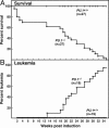
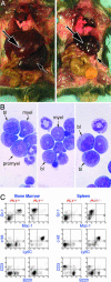
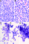
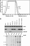
Genetically primed adult C57BL mice were deleted of exon 5 of the gene encoding the transcription factor PU.1 by IFN activation of Cre recombinase. After a 13-week delay, conditionally deleted (PU.1(-/-)) mice began dying of myeloid leukemia, and 95% of the mice surviving from early postinduction death developed transplantable myeloid leukemia whose cells were deleted of PU.1 and uniformly Gr-1 positive. The leukemic cells formed autonomous colonies in semisolid culture with varying clonal efficiency, but colony formation was enhanced by IL-3 and sometimes by granulocyte-macrophage colony-stimulating factor. Nine of 13 tumors analyzed had developed a capacity for autocrine IL-3 or granulocyte-macrophage colony-stimulating factor production, and there was evidence of rearrangement of the IL-3 gene. Acquisition of autocrine growth-factor production and autonomous growth appeared to be major events in the transformation of conditionally deleted PU.1(-/-) cells to fully developed myeloid leukemic populations.
Figures


Similar articles
-
PU.1 regulates both cytokine-dependent proliferation and differentiation of granulocyte/macrophage progenitors.EMBO J. 1998 Aug 3;17(15):4456-68. doi: 10.1093/emboj/17.15.4456. EMBO J. 1998. PMID: 9687512 Free PMC article.
-
PU.1 promotes cell cycle exit in the murine myeloid lineage associated with downregulation of E2F1.Exp Hematol. 2014 Mar;42(3):204-217.e1. doi: 10.1016/j.exphem.2013.11.011. Epub 2013 Dec 5. Exp Hematol. 2014. PMID: 24316397
-
Dose-Rate-Dependent PU.1 Inactivation to Develop Acute Myeloid Leukemia in Mice Through Persistent Stem Cell Proliferation After Acute or Chronic Gamma Irradiation.Radiat Res. 2019 Dec;192(6):612-620. doi: 10.1667/RR15359.1. Epub 2019 Sep 27. Radiat Res. 2019. PMID: 31560640
-
Stem cell fate specification: role of master regulatory switch transcription factor PU.1 in differential hematopoiesis.Stem Cells Dev. 2005 Apr;14(2):140-52. doi: 10.1089/scd.2005.14.140. Stem Cells Dev. 2005. PMID: 15910240 Review.
-
PU.1: a crucial and versatile player in hematopoiesis and leukemia.Int J Biochem Cell Biol. 2008;40(1):22-7. doi: 10.1016/j.biocel.2007.01.026. Epub 2007 Feb 4. Int J Biochem Cell Biol. 2008. PMID: 17374502 Review.
Cited by
-
The significance of low PU.1 expression in patients with acute promyelocytic leukemia.J Hematol Oncol. 2012 May 8;5:22. doi: 10.1186/1756-8722-5-22. J Hematol Oncol. 2012. PMID: 22569057 Free PMC article.
-
PRDM16s transforms megakaryocyte-erythroid progenitors into myeloid leukemia-initiating cells.Blood. 2019 Aug 15;134(7):614-625. doi: 10.1182/blood.2018888255. Epub 2019 Jul 3. Blood. 2019. PMID: 31270104 Free PMC article.
-
Radiation-induced myeloid leukemia in murine models.Hum Genomics. 2014 Jul 25;8(1):13. doi: 10.1186/1479-7364-8-13. Hum Genomics. 2014. PMID: 25062865 Free PMC article. Review.
-
Molecular characterisation of murine acute myeloid leukaemia induced by 56Fe ion and 137Cs gamma ray irradiation.Mutagenesis. 2013 Jan;28(1):71-9. doi: 10.1093/mutage/ges055. Epub 2012 Sep 17. Mutagenesis. 2013. PMID: 22987027 Free PMC article.
-
Aging is associated with functional and molecular changes in distinct hematopoietic stem cell subsets.Nat Commun. 2024 Sep 11;15(1):7966. doi: 10.1038/s41467-024-52318-1. Nat Commun. 2024. PMID: 39261515 Free PMC article.
References
Publication types
MeSH terms
Substances
Grants and funding
LinkOut - more resources
Full Text Sources
Molecular Biology Databases

