Comparative Study
. 2006 Jan 18;26(3):1045-56.
doi: 10.1523/JNEUROSCI.4499-05.2006.

Molecular and morphological heterogeneity of neural precursors in the mouse neocortical proliferative zones

Affiliations
Comparative Study

Molecular and morphological heterogeneity of neural precursors in the mouse neocortical proliferative zones

Jonathan S Gal et al. J Neurosci. .

Abstract

The proliferative ventricular zone (VZ) is the main source of projection neurons for the overlying cerebral neocortex. The number and diversity of neocortical neurons is determined, in part, by factors controlling the proliferation and specification of VZ cells during embryonic development. We used a variety of methods, including in utero electroporation with specific cellular markers, computer-assisted serial EM cell reconstruction, and time-lapse multiphoton imaging to characterize the molecular and morphological characteristics of the VZ constituents and to capture their behavior during cell division. Our analyses reveal at least two types of dividing cells in the VZ: (1) radial glial cells (RGCs) that span the entire neocortical wall and maintain contact both at the ventricular and pial surfaces throughout mitotic division, and (2) short neural precursors (SNPs) that possess a ventricular endfoot and a basal process of variable length that is retracted during mitotic division. These two precursor cell classes are present concomitantly in the VZ, but their relative number changes over the course of cortical neurogenesis. Moreover, the SNPs are morphologically, ultrastructurally and molecularly distinct from dividing RGCs. For example, SNPs are marked by their preferential expression of the tubulin alpha-1 promoter whereas RGCs instead express the glutamate-aspartate transporter and brain lipid binding protein promoters. In contrast to recent studies that suggest that RGCs are the sole type of VZ precursor, the present study indicates that the VZ in murine dorsal telencephalon is similar to that in human and nonhuman primates, because it contains multiple types of neuronal precursors.


Figures

Figure 1.
Figure 1.
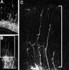
Transfected VZ cells displayed variable morphologies. A, Transfection of E/nestin:P/hsp68:EGFP elucidated nestin-expressing VZ cells. Many long RGCs were evident in the midst of shorter VZ cells (SNPs), which lacked long ascending fibers. B, Transfection using a plasmid expressing an actin-EYFP fusion protein (pactin-EYFP) also labeled SNPs (filled arrow) and RGCs (open arrow) in the VZ. The SNP in this image is mitotic and rounded at the ventricular surface and lacks an ascending process containing the fusion protein. For 3D reconstruction of this Z-stack, see supplemental movie 2 (available at www.jneurosci.org as supplemental material). C, Dividing RGCs were also evident after electroporation with pEYFP. This section was counterstained with PI (red) to label all cell nuclei. The RGC in this collapsed stack is in anaphase and has a clearly identifiable ascending process (arrows). D1, D2, SNPs are bipolar during interphase periods and possess unramified short ascending processes, which are often contained within the depth of the VZ. E1, E2, Other short VZ cells possessed ascending processes with filopodia or growth cone-like tips (arrow heads). The basal process of the cell in E1 did contact the ventricular surface, but appears displaced in the collapsed stack because of undulation of the ventricular surface. The short VZ cells with growth cone tips are likely long RGCs regrowing the basal process to the pial surface. Collapsed stacks in D1–E2 fully contained all processes of the imaged VZ cells. The dashed lines indicate the superficial boundary of the VZ.
Figure 2.
Figure 2.
Quantification of VZ cell types: retrograde DiI labeling. A, The pial surface of the telencephalic hemispheres were coated with DiI crystals. B, C, Six weeks later, many cells in the CP and IZ were labeled with DiI (red), and mitotic cells (ranging from metaphase to anaphase) at the ventricular surface were scored as DiI-labeled or -unlabeled (C, arrowheads). PI was used to counterstain the cell nuclei (blue). D, More short (DiI-unlabeled) than long (DiI-labeled) VZ cells were found at E13.5 (n = 124 total cells) and E14.5 (n = 157 total cells). *p < 0.05, t test. Error bars indicate SE.
Figure 3.
Figure 3.
Quantification of VZ cell types: counting ascending fibers. A, In this surface-rendered Z-stack from an E13.5 brain fixed 24 h after electroporation, the density of VZ cells can be compared with the number of ascending radial fibers. B, This collapsed Z-stack illustrates the VZ cell/ascending process ratio. The boxed area is shown at higher magnification in C, where ascending radial fibers are clearly evident emanating into the IZ. The white line demarcates the counting region where ascending processes were scored.
Figure 4.
Figure 4.
Quantification of VZ cell types: transfection with membrane-tagged EGFP. A, Twenty-four hours after in utero electroporation on E13.5 with pEGFP-F, numerous labeled VZ and SVZ cells are found in the neocortical wall. B1, B2, Examples from collapsed z-stacks of short metaphase cells that lacked ascending fibers (arrowheads). B3, This inset, extracted from the dotted lines in B2, contains an optical section through the middle of the identified SNP and demonstrates the membrane-labeling profile after transfection with EGFP-F. C, Z-stacks also contained mitotic long RGCs whose radial fibers could be traced to the pial surface (arrows). The arrowhead denotes a rounded SNP. D, In 3D z-stack projections, the number of mitotic SNPs (black bars) and mitotic RGCs (white bars) were counted on E13.5 (n = 234 cells), E14.5 (189 cells), E15.5 (n = 153 cells), and E16.5 (n = 203 cells). On each day, the ratio of long to short cells was nearly 50% and not statistically significant, although there were more SNPs found on E14.5 (*p < 0.05; t test). Error bars indicate SE.
Figure 5.
Figure 5.
3D reconstructions of SNPs by EM. A1, A2, This short cell in telophase was reconstructed from serial sections taken from unstained tissue from an E13.5 neocortex. The membrane of the cell is indicated with small arrows. The ventricular surface is denoted by a large arrow in the surface rendered image in A2. Adherens junctions are indicated by arrowheads. B1, This E13.5 VZ cell was first labeled by in utero electroporation with EGFP-F. Subsequent anti-EGFP immunolabeling demarcated the cell border (small arrows) with electron dense immunoperoxidase-DAB reaction end-product. Two centrioles (c) located in controversial poles of the cell body (only one of which is seen in this serial section), chromosomes (chr) in cytoplasm, and fragments of forming nuclear membranes (nm) indicate that the cell is in early telophase. The framed area in B1 is enlarged in B2. B3, 3D reconstruction from 144 contiguous serial sections demonstrates that the cell is devoid of processes. The ultrathin section in B1 and B2 is indicated by the dashed line in B3. Scale bars: A1–B3, 1 μm.
Figure 6.
Figure 6.
Light and electron micrographs of a long mitotic RGC from E14.5 dorsal telencephalic VZ. The specimen was prepared by pial placement of DiI crystals and photoconversion of the labeling into electron dense DAB precipitation. A1, Light micrograph of a long cell with the cell body situated at the ventricular surface. The process (arrows) traverses ventricular VZs and SVZs and reaches the pial surface, as suggested by DiI/DAB labeling. The border between the zones is indicated by the dashed line. A2, Specimen of the cell trimmed for ultrathin sectioning before EM investigation. The cell of interest is the only DAB-containing cell in the region. B, EM image of a profile of the DAB-containing cell with initial fragment of the radial process (arrow). Chromosomes (chr) and centriole (c) in cytoplasm indicate that the cell is in metaphase. Scale bars: A1, A2, 20 μm; B, 2 μm.
Figure 7.
Figure 7.
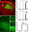
Electroporation of cell-specific promoters was used to differentially label short and long VZ cells. A–E, The Tα1 promoter construct preferentially labels short VZ cells (supplemental movie 5, available at www.jneurosci.org as supplemental material). A, Forty-eight hours after in utero electroporation with pTα1-hGFP, many neurons generated from the VZ and SVZ are found migrating radially through the IZ into the CP. B, At higher magnification, very few GFP+ ascending fibers from RGCs were found in superficial portions of the neocortical wall. C, Most neurons migrating into the CP appeared bipolar, with thin trailing processes and longer, thicker leading processes, and did not appear to be associated with GFP+ RGC fibers. D, E, Reconstructed VZ cells (arrowheads), depicted here in collapsed stacks within which they were fully contained, had short ascending processes (double arrowheads) before their entry into metaphase (D), and lacked processes when dividing at the surface of the ventricle (E). 3D-reconstructed mitotic cells were scored as short (black bars) or long cells (white bars) 24 h (F) or 48 h (G) after in utero electroporation with pGlast-EGFP, pBlbp-EGFP, or pTα1-hGFP. Of the total number of cells expressing each construct, pGlast-EGFP and pBlbp-EGFP were primarily expressed by long RGCs. Conversely, short dividing cells preferentially expressed the pTα1-hGFP construct. The percentage of unclassifiable cells is represented as N.D.(> 18,000 transfected cells were scored for the experiments in F and G). H, Cotransfection of EGFP/DsRed2 plasmid pairs demonstrated that mitotic VZ cells expressing either pGlast or pBlbp did not concurrently express pTα1, although long cells did coexpress pGlast and pBlbp. *p < 0.0001. Error bars indicate SE.
Figure 8.
Figure 8.
SNPs progress through the cell cycle. The denoted plasmid constructs were used for in utero electroporation on E14.5 followed 24 h later by cumulative labeling with BrdU or immunostaining for Ki67 antigen. A–C, Four hours of BrdU labeling identified bipolar SNPs that were labeled or unlabeled with BrdU. As depicted in A, BrdU+ cells would be present in phases S-early G1 based on cell cycle parameters specified by Takahashi et al. (1995). Correspondingly, BrdU short cells would be in mid to late G1 phase. B, b1–b3, This Tα1:hGFP expressing cell was BrdU and therefore in G1 phase, whereas the pEYFP-C2 expressing cell in C and c1–c3 was BrdU+. D, Depiction of the cell-cycle location of BrdU+ and BrdU VZ cells after 6 h labeling. E–f2, Examples of BrdU+ SNPs reconstructed after transfection with pEGFP-F and Tα1:hGFP. More SNPs were BrdU+ after 6 h of cumulative BrdU exposure compared with 4 h BrdU exposure, suggesting that SNPs progress through the cell cycle (see text for details). G–h2, Ki67 immunostaining also revealed that Tα1-expressing VZ cells are arrayed throughout the cell cycle. The higher magnification insets in H–H2 demonstrate two Tα1-expressing cells with abventricular somata that are Ki67+.

Similar articles

Cited by

References

    1. Aguirre A, Gallo V (2004) Postnatal neurogenesis and gliogenesis in the olfactory bulb from NG2-expressing progenitors of the subventricular zone. J Neurosci 24: 10530–10541. - PMC - PubMed
    1. Aguirre AA, Chittajallu R, Belachew S, Gallo V (2004) NG2-expressing cells in the subventricular zone are type C-like cells and contribute to inter-neuron generation in the postnatal hippocampus. J Cell Biol 165: 575–589. - PMC - PubMed
    1. Akamatsu W, Okano HJ, Osumi N, Inoue T, Nakamura S, Sakakibara S, Miura M, Matsuo N, Darnell RB, Okano H (1999) Mammalian ELAV-like neuronal RNA-binding proteins HuB and HuC promote neuronal development in both the central and the peripheral nervous systems. Proc Natl Acad Sci USA 96: 9885–9890. - PMC - PubMed
    1. Angevine JB, Sidman RL (1961) Autoradiographic study of cell migration during histogenesis of the cerebral cortex in the mouse. Nature 192: 766–768. - PubMed
    1. Anthony TE, Klein C, Fishell G, Heintz N (2004) Radial glia serve as neuronal progenitors in all regions of the central nervous system. Neuron 41: 881–890. - PubMed

Publication types