. 2006 Jan 16;172(2):259-68.
doi: 10.1083/jcb.200506179. Epub 2006 Jan 9.

Focal adhesion size controls tension-dependent recruitment of alpha-smooth muscle actin to stress fibers

Affiliations

Focal adhesion size controls tension-dependent recruitment of alpha-smooth muscle actin to stress fibers

Jérôme M Goffin et al. J Cell Biol. .

Abstract

Expression of alpha-smooth muscle actin (alpha-SMA) renders fibroblasts highly contractile and hallmarks myofibroblast differentiation. We identify alpha-SMA as a mechanosensitive protein that is recruited to stress fibers under high tension. Generation of this threshold tension requires the anchoring of stress fibers at sites of 8-30-microm-long "supermature" focal adhesions (suFAs), which exert a stress approximately fourfold higher (approximately 12 nN/microm2) on micropatterned deformable substrates than 2-6-microm-long classical FAs. Inhibition of suFA formation by growing myofibroblasts on substrates with a compliance of < or = 11 kPa and on rigid micropatterns of 6-microm-long classical FA islets confines alpha-SMA to the cytosol. Reincorporation of alpha-SMA into stress fibers is established by stretching 6-microm-long classical FAs to 8.1-microm-long suFA islets on extendable membranes; the same stretch producing 5.4-microm-long classical FAs from initially 4-microm-long islets is without effect. We propose that the different molecular composition and higher phosphorylation of FAs on supermature islets, compared with FAs on classical islets, accounts for higher stress resistance.


Figures
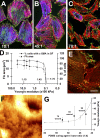
Figure 1.
Figure 1.
Matrix compliance controls FA size and α-SMA localization. REF-52 myofibroblasts were cultured for 12 h on PDMS substrates with a Young's modulus of 780 (A), 23 (B), and 9.6 kPa (C and E) and stained for vinculin (A–C, green), α-SMA (A–C, blue), and F-actin (phalloidin; A–C, red) after standard fixation. Numbers indicate PDMS base/curing agent ratio. (D) From such images, all FAs in the field were analyzed with a segmentation algorithm and FA size was plotted as a function of substrate compliance. The number of cells with clear α-SMA–positive stress fibers (SF) was manually assessed and related to the total number of cells. 10 cells were analyzed per substrate, and the experiment was performed three times (nFAs/substrate ∼3,000). Mean values (±SD) that differ significantly from control on plastic (P ≤ 0.001) are open symbols. Culture plastic is arbitrarily set to 3,000 kPa. (E) By using a detergent-free staining protocol, cytosolic α-SMA (green) was shown to disappear from β-cytoplasmic actin–positive stress fibers on 9.6 kPa substrates (red) and to accumulate in rods. (F) A force indentation profile was produced by AFM on a 100 × 100-μm area of a section of 9-d-old granulation tissue. Lumen and wall of a small vessel, surrounded by fibroblast-populated tissue, are obvious in the upper righthand corner. Bars: (A–C) 50 μm; (E and F) 20 μm. (G) The mean elastic modulus of 7–9-d-old granulation tissue (squares) is related to that of 1:40 to 1:60 PDMS (dotted line), both assessed with AFM.
Figure 2.
Figure 2.
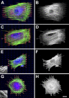
Incorporation of α-SMA into stress fibers requires FAs longer than 6 μm. Arrays of FN (50 μg/ml) islets with 6-μm spacing, 1.25-μm width, and 20- (A and B), 10- (C and D), 6- (E and F), and 2-μm (G and H) lengths were created on glass by μCP, and nonprinted regions were passivated. After a 12-h culture, REF-52 myofibroblasts were stained for F-actin (green), FN (blue), vinculin (red), and α-SMA (black and white). (E and G, insets) F-actin stress fibers at a 4× higher magnification. Representative results of one out of six independent experiments are presented. Bar, 20 μm.
Figure 3.
Figure 3.
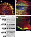
Stress fiber–derived α-SMA accumulates in detergent-soluble rods. REF-52 myofibroblasts were grown for 12 h on 4- (A) and 20-μm-long (B and D) adhesion islets and stained for α-SMA (green), β-cytoplasmic actin (red), and FN (blue). By using a detergent-free staining procedure, α-SMA was identified in cytosolic rods after growth on 4-μm-long islets (A) and in stress fibers on 20-μm-long islets of control cells (B). Inhibition of Rho-associated kinase on 20-μm-long islets leads to the neoformation of α-SMA rods (D; and Video 1). Bar, 20 μm. (C) Western blotting of the TX-100–insoluble cytoskeleton demonstrates the decrease of specific contractile proteins in the TX-100–insoluble fraction with decreasing adhesion islet size (Table I). Representative results of one out of four independent experiments are presented. MLCK, myosin light chain kinase; NM, nonmuscle; SM, smooth muscle. Video 1 is available at http://www.jcb.org/cgi/content/full/jcb.200506179/DC1.
Figure 4.
Figure 4.
Enlargement of classical to suFAs recruits α-SMA to stress fibers. REF-52 myofibroblasts were grown for 12 h on extendable PDMS membranes that have been provided with covalently bound FN islets. Cells were then subjected to a 135% linear stretch and stained after 6 h for α-SMA (A and D), vinculin (B and E), and FN (C and F). Despite enlarging 6- and 4-μm-long classical FA-sized islets by the same factor, only the resulting 8.1-μm-long suFA islets (B and C) induced formation of α-SMA–positive stress fibers (A), but not resulting 5.4-μm-long classical FA islets (D–F). The experiment was performed three times and representative results are shown. Bar, 25 μm.
Figure 5.
Figure 5.
suFAs generate higher stress than classical FAs. (A) Fibroblasts and myofibroblasts transfected with GFP-paxillin were cultured for 4 d on micropatterned deformable substrates (Video 3). Force transmission at FAs (green) leads to displacement of the fluorescent dots (red) that return to their original position (blue) after completely relaxing the cell with cytochalasin D; static dots appear pink. (B) Forces at individual FAs (black) that were identified with a segmentation algorithm were calculated from displacement maps using a regularization method and are depicted as red vectors in binary images; close-up in A corresponds to the shaded region in B. Bars: (A) 20 μm; (B) 50 μm. Force vectors, 80 nN. Forces were expressed as a function of FA area for fibroblasts (C; n cells = 12 and n FA = 1,120) and myofibroblasts (D; n cells = 11 and n FAs = 1,166); colored background highlight focal complexes (yellow), classical FAs (blue), and suFAs (red); the same color code is used for linear regressions in D. Inset in C shows fibroblast FA distribution using the same scale as in D. Video 3 is available at http://www.jcb.org/cgi/content/full/jcb.200506179/DC1.
Figure 6.
Figure 6.
Adhesion size dictates FA composition and phosphorylation. REF-52 myofibroblasts were grown for 12 h on 20-μm-long suFA islets (A–C) and 4-μm-long classical FA islets (D–F) created by negative μCP. Cells were stained for β3 integrin (A and D, green), paxillin (A and D, red), tensin (B and E, red), vinculin (B and E, green), phospho-tyrosine (C and F, red), and phospho-FAK (C and F, green). Growth on small islets led to the separation of classical FA and fibrillar adhesion markers that colocalize on large islets. (G) Reduced phosphorylation levels of FA proteins on small islets are confirmed by Western blotting of total cell extracts. Representative results of three independently performed experiments are presented. Bar, 20 μm.
Figure 7.
Figure 7.
Changes in matrix rigidity and organization control incorporation of α-SMA into stress fibers by modulating the size of adhesion. (A) Fibroblasts grown in a mechanically restrained, but compliant, three-dimensional matrix develop small adhesions and stress fibers that contain only cytoplasmic actins. (B) In the presence of TGFβ, fibroblasts start to express α-SMA, which at first remains diffusely distributed in the cytosol or organizes in transient rodlike structures. (C) Remodeling activity of cells leads to ECM fiber alignment and creates larger surfaces for adhesion formation; larger adhesions permit development of stronger stress fibers and generation of higher contractile forces. (C) Continuing ECM fiber alignment further enlarges adhesion sites and intracellularly generated tension. When adhesion sites grow to the size of suFA, intracellular tension reaches a critical level that allows incorporation of α-SMA into preexisting cytoplasmic actin stress fibers. The force generated by α-SMA–containing stress fiber is significantly higher compared with cytoplasmic actin stress fibers leading to further FA supermaturation and ECM contraction.

Similar articles

Cited by

References

    1. Arora, P.D., N. Narani, and C.A. McCulloch. 1999. The compliance of collagen gels regulates transforming growth factor-beta induction of alpha-smooth muscle actin in fibroblasts. Am. J. Pathol. 154:871–882. - PMC - PubMed
    1. Balaban, N.Q., U.S. Schwarz, D. Riveline, P. Goichberg, G. Tzur, I. Sabanay, D. Mahalu, S. Safran, A. Bershadsky, L. Addadi, and B. Geiger. 2001. Force and focal adhesion assembly: a close relationship studied using elastic micropatterned substrates. Nat. Cell Biol. 3:466–472. - PubMed
    1. Ballestrem, C., B. Hinz, B.A. Imhof, and B. Wehrle-Haller. 2001. Marching at the front and dragging behind: differential αVβ3-integrin turnover regulates focal adhesion behavior. J. Cell Biol. 155:1319–1332. - PMC - PubMed
    1. Bao, G., and S. Suresh. 2003. Cell and molecular mechanics of biological materials. Nat. Mater. 2:715–725. - PubMed
    1. Beningo, K.A., M. Dembo, I. Kaverina, J.V. Small, and Y.L. Wang. 2001. Nascent focal adhesions are responsible for the generation of strong propulsive forces in migrating fibroblasts. J. Cell Biol. 153:881–888. - PMC - PubMed

Publication types