. 2006 Jan;80(2):794-801.
doi: 10.1128/JVI.80.2.794-801.2006.

Involvement of template-activating factor I/SET in transcription of adenovirus early genes as a positive-acting factor

Affiliations

Involvement of template-activating factor I/SET in transcription of adenovirus early genes as a positive-acting factor

Hirohito Haruki et al. J Virol. 2006 Jan.

Abstract

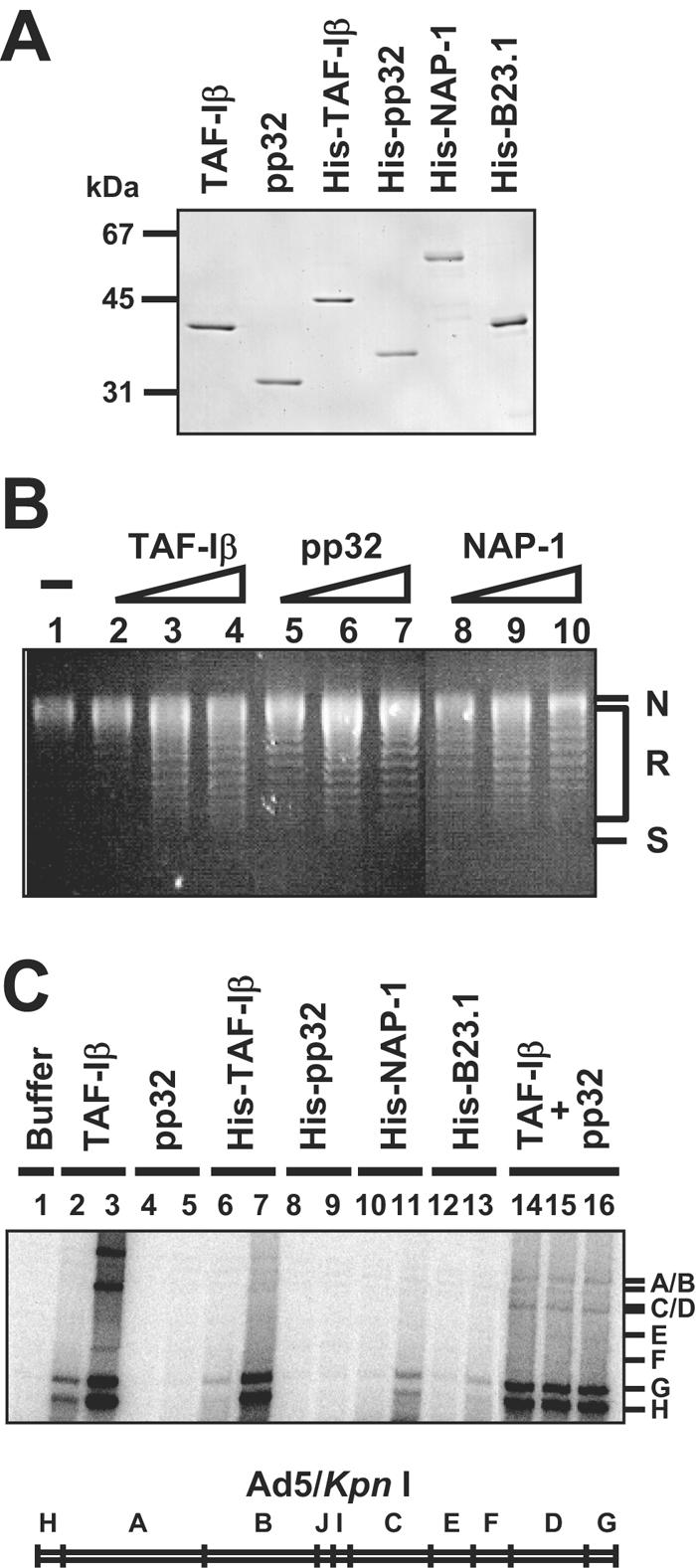
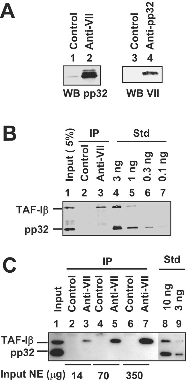
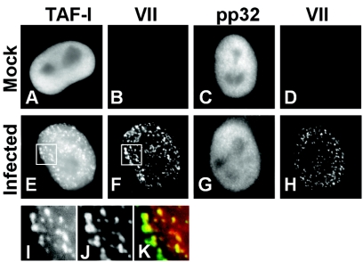
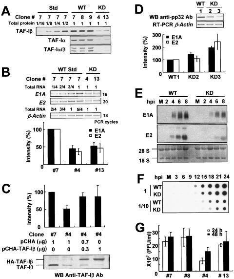
The adenovirus genome complexed with viral core protein VII (adenovirus DNA-protein VII complex) at least is the bona fide template for transcription of adenovirus early genes. It is believed that the highly basic protein VII, like cellular histones, is a negative regulator for genome functions. Analyses with in vitro replication and transcription systems using the adenovirus DNA-protein VII complex have revealed that remodeling of the complex is crucial for efficient DNA replication and transcription. We identified host acidic proteins, template-activating factor I (TAF-I), TAF-II, and TAF-III as stimulatory factors for replication from the adenovirus DNA-protein VII complex. Recently, it was reported that the adenovirus DNA interacts with TAF-I and pp32, another host acidic protein (Y. Xue, J. S. Johnson, D. A. Ornelles, J. Lieberman, and D. A. Engel, J. Virol. 79:2474-2483, 2005). We found that TAF-I interacts and colocalizes with protein VII in adenovirus-infected cells during the early phases of infection, but pp32 does not. Although pp32 had the potential ability to interact with protein VII, pp32 did not remodel the adenovirus DNA-protein VII complex in vitro. Small interfering RNA-mediated knockdown of TAF-I expression leads to the delay of the transcription timing of early genes. These results provide evidence that TAF-I plays an important role in the early stages of the adenovirus infection cycle.


Figures

FIG. 1.
FIG. 1.
Stimulatory activity for DNA replication of the Ad DNA-protein VII complex. (A) Purified recombinant proteins. Recombinant TAF-Iβ and pp32 and recombinant His-tagged TAF-Iβ, pp32, NAP-1, and B23.1 (200 ng each) generated by bacterial expression systems were separated by SDS-10% PAGE and visualized by staining with Coomassie brilliant blue. (B) Supercoiling assay. Core histones (200 ng) preincubated without (lane 1) or with 100 ng (lanes 2, 5, and 8), 200 ng (lanes 3, 6, and 9), or 500 ng (lanes 4, 7, and 10) of His-tagged TAF-Iβ (lanes 2 through 4), pp32 (lanes 5 through 7), or NAP-1 (lanes 8 through 10) were mixed with closed circular DNA (200 ng) relaxed by topoisomerase I (1 U; TaKaRa) and further incubated at 37°C for 1 h. The DNA was purified and separated by electrophoresis on a 1% agarose gel and visualized by staining with ethidium bromide. Positions of relaxed and partially supercoiled (R), supercoiled (S), or nicked (N) circular plasmid DNA are indicated. (C) Cell-free DNA replication of the Ad DNA-protein VII complex. Cell-free Ad DNA replication assays using the Ad DNA-protein VII complex as template were performed in the absence (lane 1) or presence of 50 ng (lanes 2, 4, 6, 8, 10, and 12) or 200 ng (lanes 3, 5, 7, 9, 11, and 13) of TAF-Iβ (lanes 2 and 3), pp32 (lanes 4 and 5), His-tagged TAF-Iβ (lanes 6 and 7), His-tagged pp32 (lanes 8 and 9), His-tagged NAP-1 (lanes 10 and 11), or His-tagged B23.1 (lanes 12 and 13). For lanes 14 through 16, either 50 ng (lane 14), 100 ng (lane 15), or 200 ng (lane 16) of pp32 in the presence of 100 ng of TAF-Iβ was added to the reaction mixture. Replication products labeled with [α-32P]dCTP were purified, digested with KpnI, separated on a 0.8% agarose gel, and visualized by autoradiography. KpnI fragments of HAdV5 DNA are indicated underneath.
FIG. 2.
FIG. 2.
Interaction of protein VII with TAF-I and pp32. (A) Interaction between recombinant pp32 and protein VII. Protein VII (100 ng) was incubated with pp32 (200 ng). Next, the samples were subjected to immunoprecipitation with control (lanes 1 and 3), anti-protein VII (lane 2), or anti-pp32 (lane 4) antibodies. Immunoprecipitated proteins were separated by SDS-10% PAGE and detected by Western blotting (WB) using anti-pp32 (lanes 1 and 2) or anti-protein VII (lanes 3 and 4) antibodies. (B) Immunoprecipitation analysis using infected cell extracts. Extracts were prepared from infected cells at 4 h p.i. as described in Materials and Methods and subjected to immunoprecipitation with control (lane 2) or anti-protein VII (lane 3) antibodies. Input (5%) (lane 1), immunoprecipitated proteins (lanes 2 and 3), and indicated amounts of recombinant TAF-I and pp32 as standards (Std) (lanes 4 through 7) were separated by SDS-10% PAGE and detected by Western blotting using a mixture of anti-TAF-Iβ and anti-pp32 antibodies. (C) Immunoprecipitation analysis using uninfected nuclear extracts (NE) and protein VII. Nuclear extracts prepared from uninfected HeLa cells (14, 70, and 350 μg for lanes 2 and 3, 4 and 5, and 6 and 7, respectively) were mixed with purified protein VII (100 ng) and then subjected to immunoprecipitation with control (lanes 2, 4, and 6) or anti-protein VII (lanes 3, 5, and 7) antibodies. Lane 1 indicates input extracts (35 μg), and lanes 8 and 9 show recombinant pp32 and TAF-I.
FIG. 3.
FIG. 3.
Colocalization of TAF-I with protein VII in infected cells during early phases of infection. Cells were mock infected (A through D) or infected (E through H) with HAdV5 at an MOI of 250 and fixed at 4 h p.i. TAF-Iβ (A and E) and protein VII (B and F) or pp32 (C and G) and protein VII (D and H) were stained by the indirect immunofluorescence method with specific antibody for each protein. Panels I and J show higher-magnified images of the regions marked by squares in panels E and F, respectively. Panel K is a merged image of panels I and J visualized with red and green, respectively. The dots inside the nucleus contain both protein VII and TAF-I, while some portion of protein VII is localized at the periphery of the nucleus.
FIG. 4.
FIG. 4.
Knockdown of TAF-I expression. (A) The level of TAF-I in TAF-I KD cell lines derived from HeLa cells. Cell extracts prepared from WT (clones 7, 8, and 9) and TAF-I KD (clones 4 and 13) cell lines were separated by SDS-10% PAGE, and TAF-I proteins were detected by immunoblotting with anti-TAF-Iβ (top gel), anti-TAF-Iα (middle gel), or anti-TAF-Iα/β common (bottom gel) antibodies. Std, standard. (B) Semiquantitative RT-PCR. WT (clone 7) and TAF-I KD (clones 4 and 13) cell lines were infected with HAdV5 at an MOI of 100 and collected at 4 h p.i. Total RNA was isolated and analyzed by RT-PCR for semiquantitative detection of transcripts from β-actin, E1A, and E2 genes. PCR products amplified by 18, 16, and 20 PCR cycles for β-actin, E1A, and E2, respectively, were loaded onto a 6% polyacrylamide gel. After separation, PCR products were visualized by staining with SYBR Gold (upper panel). The intensity of the band was measured using NIH Image software. Mean values ± standard deviations from four independent experiments are summarized in the lower panel, where the level of WT is set as 100%. (C) Rescue experiments. WT (clone 7) and TAF-I KD (clone 4) cells were transfected with indicated amounts of pCHA and pCHA-TAF-Iβ vectors, infected with HAdV5 at an MOI of 100 at 48 h after transfection, and collected at 4 h p.i. The level of E1A RNA was analyzed as shown in panel B (upper part of panel). Data are mean values ± standard deviations from five independent experiments. The levels of HA-TAF-Iβ and endogenous TAF-Iβ proteins were examined by Western blotting using anti TAF-Iβ antibody (bottom of panel). (D) KD of pp32. The level of pp32 protein in HeLa pp32 KD cell lines was examined by Western blotting (WB; top gel). Semiquantitative RT-PCR was performed as described above for panel B using pp32 KD cell lines (bottom gel for β-actin; graph for E1A and E2). Data are mean values ± standard deviations from three independent experiments. (E) Northern blot analysis. WT and TAF-I KD cells were mock infected (M) or infected with HAdV5 at an MOI of 100 and collected at indicated h p.i. Total RNA was isolated and analyzed by Northern blot hybridization using probes complementary to a part of E1A (top gel) or E2 (middle gel) RNA. Ribosomal RNAs were also visualized by staining with ethidium bromide (bottom gel). (F) Time course of viral DNA synthesis in WT and TAF-I KD cells. WT and TAF-I KD cells were mock infected (M) or infected with HAdV5 at an MOI of 100 and collected every 3 h after infection. Total DNA was purified, dot blotted, and hybridized with 32P-labeled probe complementary to the HAdV5 genome DNA. (G) One-step virus growth assay. WT (clones 7 and 8) and TAF-I KD (clones 4 and 13) cell lines were infected with HAdV5 at an MOI of 100. Infected cells and their culture media were collected at 24 and 48 h p.i., and the virus titer was measured by plaque assay.

Similar articles

Cited by

References

    1. Anderson, C. W., M. E. Young, and S. J. Flint. 1989. Characterization of the adenovirus 2 virion protein, mu. Virology 172:506-512. - PubMed
    1. Beresford, P. J., D. Zhang, D. Y. Oh, Z. Fan, E. L. Greer, M. L. Russo, M. Jaju, and J. Lieberman. 2001. Granzyme A activates an endoplasmic reticulum-associated caspase-independent nuclease to induce single-stranded DNA nicks. J. Biol. Chem. 276:43285-43293. - PubMed
    1. Black, B. C., and M. S. Center. 1979. DNA-binding properties of the major core protein of adenovirus 2. Nucleic Acids Res. 6:2339-2353. - PMC - PubMed
    1. Brennan, C. M., I. E. Gallouzi, and J. A. Steitz. 2000. Protein ligands to HuR modulate its interaction with target mRNAs in vivo. J. Cell Biol. 151:1-14. - PMC - PubMed
    1. Chatterjee, P. K., M. E. Vayda, and S. J. Flint. 1986. Adenoviral protein VII packages intracellular viral DNA throughout the early phase of infection. EMBO J. 5:1633-1644. - PMC - PubMed

Publication types

MeSH terms

LinkOut - more resources