. 2006 Jan;4(1):e9.
doi: 10.1371/journal.pbio.0040009.

Activation of NK cells by an endocytosed receptor for soluble HLA-G

Affiliations

Activation of NK cells by an endocytosed receptor for soluble HLA-G

Sumati Rajagopalan et al. PLoS Biol. 2006 Jan.

Abstract

Signaling from endosomes is emerging as a mechanism by which selected receptors provide sustained signals distinct from those generated at the plasma membrane. The activity of natural killer (NK) cells, which are important effectors of innate immunity and regulators of adaptive immunity, is controlled primarily by receptors that are at the cell surface. Here we show that cytokine secretion by resting human NK cells is induced by soluble, but not solid-phase, antibodies to the killer cell immunoglobulin-like receptor (KIR) 2DL4, a receptor for human leukocyte antigen (HLA)-G. KIR2DL4 was constitutively internalized into Rab5-positive compartments via a dynamin-dependent process. Soluble HLA-G was endocytosed into KIR2DL4-containing compartments in NK cells and in 293T cells transfected with KIR2DL4. Chemokine secretion induced by KIR2DL4 transfection into 293T cells occurred only with recombinant forms of KIR2DL4 that trafficked to endosomes. The profile of genes up-regulated by KIR2DL4 engagement on resting NK cells revealed a proinflammatory/proangiogenic response. Soluble HLA-G induced secretion of a similar set of cytokines and chemokines. This unique stimulation of resting NK cells by soluble HLA-G, which is endocytosed by KIR2DL4, implies that NK cells may provide useful functions at sites of HLA-G expression, such as promotion of vascularization in maternal decidua during early pregnancy.


Figures

Figure 1
Figure 1. IFN-γ Secretion by Resting NK Cells Is Induced by Soluble Antibodies to KIR2DL4
(A) KIR2DL4 expression on the surface of NK cells immediately after isolation (day 0) or after 7, 14, or 21 d in culture with autologous feeder cells and rIL-2. Cells were stained with mAb 33. (B) Resting NK cells were incubated with soluble (10 μg/ml), plate-coated (5 μg/0.1 ml/well), or bead-bound (4 beads/cell) antibodies to KIR2DL4 (mAb 33) or CD16 (mAb 3G8). After 24 h, culture supernatants were tested by ELISA for IFN-γ production.
Figure 2
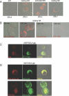
Figure 2. KIR2DL4 Is Endocytosed into Intracellular Vesicles
(A) The B cell line 721.221 (221), resting NK cells, and activated NK cells were fixed, permeabilized, and stained with Cy3-conjugated mAb 33. In the image on the right, resting NK cells were fixed, permeabilized, and stained with a polyclonal antibody specific for the tail of KIR2DL1, followed by Alexa-568–conjugated secondary antibodies. (B) Resting NK cells were incubated at 37 °C for 30 min or 120 min with KIR2DL4-specific mAb 33-Cy3, with a Fab of control mAb 2A8 or with a Fab of anti-KIR2DL4 mAb 33, as indicated. Cells were then fixed and analyzed by confocal microscopy. (C) The 293T cells stably transfected with KIR2DL4-gfp (293T-2DL4-gfp) were fixed, permeabilized, and stained with anti-KIR2DL4 mAb 33 followed by Alexa-568–conjugated secondary antibodies to show co-localization with KIR2DL4-gfp. Single confocal sections are shown. (D) The 293T-2DL4-gfp cells were incubated at 37 °C for 120 min with anti-KIR2DL4 mAb 33, either intact (33 whole Ab) or as a Fab (33-Fab). Cells were fixed, stained with Alexa-568–conjugated secondary antibodies, and analyzed by confocal microscopy. Single confocal sections are shown.
Figure 3
Figure 3. Internalization of KIR2DL4 Is Dynamin Dependent
The 293T cells transfected with HA-tagged KIR2DL4 together with either wild-type dynamin-gfp (Dynamin Egfp-WT) or a dominant-negative mutant of dynamin (dynamin Egfp-K44A) were loaded with KIR2DL4-specific Cy3-conjugated mAb 33 for 120 min and fixed. Individual confocal sections are shown.
Figure 4
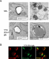
Figure 4. Immunolocalization of Endocytosed KIR2DL4 in NK Cells by Electron Microscopy
(A) The NK cell lines NKL and YTS-2DL4-gfp were loaded with anti-KIR2DL4 mAb 33 for 120 min. Endocytosed receptor was detected using HRP-conjugated sheep anti-F(ab′)2 mouse IgG. The HRP reaction product visible as a dark stain identifies the location of endocytosed KIR2DL4. Vesicular structures positive for KIR2DL4 ranged between 250 and 500 nm in size. (B) The NK cell line YTS, stably transfected with KIR2DL4-gfp (YTS-2DL4-gfp), was fixed, permeabilized, and stained with antibody to perforin followed by Alexa-568–conjugated secondary antibodies. Single confocal sections are shown.
Figure 5
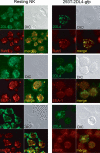
Figure 5. KIR2DL4 Resides in Endocytic Compartments
Resting NK cells and 293T-2DL4-gfp cells were fixed, permeabilized, and stained with antibodies against Rab5, EEA-1, and M6PR, followed by Alexa-568–conjugated secondary antibodies. NK cells were further stained with mAb 33 coupled to Alexa-488 to detect KIR2DL4.
Figure 6
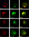
Figure 6. KIR2DL4 Localizes to a Subset of Endosomes Containing Rab5
The 293T cells were transfected with HA-tagged KIR2DL4 and gfp-tagged versions of Rab4, Rab5, Rab7, and Rab11. Forty hours after transfection, cells were fixed and stained with anti-HA mAb, followed by Alexa-568–conjugated secondary antibodies to detect KIR2DL4. Single confocal sections are shown.
Figure 7
Figure 7. Binding of KIR2DL4-Ig Fusion Proteins to HLA-G–Expressing Cells Is Blocked by Anti-KIR2DL4 and Anti-HLA Class I mAbs
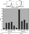
(Top) 221-G cells were stained with mAb DX17 (pan–HLA class I mAb) and mAb G233 (HLA-G–specific mAb) (solid lines). Staining with secondary antibody alone is also shown (dotted lines). (Bottom) The 221 and 221-G cells were incubated with 50 μg/ml KIR2DL4-Ig fusion protein in the presence of 20 μg/ml of either isotype-matched control Abs or mAbs specific for class I (DX17), HLA-G (G233), or KIR2DL4 (33). Cells were then stained with goat anti-human IgG1 secondary antibodies and assessed by flow cytometry. The data are expressed as mean fluorescence intensity (MFI).
Figure 8
Figure 8. Cell Surface Shed and Secreted, Soluble HLA-G Is Endocytosed into KIR2DL4-Containing Vesicles
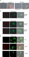
(A) Endocytosis of soluble HLA-G in resting NK cells. The 221 cells and 221 cells transfected with HLA-Cw3 (221-Cw3) were fixed, permeabilized, and stained with mAb F4/326. Resting NK cells were incubated at 37 °C for 120 min with soluble, refolded HLA-C or HLA-G. Cells were then fixed, permeabilized, and stained with reagents to detect HLA-C (F4/326) or HLA-G (G233) as indicated. (B) The NK cell line YTS-2DL4-gfp was loaded at 37 °C for 120 min with refolded HLA-G. Cells were fixed, permeabilized, and stained with mAb G233 to detect co-localization of soluble HLA-G with gfp-tagged KIR2DL4. (C) Recombinant soluble molecules of HLA-G but not HLA-C are endocytosed into 293T-2DL4-gfp cells. Refolded HLA-G and HLA-C were incubated with 293T-2DL4-gfp cells for 2 h. Cells were then fixed, permeabilized, and stained with either mAb G233 (to detect endocytosed HLA-G; upper) or mAb F4/326 (to detect endocytosed HLA-C; middle). (D) The 293T-2DL4-gfp cells were co-cultured with an equal number of 221 cells, 221 cells expressing transmembrane HLA-G (221-G), and 221 cells expressing a soluble isoform of HLA-G (221-sG) for 48 h. Adherent 293T-2DL4-gfp cells were fixed, permeabilized, and stained with mAb G233 followed by Alexa-568–conjugated secondary antibodies prior to acquisition of confocal images. Two 221-G cells are visible in the middle panel.
Figure 9
Figure 9. Endocytosis of Soluble HLA-G into 293T-2DL4-gfp Cells Is Blocked by Anti-KIR2DL4 mAb and by Soluble KIR2DL4
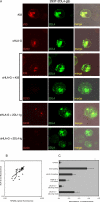
(A) Recombinant, soluble, sHLA-G at 50 μg/ml or mAb 33 (50 μg/ml) was incubated with 293T-2DL4-gfp cells. sHLA-G was also incubated together with 50 μg/ml mAb 33, 50 μg/ml KIR2DL1-Ig, or 50 μg/ml KIR2DL4-Ig, as indicated on the left. Cells were fixed, permeabilized, and stained with either Alexa-568–conjugated secondary antibodies to detect mAb 33 or anti-HLA-G mAb G233, as indicated. Individual confocal sections are shown. (B) Uptake of sHLA-G into 293T-2DL4-gfp cells correlates with level of KIR2DL4 expression. Red fluorescence intensity of G233 staining and green fluorescence intensity of gfp were quantified in 38 individual cells and plotted on a log scale. A best-fit line was generated by linear regression analysis using EXCEL data analysis software. (C) Ratio of red to green fluorescence was quantified for each loading condition as indicated. Average of 10 cells is shown, and standard deviation is shown as bars.
Figure 10
Figure 10. Binding of Soluble mAb to KIR2DL4 Up-regulates Multiple Genes in Resting NK Cells
(A) All genes exhibiting greater than 2-fold up-regulation in microarray experiments with resting NK cells of at least two of three different individuals are listed. (B) Semiquantitative RT-PCR was performed on total RNA isolated at different time points from resting NK cells stimulated with either control IgM mAbs or anti-KIR2DL4 IgM mAbs 36 and 64. (C) Time course of TNF-α, IL-1β, and IFN-γ secretion by resting NK cells stimulated with anti-KIR2DL4 mAbs 36 and 64 (open symbols) or control IgM mAbs (closed symbols). Squares and triangles represent data obtained from two different donors. Protein secretion was detected by ELISA.
Figure 11
Figure 11. Cytokine/Chemokine Synthesis Induced by Soluble HLA-G in Resting NK Cells
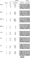
Resting NK cells (5 × 10⁵ cells/well) from three different donors were incubated separately for 48 h with either soluble, control IgM Ab (cIg), soluble anti-KIR2DL4 IgM mAb 36 (anti-2DL4), soluble HLA-G produced in CHO cells (sHLA-G), or beads coated with control anti-HA IgG1 mAb 16B12 (cIg) or with anti-CD16 IgG1 mAb 3G8 (3G8), as indicated. sHLA-G was used together with control IgG2a mAb (cIg) or with anti-HLA-G IgG2a mAb G233 (G233), as indicated. Secretion of the cytokines/chemokines listed on the left is given in pg/ml for each donor separately. Secretion induced by sHLA-G and by Ab-coated beads was compared to that induced by anti-KIR2DL4 mAb for each donor separately and is expressed as a percentage of the anti-KIR2DL4 response. The graphs represent the average ± standard deviation from three experiments.
Figure 12
Figure 12. IL-8 Secretion Induced by KIR2DL4 Is Independent of the Transmembrane Arginine Residue and Requires Trafficking to Intracellular Vesicles
(A) Expression of KIR2DL4 in 293T cells induces IL-8 secretion. The 293T cells were transfected with HA-tagged 2B4, gp49B, and KIR2DL4. After 48 h, culture supernatants were tested for IL-8 by ELISA. (B) Schematic representation of KIR2DL4 variants used in this study. Receptor localization was determined by confocal analysis of immunofluorescence staining of 293T cells transfected with the indicated constructs for 48 h. (C) IL-8 secretion induced by KIR2DL4 mutants in 293T cells. The 293T cells were transfected with HA-tagged KIR2DL4, KIR2DL4(RY-GT), and KIR2DL4-TR. After 48 h, culture supernatants were tested for IL-8 by ELISA. Transfection efficiency was verified by monitoring HA-positive cells by confocal microscopy. (D) Cell surface–expressed gp49B/2DL4 chimera does not induce IL-8 secretion. The 293T cells were transfected with HA-tagged 2B4, KIR2DL4, and gp49B/2DL4 chimera, as indicated. After 48 h, beads coated with control IgG (solid bars) or anti-HA mAb (hatched bars) were added at four beads per cell. Twelve hours later, culture supernatants were tested for IL-8 secretion. The percentage of 293T cells expressing receptor at the cell surface was monitored by HA staining and flow cytometry, and was as follows: vector control, 2%; 2B4, 39%; KIR2DL4, 9%; gp49B, 31%; gp49B/2DL4, 26%.

Similar articles

Cited by

References

    1. Moretta L, Moretta A. Unravelling natural killer cell function: Triggering and inhibitory human NK receptors. EMBO J. 2004;23:255–259. - PMC - PubMed
    1. Lanier LL. NK cell recognition. Annu Rev Immunol. 2005;23:225–274. - PubMed
    1. Cerwenka A, Lanier LL. Natural killer cells, viruses, and cancer. Nat Rev Immunol. 2001;1:41–49. - PubMed
    1. Gasser S, Orsulic S, Brown EJ, Raulet DH. The DNA damage pathway regulates innate immune system ligands of the NKG2D receptor. Nature. 2005;436:1186–1190. - PMC - PubMed
    1. Moffett-King A. Natural killer cells and pregnancy. Nat Rev Immunol. 2002;2:656–663. - PubMed

Publication types

Associated data