Comparative Study
. 2005 Dec 19;202(12):1703-13.
doi: 10.1084/jem.20051047.

Carbon monoxide ameliorates chronic murine colitis through a heme oxygenase 1-dependent pathway

Affiliations
Comparative Study

Carbon monoxide ameliorates chronic murine colitis through a heme oxygenase 1-dependent pathway

Refaat A F Hegazi et al. J Exp Med. .

Abstract

Heme oxygenase (HO)-1 and its metabolic product carbon monoxide (CO) play regulatory roles in acute inflammatory states. In this study, we demonstrate that CO administration is effective as a therapeutic modality in mice with established chronic colitis. CO administration ameliorates chronic intestinal inflammation in a T helper (Th)1-mediated model of murine colitis, interleukin (IL)-10-deficient (IL-10(-/-)) mice. In Th1-mediated inflammation, CO abrogates the synergistic effect of interferon (IFN)-gamma on lipopolysaccharide-induced IL-12 p40 in murine macrophages and alters IFN-gamma signaling by inhibiting a member of the IFN regulatory factor (IRF) family of transcription factors, IRF-8. A specific signaling pathway, not previously identified, is delineated that involves an obligatory role for HO-1 induction in the protection afforded by CO. Moreover, CO antagonizes the inhibitory effect of IFN-gamma on HO-1 expression in macrophages. In macrophages and in Th1-mediated colitis, pharmacologic induction of HO-1 recapitulates the immunosuppressive effects of CO. In conclusion, this study begins to elucidate potential etiologic and therapeutic implications of CO and the HO-1 pathway in chronic inflammatory bowel diseases.


Figures

Figure 1.
Figure 1.
CO abrogates colitis in IL-10 /mice. IL-10 / mice were housed in ambient air (n = 8) or a chamber maintaining a constant concentration of 250 ppm CO (n = 12) from 8 through 12 wk of life. (A) Representative hematoxylin and eosin staining and histologic colitis of IL-10 / colonic tissue. Left, ambient air; right, CO-treated. (B) Colitis scores were significantly decreased in CO-exposed mice compared with control mice. *, P < 0.01; **, P < 0.001. (C) Colitis scores of 8-wk-old IL-10 / mice fed piroxicam-supplemented chow for 5 d and then exposed to ambient air or 250 ppm CO for 2 wk. *, P < 0.01; **, P < 0.001. (D) Spontaneous IL-12 p40 and TNF protein secretion was determined in 24-h supernatants from intestinal explants from CO- and air-exposed IL-10 / mice. Each result represents the mean ± SD of triplicate assays and is representative of three independent experiments. (E) 10-wk-old IL-10 / mice were exposed to 250 ppm CO or ambient air for 1, 4, or 7 d (four mice per group). Intestinal explants were cultured for 24 h, and spontaneous IL-12 p40 and p70 as well as IL-23 protein secretion was determined. Results represent the mean of triplicate samples of pooled intestinal explant cultures from each group.
Figure 1.
Figure 1.
CO abrogates colitis in IL-10 /mice. IL-10 / mice were housed in ambient air (n = 8) or a chamber maintaining a constant concentration of 250 ppm CO (n = 12) from 8 through 12 wk of life. (A) Representative hematoxylin and eosin staining and histologic colitis of IL-10 / colonic tissue. Left, ambient air; right, CO-treated. (B) Colitis scores were significantly decreased in CO-exposed mice compared with control mice. *, P < 0.01; **, P < 0.001. (C) Colitis scores of 8-wk-old IL-10 / mice fed piroxicam-supplemented chow for 5 d and then exposed to ambient air or 250 ppm CO for 2 wk. *, P < 0.01; **, P < 0.001. (D) Spontaneous IL-12 p40 and TNF protein secretion was determined in 24-h supernatants from intestinal explants from CO- and air-exposed IL-10 / mice. Each result represents the mean ± SD of triplicate assays and is representative of three independent experiments. (E) 10-wk-old IL-10 / mice were exposed to 250 ppm CO or ambient air for 1, 4, or 7 d (four mice per group). Intestinal explants were cultured for 24 h, and spontaneous IL-12 p40 and p70 as well as IL-23 protein secretion was determined. Results represent the mean of triplicate samples of pooled intestinal explant cultures from each group.
Figure 2.
Figure 2.
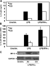
CO inhibits LPS/IFN-γ–induced IL-12 p40 expression in murine macrophages and alters IFN-γ signaling. (A) BM-derived murine macrophages were cultured in 250 ppm CO or ambient air. After activation with 1 μg/ml LPS and/or 10 ng/ml IFN-γ, IL-12 p40 protein was assayed from supernatants at 24 h. (B) IL-12 p40 mRNA expression was determined at 4 h by real-time RT-PCR. (C) IL-12 p70 and IL-23 protein secretion was assayed by ELISA at 24 h. (D) J774 murine macrophages were transiently transfected with an IL-12 p40 promoter-luciferase reporter construct or (E) a multimerized ISRE-luciferase reporter plasmid. Cells were cultured in 250 ppm CO after the addition of 1 μg/ml LPS or 1 μg/ml LPS plus 10 ng/ml IFN-γ for 18 h. Results are expressed as relative light units normalized to activity from a cotransfected constitutively active β-galactosidase reporter plasmid.
Figure 3.
Figure 3.
CO selectively inhibits LPS/IFN-γ–induced IRF-8 in murine macrophages. (A) Western blot analysis of IRF family members in J774 murine macrophages. Lanes 1–4 represent ambient air-exposed cells, and lanes 5–8 represent cells exposed to CO. Lanes 1 and 5, unstimulated; lanes 2 and 6, LPS/IFN-γ stimulated for 4 h; lanes 3 and 7, LPS/IFN-γ stimulated for 6 h; lanes 4 and 8, LPS/IFN-γ stimulated for 12 h; lane 9, nuclear extracts of RAW264.7 cells stimulated with LPS/IFN-γ. (B) Western blot analysis of IRF family members or phospho–STAT-3 in murine splenocytes. Lane 1, unstimulated; lanes 2 and 3 stimulated with LPS/IFN-γ for 8 h in the absence or presence of 250 ppm CO, respectively. (C) BM-derived macrophages were transduced with retrovirus IRF-8 or empty virus as control. 48 h after infection, cells were seeded in 24-well plates at 106 cells/ml, stimulated with 1 μg/ml LPS or LPS and 10 ng/ml IFN-γ, and incubated for 24 h in the absence or presence of 250 ppm CO. Supernatants were then collected, and IL-12 p40 protein secretion was measured by ELISA. IRF-8 protein expression was determined by Western blot in nuclear extracts from unstimulated retrovirus IRF-8–infected macrophages and control virus–infected cells (inset). Each result is representative of three independent experiments.
Figure 4.
Figure 4.
CO inhibits LPS/IFN-γ–induced IL-12 p40 expression independent of IL-10. (A) BM-derived macrophages from IL-10 / mice were cultured in 250 ppm CO or ambient air before activation with 1 μg/ml LPS or 1 μg/ml LPS plus 10 ng/ml IFN-γ. IL-12 p40 protein was assayed from supernatants at 24 h. (B) IL-12 p40 mRNA expression was determined at 4 h by real-time RT-PCR. Each result represents the mean ± SD of triplicate assays and is representative of three independent experiments. (C) IL-10 / BM-derived macrophages were either unstimulated or activated with LPS/IFN-γ for 8 h in the absence or presence of 250 ppm CO. IRF-8 protein expression was determined in nuclear extracts by Western blot (representative of three experiments).
Figure 5.
Figure 5.
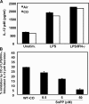
Inhibition of LPS/IFN-γ–induced IL-12 p40 production by CO is HO-1 dependent. (A) BM-derived macrophages from hmox / mice were activated with 1 μg/ml LPS and 10 ng/ml IFN-γ in the presence or absence of 250 ppm CO. IL-12 p40 protein was measured in supernatants at 24 h. Each result represents the mean ± SD of triplicate assays and is representative of three independent experiments. (B) BM-derived murine macrophages were cultured with or without an HO-1 inhibitor (SnPP) for 1 h before stimulation with LPS/IFN-γ in the presence or absence of 250 ppm CO. Results are presented as percent inhibition of LPS/IFN-γ–stimulated IL-12 p40 secretion by CO at 24 h. (C) BM-derived macrophages were incubated with 25 and 50 μM of the HO-1 inhibitor SnPP for 1 h before stimulation with 1 μg/ml LPS plus 10 ng/ml IFN-γ in the absence or presence of 250 ppm CO for 8 h. IRF-8 protein expression in nuclear extracts was determined by Western blot. This result is representative of three independent experiments. (D) BM-derived macrophages from hmox / mice were stimulated with 1 μg/ml LPS plus 10 ng/ml IFN-γ in the absence or presence of 250 ppm CO for 8 h. IRF-8 in nuclear extracts was determined by Western blot. This result is representative of three independent experiments.
Figure 5.
Figure 5.
Inhibition of LPS/IFN-γ–induced IL-12 p40 production by CO is HO-1 dependent. (A) BM-derived macrophages from hmox / mice were activated with 1 μg/ml LPS and 10 ng/ml IFN-γ in the presence or absence of 250 ppm CO. IL-12 p40 protein was measured in supernatants at 24 h. Each result represents the mean ± SD of triplicate assays and is representative of three independent experiments. (B) BM-derived murine macrophages were cultured with or without an HO-1 inhibitor (SnPP) for 1 h before stimulation with LPS/IFN-γ in the presence or absence of 250 ppm CO. Results are presented as percent inhibition of LPS/IFN-γ–stimulated IL-12 p40 secretion by CO at 24 h. (C) BM-derived macrophages were incubated with 25 and 50 μM of the HO-1 inhibitor SnPP for 1 h before stimulation with 1 μg/ml LPS plus 10 ng/ml IFN-γ in the absence or presence of 250 ppm CO for 8 h. IRF-8 protein expression in nuclear extracts was determined by Western blot. This result is representative of three independent experiments. (D) BM-derived macrophages from hmox / mice were stimulated with 1 μg/ml LPS plus 10 ng/ml IFN-γ in the absence or presence of 250 ppm CO for 8 h. IRF-8 in nuclear extracts was determined by Western blot. This result is representative of three independent experiments.
Figure 6.
Figure 6.
CO abrogates the inhibitory effect of IFN-γ on basal and LPS-induced HO-1 protein expression in macrophages and up-regulates intestinal HO-1 expression in IL-10 / mice. (A) BM-derived macrophages were stimulated with 1 μg/ml LPS, 10 ng/ml IFN-γ, or both for 8 h and cultured in the presence or absence of CO. Western blotting was performed on whole cell extracts for HO-1 protein expression. Results are representative of three independent experiments (B) Immunohistochemical analysis of intestinal HO-1 protein expression in CO-treated and control air–exposed IL-10 −/− mice (exposed from 8 to 12 wk of age). Depicted intestinal sections were matched for similar histologic colitis activity.
Figure 7.
Figure 7.
HO-1 induction inhibits LPS/IFN-γ–activated IRF-8 and IL-12 p40 expression in macrophages. (A) BM-derived macrophages were cultured with or without the HO-1 inducer hemin for 3 h before stimulation with 1 μg/ml LPS or 1 μg/ml LPS plus 10 ng/ml IFN-γ. IL-12 p40 protein was measured in 24-h supernatants. Results are presented as percent inhibition of LPS/IFN-γ–stimulated IL-12 p40 secretion by hemin and represent the mean ± SD from three independent experiments. (B) BM-derived macrophages were cultured with or without hemin for 3 h before stimulation with LPS/IFN-γ for 8 h. IRF-8 and HO-1 protein expression was measured in 20 μg of nuclear extract by Western blot. Equal loading of lanes was confirmed by equivalent expression of GAPDH between lanes (not depicted). Results are representative of three independent experiments.
Figure 7.
Figure 7.
HO-1 induction inhibits LPS/IFN-γ–activated IRF-8 and IL-12 p40 expression in macrophages. (A) BM-derived macrophages were cultured with or without the HO-1 inducer hemin for 3 h before stimulation with 1 μg/ml LPS or 1 μg/ml LPS plus 10 ng/ml IFN-γ. IL-12 p40 protein was measured in 24-h supernatants. Results are presented as percent inhibition of LPS/IFN-γ–stimulated IL-12 p40 secretion by hemin and represent the mean ± SD from three independent experiments. (B) BM-derived macrophages were cultured with or without hemin for 3 h before stimulation with LPS/IFN-γ for 8 h. IRF-8 and HO-1 protein expression was measured in 20 μg of nuclear extract by Western blot. Equal loading of lanes was confirmed by equivalent expression of GAPDH between lanes (not depicted). Results are representative of three independent experiments.
Figure 8.
Figure 8.
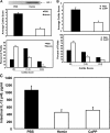
HO-1 induction ameliorates colitis in IL-10 /mice. (A) 10-wk-old IL-10 / mice were treated with i.p. injection of hemin (25 mg/kg twice weekly for 2 wk), and control mice were treated i.p. with PBS vehicle (n = 7 per group). Induction of intestinal HO-1 in hemin-treated mice was detected by Western blot (top). Colitis scores were significantly lower in the hemin-treated mice. *, P < 0.01; **, P < 0.001. Histologic improvement in colitis is presented as average colitis scores (middle) and percent of histologic field that demonstrate no inflammation, mild to moderate inflammation, and severe inflammation as described in Results (bottom). (B) 10-wk-old IL-10 / mice were treated with i.p. injection of CoPP (5 mg/kg twice weekly for 2 wk), and control mice were treated i.p. with PBS vehicle (n = 7 per group). Colitis scores, presented as in A, were significantly lower in CoPP-treated mice. *, P < 0.01; **, P < 0.001. (C) Intestinal explants from vehicle-, hemin-, or CoPP-treated IL-10 / mice were cultured for 24 h for measurement of spontaneous IL-12 p40 secretion by ELISA. Results represent mean ± SD of triplicate assays of pooled intestinal explants (n = 7 mice per group).
Figure 9.
Figure 9.
Model for the regulation of chronic intestinal inflammation by CO and HO-1. During chronic inflammation upon exposure to enteric bacteria and IFN-γ, intestinal macrophages secrete IL-12 p40. CO antagonizes this synergism through inhibition of IRF-8. IFN-γ also inhibits both basal and LPS-induced HO-1 expression, and CO abrogates this inhibitory effect. Therefore, HO-1 acts downstream of CO to ameliorate Th1-mediated chronic intestinal inflammation.

Similar articles

Cited by

References

    1. Elson, C.O., R.B. Sartor, S.R. Targan, and W.J. Sandborn. 2003. Challenges in IBD research: updating the scientific agendas. Inflamm. Bowel Dis. 9:137–153. - PubMed
    1. Plevy, S., and L. Mayer. 2003. Meeting summary: signal transduction pathways in immune and inflammatory cells. November 30-December 3, 2000, Amelia Island, Florida, U.S.A. Inflamm. Bowel Dis. 9:28–33. - PubMed
    1. Jick, H., and A.M. Walker. 1983. Cigarette smoking and ulcerative colitis. N. Engl. J. Med. 308:261–263. - PubMed
    1. Boyko, E.J., T.D. Koepsell, D.R. Perera, and T.S. Inui. 1987. Risk of ulcerative colitis among former and current cigarette smokers. N. Engl. J. Med. 316:707–710. - PubMed
    1. Otterbein, L.E., J.K. Kolls, L.L. Mantell, J.L. Cook, J. Alam, and A.M. Choi. 1999. Exogenous administration of heme oxygenase-1 by gene transfer provides protection against hyperoxia-induced lung injury. J. Clin. Invest. 103:1047–1054. - PMC - PubMed

Publication types

MeSH terms