. 2006 Mar 1;173(5):540-7.
doi: 10.1164/rccm.200504-538OC. Epub 2005 Dec 15.

Resident pleural macrophages are key orchestrators of neutrophil recruitment in pleural inflammation

Affiliations

Resident pleural macrophages are key orchestrators of neutrophil recruitment in pleural inflammation

Jean François Cailhier et al. Am J Respir Crit Care Med. .

Abstract

Rationale: The role played by resident pleural macrophages in the initiation of pleural inflammation is currently unclear.

Objective: To evaluate the role of resident pleural macrophages in the initiation of inflammation.

Methods: We have used a conditional macrophage ablation strategy to determine the role of resident pleural macrophages in the regulation of neutrophil recruitment in a murine model of experimental pleurisy induced by the administration of carrageenan and formalin- fixed Staphylococcus aureus.

Measurements and main results: Conditional macrophage ablation mice express the human diphtheria toxin receptor under the control of the CD11b promoter such that the administration of diphtheria toxin induces ablation of nearly 97% of resident macrophages. Ablation of resident pleural macrophages before the administration of carrageenan or S. aureus dramatically reduced neutrophil influx into the pleural cavity. In the carrageenan model, the reduction in neutrophil infiltration was associated with marked early reduction in the level of macrophage inflammatory protein 2 as well as reduced levels of various cytokines, including tumor necrosis factor alpha, interleukin 6, and interleukin 10. Adoptive transfer of nontransgenic macrophages partially restored neutrophil infiltration. We also stimulated macrophage-depleted and nondepleted pleural cell populations with carrageenan in vitro and determined the production of chemokines and cytokines. Chemokine and cytokine production was markedly reduced by macrophage depletion, reinforcing the role of resident pleural macrophages in the generation of mediators that initiate acute inflammation.

Conclusion: These studies indicate a critical role for resident pleural macrophages in sensing perturbation to the local microenvironment and orchestrating subsequent neutrophil infiltration.


Figures
<b>Figure 1.</b>
Figure 1.
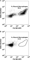
Administration of DT 24 h before performing pleural lavage results in ablation of pleural F4/80-positive macrophages (Mφ). CD11b-DTR and FVB/N wild-type (WT) mice were treated with diphtheria toxin (DT) intraperitoneally at a dose of 25 ng/g body weight. Pleural lavage was performed 24 h later. Cells were stained for the Mφ surface marker F4/80 and analyzed by flow cytometry. (A) Representative flow cytometry dot plot indicating that over 50% of pleural cells retrievable by pleural lavage 24 h after DT administration in FVB/N mice are F4/80 positive. (B) Administration of DT results in marked ablation of resident F4/80 positive pleural Mφ in CD11b-DTR mice. DT administration ablated 96.1 ± 0.8% of the resident Mφ population compared with baseline Mφ numbers (n = 9 mice, p < 0.0001). APC = allophycocyanin.
<b>Figure 2.</b>
Figure 2.
Resident Mφ ablation 24 h before administration of carrageenan blunts neutrophil (PMN) recruitment. 0.1 ml of 1% carrageenan was administered to CD11b-DTR and FVB/N WT mice 24 h after DT treatment. Pleural lavage was performed at 0, 6, 24, and 72 h after carrageenan administration. Lavaged cells were stained for GR1 and counted by flow cytometry (*p < 0.05 vs. CD11b-DTR group; n = 4–5 mice/group).
<b>Figure 3.</b>
Figure 3.
Adoptive transfer of Mφ-rich pleural cells and purified pleural macrophages partially restores PMN infiltration in carrageenan-induced pleurisy. FVB/N WT and six groups of CD11b-DTR mice were injected with DT (25 ng/g body weight) 24 h before carrageenan injection. Three groups of Mφ-depleted CD11b-DTR mice were reconstituted with (1) purified Mφ isolated by negative selection (90% pure, designated Mφ), (2) Mφ-rich pleural cells (designated MφR), or (3) Mφ-depleted pleural cells (designated MφD) at the same time as the administration of carrageenan. Mice underwent pleural lavage 6 h after the induction of inflammation. Controls comprised the adoptive transfer of either (1) purified Mφ or (2) Mφ-rich pleural cell populations to DT-treated CD11b-DTR mice in the absence of carrageenan. DT-treated CD11b-DTR mice exhibited a marked reduction in PMN infiltration in response to carrageenan, whereas reconstitution of Mφ-depleted mice with either purified Mφ or a Mφ-rich pleural cell population partially restored PMN infiltration. The adoptive transfer of an Mφ-depleted pleural cell population did not increase PMN infiltration. The adoptive transfer of either purified Mφ or an Mφ-rich pleural cell population alone did not induce significant PMN infiltration compared with DT-treated CD11b-DTR mice (n = 8–10 mice/group; *p < 0.05 vs. DT-treated CD11b-DTR mice that received carrageenan).
<b>Figure 4.</b>
Figure 4.
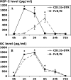
Resident Mφ ablation attenuates chemokine production in carrageenan-induced pleurisy. CD11b-DTR and FVB/N WT mice were injected with DT (25 ng/g body weight) 24 h before administration of carrageenan. Pleural lavage was performed 1, 3, 6, 24, and 72 h after the induction of pleurisy. The levels of macrophage inflammatory protein 2 (MIP-2; A) and keratinocyte-derived chemokine (KC; B) were determined in the pleural lavage supernatant by specific ELISA. The level of macrophage chemoattractant protein 1 (MCP-1; C) in the pleural lavage supernatant was determined by cytometric bead array (CBA) analysis (*p < 0.05 vs. CD11b-DTR group; n = 5 mice/group).
<b>Figure 5.</b>
Figure 5.
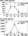
Resident Mφ ablation attenuates cytokine production in carrageenan-induced pleurisy. CD11b-DTR and FVB/N WT mice were injected with DT (25 ng/g body weight) 24 h before carrageenan injection. Pleural lavage was performed 1, 3, 6, 24, and 72 h after the induction of pleurisy. The level of tumor necrosis factor α (TNF-α; A) in the pleural lavage supernatant was determined by specific ELISA, whereas the levels of interleukin 6 (IL-6; B) and IL-10 (C) were determined by CBA analysis (*p < 0.05 vs. CD11b-DTR group; n = 5 mice/group).
<b>Figure 6.</b>
Figure 6.
In vitro production of MIP-2 and KC after carrageenan stimulation is Mφ dependent. Resident pleural cells were harvested and immunodepleted of resident pleural Mφ by passage over a magnetic column. Equivalent numbers of cells were plated and stimulated with 0.25% carrageenan or normal medium for 6 h. Supernatants were harvested and analyzed by specific ELISA for MIP-2 and KC (*p < 0.05 vs. all cells with medium; n = 4 wells/condition).
<b>Figure 7.</b>
Figure 7.
In vitro production of the cytokines TNF-α, IL-10, and IL-6 after carrageenan stimulation is Mφ dependent. Resident pleural cells were harvested and immunodepleted of resident pleural Mφ by passage over a magnetic column. Equivalent numbers of cells were stimulated with 0.25% carrageenan or normal medium for 6 h. Supernatants were harvested and analyzed by specific ELISA for TNF-α and by CBA for IL-10 and IL-6 (*p < 0.05 all cells vs. Mφ-depleted for their respective condition, i.e., Mφ with medium or Mφ with carrageenan). n = 4 wells/condition.
<b>Figure 8.</b>
Figure 8.
Resident Mφ ablation 24 h before the administration of formalin-fixed Staphylococcus aureus significantly blunts PMN recruitment. (A) A total of 3 × 10⁶ formalin-fixed, fluorescently labeled S. aureus were instilled into the pleural cavity of CD11b-DTR and FVB/N WT mice 24 h after DT treatment with phosphate buffered saline (PBS)–treated CD11b-DTR serving as an additional control. Pleural lavage was performed at 4 h after the administration of S. aureus. Lavaged cells were stained for GR1 and counted by flow cytometry (*p < 0.05 vs. DT-treated CD11b-DTR group; n = 4 mice/group). (B) Photomicrographs of Diffquick-stained (A and C) or Hoechst-stained (B and D) cytospin preparations of pleural lavage cells from either DT-treated FVB/N WT mice (A and B) or DT-treated CD11b-DTR mice (C and D) 4 h after the administration of 3 × 10⁶ formalin-fixed, fluorescently labeled S. aureus. PMNs may be readily distinguished from Mφ by their smaller size and the characteristic lobulated or circular nuclear morphology. Note that in B, the cell indicated with an arrow is the only Mφ present in the field and exhibits a large, rounded nucleus, whereas the remaining smaller PMNs exhibit a polylobular nuclear morphology. There are no Mφ present in C and D. Prominent ingestion of S. aureus particles by Mφ is evident in control DT-treated FVB/N WT mice (examples shown with arrows in A and B), with very limited uptake by PMNs. In contrast, in the absence of Mφ, DT-treated CD11b-DTR mice exhibit marked ingestion of S. aureus particles by PMNs (examples shown with arrows in C and D).

Similar articles

Cited by

References

    1. Kroegel C, Antony VB. Immunobiology of pleural inflammation: potential implications for pathogenesis, diagnosis and therapy. Eur Respir J 1997;10:2411–2418. - PubMed
    1. Antony VB. Immunological mechanisms in pleural disease. Eur Respir J 2003;21:539–544. - PubMed
    1. Antony VB, Hott JW, Kunkel SL, Godbey SW, Burdick MD, Strieter RM. Pleural mesothelial cell expression of C-C (monocyte chemotactic peptide) and C-X-C (interleukin 8) chemokines. Am J Respir Cell Mol Biol 1995;12:581–588. - PubMed
    1. Mohammed KA, Nasreen N, Ward MJ, Mubarak KK, Rodriguez-Panadero F, Antony VB. Mycobacterium-mediated chemokine expression in pleural mesothelial cells: role of C-C chemokines in tuberculous pleurisy. J Infect Dis 1998;178:1450–1456. - PubMed
    1. Mohammed KA, Nasreen N, Ward MJ, Antony VB. Macrophage inflammatory protein-1alpha C-C chemokine in parapneumonic pleural effusions. J Lab Clin Med 1998;132:202–209. - PubMed

Publication types