. 2006 Jan;26(1):343-53.
doi: 10.1128/MCB.26.1.343-353.2006.

Rhp51-dependent recombination intermediates that do not generate checkpoint signal are accumulated in Schizosaccharomyces pombe rad60 and smc5/6 mutants after release from replication arrest

Affiliations

Rhp51-dependent recombination intermediates that do not generate checkpoint signal are accumulated in Schizosaccharomyces pombe rad60 and smc5/6 mutants after release from replication arrest

Izumi Miyabe et al. Mol Cell Biol. 2006 Jan.

Abstract

The Schizosaccharomyces pombe rad60 gene is essential for cell growth and is involved in repairing DNA double-strand breaks. Rad60 physically interacts with and is functionally related to the structural maintenance of chromosomes 5 and 6 (SMC5/6) protein complex. In this study, we investigated the role of Rad60 in the recovery from the arrest of DNA replication induced by hydroxyurea (HU). rad60-1 mutant cells arrested mitosis normally when treated with HU. Significantly, Rad60 function is not required during HU arrest but is required on release. However, the mutant cells underwent aberrant mitosis accompanied by irregular segregation of chromosomes, and DNA replication was not completed, as revealed by pulsed-field gel electrophoresis. The deletion of rhp51 suppressed the aberrant mitosis of rad60-1 cells and caused mitotic arrest. These results suggest that Rhp51 and Rad60 are required for the restoration of a stalled or collapsed replication fork after release from the arrest of DNA replication by HU. The rad60-1 mutant was proficient in Rhp51 focus formation after release from the HU-induced arrest of DNA replication or DNA-damaging treatment. Furthermore, the lethality of a rad60-1 rqh1Delta double mutant was suppressed by the deletion of rhp51 or rhp57. These results suggest that Rad60 is required for recombination repair at a step downstream of Rhp51. We propose that Rhp51-dependent DNA structures that cannot activate the mitotic checkpoints accumulate in rad60-1 cells.


Figures
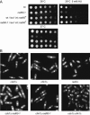
FIG. 1.
FIG. 1.
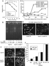
rad60-1 cells are sensitive to HU and proficient in arresting cell cycle progression in response to HU. (A) Spot assays to examine HU and the temperature sensitivities of rad60-1 cells. leu1 int::rad60+ represents the rad60+ gene integrated at the leu1 locus. Five serial dilutions of the indicated strains were spotted on YES medium plates with or without HU and incubated at the indicated temperatures for 3 days. (B) Analysis of cell cycle checkpoint. Cells of the indicated mutants were incubated in YES medium containing 15 mM HU at 30°C for 4 h. Cells were fixed with 70% ethanol and stained with DAPI and calcofluor white. wt, wild type.
FIG. 2.
FIG. 2.
Rad60's function is required for normal chromosome segregation after release from HU treatment. (A) Aberrant mitosis of rad60-1 cells after release from HU treatment. Exponentially growing wild-type and rad60-1 cells were incubated in YES medium containing 15 mM HU at 30°C for 4 h, released into fresh YES medium without HU, and further incubated at 30°C for 2 h. Cells were stained with DAPI and calcofluor white after fixation. Arrows indicate cells undergoing aberrant mitosis. (B, C, and D) Frequency of aberrant mitosis of rad60-1 and wild-type cells. (B) Exponentially growing wild-type and rad60-1 cells were incubated in YES medium containing 15 mM HU at 30°C for 4 h, released into fresh YES medium without HU, and further incubated at 30°C. Wild-type or rad60-1 cells that septated and were undergoing aberrant mitosis were counted at 20-min intervals. (C) Wild-type or rad60-1 cells were treated as in panel B, except that they were incubated at 36°C after release from HU. (D) rad60-1 cells were incubated in YES medium containing 15 mM HU at 30°C for 4 h, with or without subsequent incubation at 36°C for 1 h. The cells were then released into HU-free medium and incubated at 30°C or 36°C for 120 min. wt, wild type.
FIG. 3.
FIG. 3.
DNA synthesis and aberrant DNA structure after release from HU arrest. Exponentially growing wild-type and rad60-1 cells were incubated in YES medium containing 15 mM HU for 4 h at 30°C. Cells were then incubated at 36°C after release from HU treatment. (A) FACS analysis of DNA contents of rad60-1 cells after release from HU treatment. Cells were fixed at the indicated time points, stained with propidium iodide, and analyzed by FACS to determine the DNA content. (B) Analysis of chromosomes of rad60-1 cells after release from HU treatment. Genomic DNA was subjected to pulsed-field gel electrophoresis. Samples were taken at the indicated time points. wt, wild type.
FIG. 4.
FIG. 4.
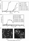
Phenotypes of rad60-1 cells in response to HU treatment are dependent on Rhp51 function. (A) Sensitivity of rad60-1 and rhp51Δ cells to HU treatment. Exponentially growing cells of the indicated strains were incubated in YES medium containing 15 mM HU at 30°C. Samples were taken at 2-h intervals. Relative numbers of CFU were calculated as described in Materials and Methods. (B) Analysis of chromosomes of rhp51Δ and rhp51Δ rad60-1 cells. Genomic DNA was subjected to pulsed-field gel electrophoresis. Samples were taken at the indicated time points after release from 15 mM HU treatment at 30°C. (C) Cellular morphology of rhp51Δ and rhp51Δ rad60-1 cells. rhp51Δ and rhp51Δ rad60-1 cells were fixed after the indicated treatments and stained with DAPI and calcofluor white. (D) Time course analysis of septum formation and aberrant mitosis of rad60-1 and rhp51Δ cells. Exponentially growing rhp51Δ and rhp51Δ rad60-1 cells were incubated in YES medium containing 15 mM HU at 30°C for 4 h, released into fresh YES medium without HU, and further incubated at 36°C. rhp51Δ or rhp51Δ rad60-1 cells that were septated or undergoing aberrant mitosis were counted at 30-min intervals. The results for the rad60-1 single mutant were the same as those shown in Fig. 2C. (E) Aberrant mitosis of smc6-74 cells after release from HU treatment. Exponentially growing smc6-74 and sm6-74 rhp51Δ cells were incubated in YES medium containing 15 mM HU at 30°C for 4 h, released into fresh YES medium without HU, and further incubated at 30°C for 2 h. Cells were stained with DAPI and calcofluor white after fixation. (F) Proportion of Rhp51 focus-forming cells after HU and UV treatment. Exponentially growing cells, cells incubated in YES medium containing 15 mM HU 4 h at 30°C, cells incubated in YES medium containing 15 mM HU 4 h at 30°C and released in HU-free medium at 36°C for 30 min, and cells irradiated with 50 J/m2 UV and incubated at 30°C 1 h were processed for indirect immunofluorescence using anti-Rhp51 antibody. The number of cells containing Rhp51 foci was counted and divided by the number of total cells to calculate the percentage. wt, wild type.
FIG. 5.
FIG. 5.
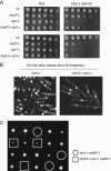
rad60-1 cells enter Rhp51-dependent aberrant mitosis after UV irradiation in G2. Wild-type and rad60-1 cells (A) and rhp51Δ and rhp51Δ rad60-1 cells (B) were synchronized in G2 by a lactose gradient procedure and irradiated with 50 J/m2 UV or mock irradiated. Cultures were incubated at 30°C. Cells that septated and were undergoing aberrant mitosis were counted at 20-min intervals. (C) Cells stained with DAPI and calcofluor white after fixation at the indicated time point were photographed. wt, wild type.
FIG. 6.
FIG. 6.
rqh1Δ cells show phenotypes similar to but distinct from those of rad60-1 cells. (A) Sensitivity to HU. Spot assay to show HU sensitivities of the indicated strains. Plates were incubated at 30°C for 2 days. (B) Cellular morphology of rqh1Δ and rhp51Δ rqh1Δ cells after release from HU treatment. rqh1Δ and rhp51Δ rqh1Δ cells were incubated for 120 min after release from HU treatment for 4 h, fixed, and stained with DAPI and calcofluor white. Arrows and arrowheads indicate cells undergoing aberrant mitosis and cells undergoing elongation without mitosis, respectively. (C) Suppression of the lethality of rqh1Δ rad60-1 cells by the deletion of rhp57. rhp57Δ rad60-1 and rqh1Δ cells were crossed, and the resulting spores were subjected to tetrad analysis. Dissected spores were grown at 26°C for 5 days on YES medium plates. Segregants enclosed by circles and squares indicate rqh1Δ rad60-1 and rhp57Δ rqh1Δ rad60-1 cells, respectively. wt, wild type.

Similar articles

Cited by

References

    1. Akamatsu, Y., D. Dziadkowiec, M. Ikeguchi, H. Shinagawa, and H. Iwasaki. 2003. Two different Swi5-containing protein complexes are involved in mating-type switching and recombination repair in fission yeast. Proc. Natl. Acad. Sci. USA 100:15770-15775. - PMC - PubMed
    1. al-Khodairy, F., and A. M. Carr. 1992. DNA repair mutants defining G2 checkpoint pathways in Schizosaccharomyces pombe. EMBO J. 11:1343-1350. - PMC - PubMed
    1. Aylon, Y., and M. Kupiec. 2004. DSB repair: the yeast paradigm. DNA Repair (Amsterdam) 3:797-815. - PubMed
    1. Bjergbaek, L., J. A. Cobb, M. Tsai-Pflugfelder, and S. M. Gasser. 2005. Mechanistically distinct roles for Sgs1p in checkpoint activation and replication fork maintenance. EMBO J. 24:405-417. - PMC - PubMed
    1. Boddy, M. N., B. Furnari, O. Mondesert, and P. Russell. 1998. Replication checkpoint enforced by kinases Cds1 and Chk1. Science 280:909-912. - PubMed

Publication types

MeSH terms

LinkOut - more resources