. 2006 Jan;26(1):117-30.
doi: 10.1128/MCB.26.1.117-130.2006.

PTF1 is an organ-specific and Notch-independent basic helix-loop-helix complex containing the mammalian Suppressor of Hairless (RBP-J) or its paralogue, RBP-L

Affiliations

PTF1 is an organ-specific and Notch-independent basic helix-loop-helix complex containing the mammalian Suppressor of Hairless (RBP-J) or its paralogue, RBP-L

Thomas M Beres et al. Mol Cell Biol. 2006 Jan.

Abstract

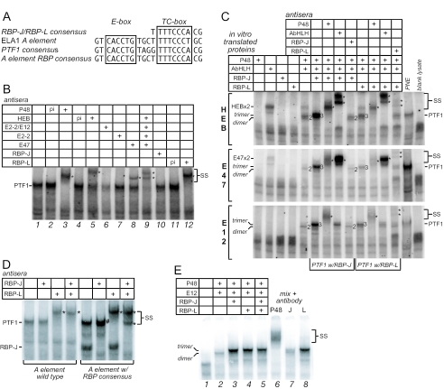
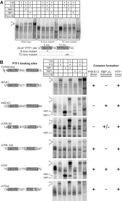
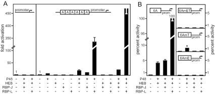
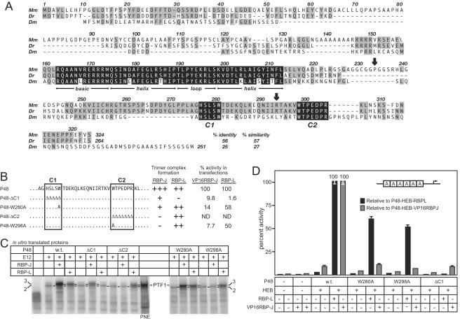
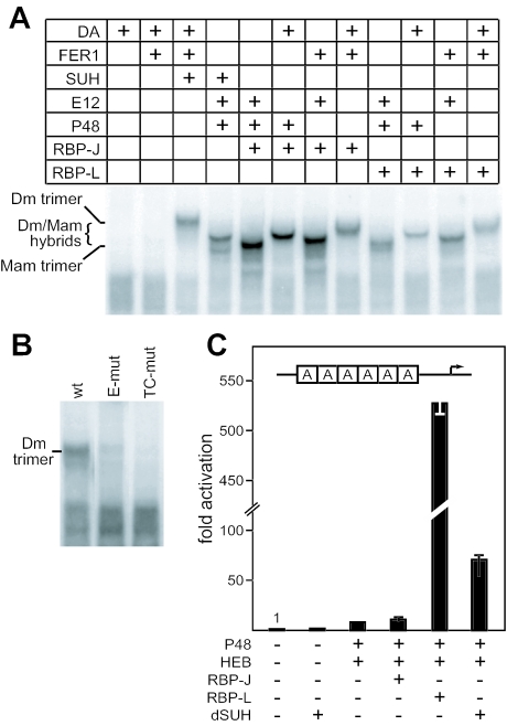
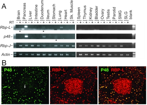
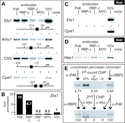
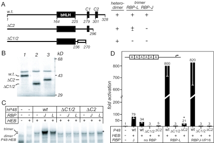
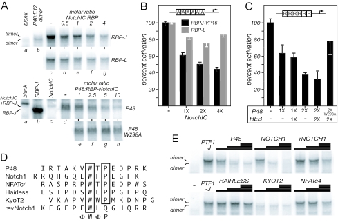
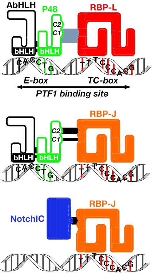
PTF1 is a trimeric transcription factor essential to the development of the pancreas and to the maintenance of the differentiated state of the adult exocrine pancreas. It comprises a dimer of P48/PTF1a (a pancreas and neural restricted basic helix-loop-helix [bHLH] protein) and a class A bHLH protein, together with a third protein that we show can be either the mammalian Suppressor of Hairless (RBP-J) or its paralogue, RBP-L. In mature acinar cells, PTF1 exclusively contains the RBP-L isoform and is bound to the promoters of acinar specific genes. P48 interacts with the RBP subunit primarily through two short conserved tryptophan-containing motifs, similar to the motif of the Notch intracellular domain (NotchIC) that interacts with RBP-J. The transcriptional activities of the J and L forms of PTF1 are independent of Notch signaling, because P48 occupies the NotchIC docking site on RBP-J and RBP-L does not bind the NotchIC. Mutations that delete one or both of the RBP-interacting motifs of P48 eliminate RBP-binding and are associated with a human genetic disorder characterized by pancreatic and cerebellar agenesis, which indicates that the association of P48 and RBPs is required for proper embryonic development. The presence of related peptide motifs in other transcription factors indicates a broader Notch-independent function for RBPJ/SU(H).


Figures

FIG. 1.
FIG. 1.
RBP-L is a subunit of the PTF1 complex. (A) The 21-bp Ela1 A element contains a TC-box similar to an RBP-J/L binding site one turn away from an E-box. Shown are the A element, the consensus RBP binding site, the PTF1-binding site consensus, and the Ela1 A element modified to contain the RBP consensus. (B) EMSA supershift analyses of complexes with nuclear extracts from rat pancreas with antisera to P48, HEB, E12, E47, E2.2, RBP-J, or RBP-L. p.i., preimmune; *, antibody-supershifted complexes (also indicated by SS). (C) IVT P48 forms heterodimeric complexes with HEB, E47, or E12, and trimeric complexes with the addition of RBP-L or RBP-J. Antibodies that recognize each component confirm their presence in the complex. (D) RBP-J is in pancreatic nuclear extract, but not as part of a PTF1 complex. Complexes from nuclear extracts, bound to either the wild-type Ela1 PTF1-binding site or a site in which the TC-box was changed to the consensus sequence for RBP-J, were incubated with antibody to either RBP-J or -L. RBP-L was detected only as part of the PTF1 complex. *, antibody-supershifted complexes; #, supershifted RBP-J monomer migrating with a slightly slower mobility than the authentic PTF1 complex. (E) RBP-J forms the PTF1 complex much more effectively than RBP-L does. PTF1 trimers were formed by mixing equimolar amounts of IVT P48, E12, RBP-J, and RBP-L. The relative amounts of trimer with P48 and either RBP-J or RBP-L were estimated from the amount of PTF1-band depletion with subunit-specific antibodies.
FIG. 2.
FIG. 2.
PTF1 has unique binding requirements for both an E-box and a TC-box. (A) Binding of reconstituted and authentic PTF1 complexes to wild-type and mutant Ela1 PTF1 binding sites; (B) reconstituted and authentic PTF1 bind to PTF1 binding sites from the promoter regions of several digestive enzyme genes. The amount of complex formation depends on the particular sequence of the E-box or TC-box, but all bind the PTF1 trimer. 3, PTF1 trimer; 2, P48-E12 dimer.
FIG. 3.
FIG. 3.
RBP-L is the critical subunit of PTF1 for high-level transcriptional activation of the Ela1 A element. (A) The relative activity of the 6A.EIp.luc reporter gene in 293 cells was assayed in the presence or absence of cotransfected P48, HEB, RBP-J, or RBP-L, individually or in various combinations as indicated. Transcription from the EIp.luc plasmid (containing only the Ela1 minimal promoter from −92 to +8 inserted upstream of the luciferase gene) was not affected by the addition of transcription factors via cotransfection. All values are the mean of at least four transfections ± the standard error of the mean (SEM). (B) Activation of the 6A reporter is dependent on both the E-box and the TC-box. Activation of wild-type A element (6A) was compared to activation of the E-box mutant (6AmE), the TC-box mutant (6AmT), or a mutant with both the E-box and TC-box mutated (6AmET). The asterisk in the 6AmT panel highlights the reduced ability of transfected P48 and HEB (with endogenous RBP-J) to activate the reporter in the absence of a TC-box. Values represent the percentage of wild-type activation and are the mean of least three transfections; error bars indicate the SEMs.
FIG. 4.
FIG. 4.
Two conserved peptide motifs near the C terminus of P48 mediate the interaction between P48 and the RBP-J/L. (A) Alignment of the sequences of the mouse (Mm) and zebra fish (Dr) P48s and FER1 from Drosophila (Dm) shows conservation of the bHLH domain (black shading) required for heterodimerization with class A bHLH proteins and DNA binding. The other significant conservation among all three is the two peptides (C1 and C2; black shading) near the C terminus. Gray shading highlights regions of lower sequence conservation, including the vertebrate-specific conservation between positions 246 and 275. The arrows indicate the relative positions of the human P48 mutations (see Fig. 8). (B) The sequences of the wild-type and mutant C1 and C2 regions of P48. The results from panels C and D are summarized at the right. (C) Ability of IVT wild-type and mutant P48 to form DNA-binding heterodimers with E12 or trimers with E12 plus RBP-L or RBP-J. All P48 mutants formed the heterodimer as effectively as wild-type P48. (D) Transcriptional activation of by PTF1 requires the interaction of P48 and RBP-L or RBP-J. The relative activity of the 6A-EIp.luc reporter construct in 293 cells was assayed in the presence or absence of cotransfected HEB, VP16RBP-J, or RBP-L, and wild-type or mutant P48, individually or in various combinations as indicated. All values are the means of at least three transfections ± the SEM.
FIG. 5.
FIG. 5.
Structure and function of PTF1 is conserved between mammals and flies. (A) Formation of DNA-binding heterodimeric and heterotrimeric complexes using mammalian IVT P48, E12, and either RBP-J or RBP-L, Drosophila FER1, DA, and SU(H), or various combinations as indicated. The mobilities of the complexes depend on which mammalian or Drosophila proteins are used in the binding reactions. (B) Binding of the Drosophila protein complex is sensitive to same nucleotide changes in the E- and TC-boxes as the mammalian PTF1 complex. (C) The relative activity of the 6A.EIp.luc reporter gene in 293 cells was assayed in the presence or absence of cotransfected P48, HEB, RBP-J, RBP-L, or SU(H) in the combinations indicated. All values are the means of four transfections ± the SEM.
FIG. 6.
FIG. 6.
Sites of Rbp-L and p48 expression. (A) Rbp-L and p48 transcripts are present at high levels selectively in the pancreas. Amplified products from RT-PCR assays with RNA isolated from 19 adult mouse organs shows that Rbp-L transcripts, like p48 transcripts, are present at high levels in the pancreas. *, low level of Rbp-L mRNA in the duodenum and brain and p48 mRNA in the stomach. (B) Immunofluorescence with anti-P48 and anti-RBPL antibodies shows that P48 and RBP-L are specifically colocalized in the acini of the adult pancreas. In the left panel, green immunofluorescence indicates P48 is in acinar nuclei only. i, islet. In the center panel, red indicates RBP-L is in the nuclei of the acinar and islet cells. In the right panel, the merged image shows colocalization (yellow) only in acinar nuclei.
FIG. 7.
FIG. 7.
PTF1 components P48 and RBP-L are bound to acinar-specific promoters in vivo. (A) Fragments of pancreatic chromatin containing the PTF1 binding sites of the promoters for acinar-specific genes (Ela1; Amy2, amylase 2; Ctrb, chymotrypsinogen B; Cpa1, carboxypeptidase A1) were enriched with antibodies to P48 and RBP-L but not with antibodies to RBP-J or c-MYC. Schematic representations of the promoters show the locations of the PTF1 binding sites. cMYC antibody was used as a measure of nonspecific immunoprecipitation. (B) Fold enrichment (from panel A) of the Ela1 promoter upon immunoprecipitation of pancreas chromatin with P48, RBP-L, RBP-J, and cMYC antibodies and quantified by real-time PCR. Note that, relative to the anti-cMYC control, the enrichments with anti-P48 and anti-RBP-L were 32- and 22-fold, respectively. (C) ChIP of the Ela1 and Cpa1 promoters from liver chromatin with P48, RBP-L, RBP-J, and c-MYC antibodies. (D) The efficacy of the RBP-J antibody was verified by its ability to enrich the Hes1 promoter, a known Notch target, by ChIP of liver chromatin. The Hes1 promoter contains a pair of RBP-J consensus binding sites at −78 and −92. (E) Sequential ChIPs show that P48 and RBPL are present concurrently on the Ela1 promoter in chromatin from adult mouse pancreas. The numbers below the images of the PCR products from conventional PCR indicate the fold enrichment of the Ela1 promoter measured by real-time PCR after each of the two rounds of ChIP.
FIG. 8.
FIG. 8.
Human disease truncations of the human P48 disrupt binding to RBP-J and RBP-L. (A) The truncated proteins resulting from the human R296X (ΔC2) and P236fsX270 (ΔC1/2) mutations. The open rectangle represents a 24-amino-acid frameshift extension. The ability of the wild-type and mutant human P48 proteins to form heterodimers with E12 and trimers with an RBP are summarized from the data in panel C. (B) Sodium dodecyl sulfate-polyacrylamide gel analysis of IVT human P48 proteins labeled by [35S]methionine incorporation. (C) Ability of the mutant human P48 proteins to form heterodimers with E12 and trimers with E12 and either RBP-J or RBP-L that bind the Ela1 PTF1-site. The asterisk indicates a small amount of PTF1-trimer formed with P48-ΔC2. (D) The human mutant P48 proteins cannot activate transcription of a PTF1 reporter gene in transfected 293 cells. The asterisk denotes a small amount of PTF1 activity with P48-ΔC2. Error bars indicate the SEM for three experiments.
FIG. 9.
FIG. 9.
The NotchIC and peptides related to the Notch ΦWΦP-motif compete with P48 for binding to RBP-J. (A, top) NotchIC disrupts the trimeric PTF1 complex formed with RBP-J (upper set) much more than that with RBP-L (lower set). P48:E12:RBP complexes were formed with 1:1:1 molar ratios of the IVT proteins on the Ela1 PTF1 site alone (lane c) or in the presence of increasing IVT NotchIC (lanes d to g). Lane a, blank IVT reaction; lane b, dimer formation with 1:1 IVT P48 and E12. (A, bottom) P48 (upper set), but not the P48W298A mutant (lower set), disrupts the DNA-bound complex of RBP-J and NotchIC. RBP-J:NotchIC complexes were formed with 1:1 molar ratios of the IVT proteins on the consensus RBP-J binding-site (see Materials and Methods) alone (lane d) or in the presence of increasing P48 (lanes e to h). Lane a, blank IVT reaction; lane b, IVT RBP-J; lane c, IVT NotchIC. (B) The NotchIC inhibits the transcriptional activity of PTF1. Effects of increasing amounts of transfected NotchIC expression plasmid on the activation of the PTF1 reporter gene (6A.EIp.luc) in 293 cells. Error bars indicate the SEM for four transfections. (C) Conversely, P48 inhibits NotchIC-enhanced RBP-J activity. NotchIC-RBP-J activity was monitored with a luciferase reporter driven by six repeats of a consensus RBP-binding site (6R.EIp.Luc). (D) C2-like peptides from transcription factors containing the ΦWΦP-motif. RevNotch1 is a peptide with the Notch1 sequence reversed. (E) Inhibition of PTF1 complex formation with increasing concentrations (0.02, 0.2, and 2 mM) of various synthetic C2-like peptides. The relative inhibitory effects were as follows: KYOT2 ≫ NOTCH1 > P48 > revNOTCH1 > NFATc4 ≥ HAIRLESS. The P48:E12 dimer was not affected.
FIG. 10.
FIG. 10.
RBP-L and RBP-J in the PTF1 complex or associated with the NotchIC.

Similar articles

Cited by

References

    1. Apelqvist, A., H. Li, S. L., P. Beatus, D. J. Anderson, T. Honjo, M. Hrabe de Angelis, U. Lendahl, and H. Edlund. 1999. Notch signalling controls pancreatic cell differentiation. Nature 400:877-881. - PubMed
    1. Artavanis-Tsakonas, S., M. D. Rand, and R. J. Lake. 1999. Notch signaling: cell fate control and signal integration in development. Science 284:770-776. - PubMed
    1. Barolo, S., R. G. Walker, A. D. Polyanovsky, G. Freschi, T. Keil, and J. W. Posakony. 2000. A Notch-independent activity of Suppressor of Hairless is required for normal mechanoreceptor physiology. Cell 103:957-969. - PubMed
    1. Blackwell, T. K., and H. Weintraub. 1990. Differences and similarities in DNA-binding preferences of MyoD and E2A protein complexes revealed by binding site selection. Science 250:1104-1110. - PubMed
    1. Chaya, D., and K. S. Zaret. 2003. Sequential chromatin immunoprecipitation from animal tissues. Methods Enzymol. 376:361-372. - PubMed

Publication types

MeSH terms