. 2006 Jan;80(1):296-305.
doi: 10.1128/JVI.80.1.296-305.2006.

Neither the RNA nor the proteins of open reading frames 3a and 3b of the coronavirus infectious bronchitis virus are essential for replication

Affiliations

Neither the RNA nor the proteins of open reading frames 3a and 3b of the coronavirus infectious bronchitis virus are essential for replication

Teri Hodgson et al. J Virol. 2006 Jan.

Abstract

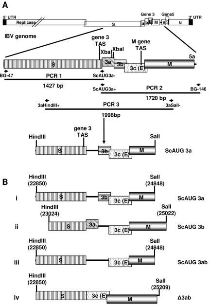
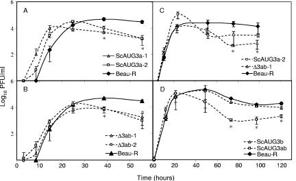
Gene 3 of infectious bronchitis virus is tricistronic; open reading frames (ORFs) 3a and 3b encode two small nonstructural (ns) proteins, 3a and 3b, of unknown function, and a third, structural protein E, is encoded by ORF 3c. To determine if either the 3a or the 3b protein is required for replication, we first modified their translation initiation codons to prevent translation of the 3a and 3b proteins from recombinant infectious bronchitis viruses (rIBVs). Replication in primary chick kidney (CK) cells and in chicken embryos was not affected. In chicken tracheal organ cultures (TOCs), the recombinant rIBVs reached titers similar to those of the wild-type virus, but in the case of viruses lacking the 3a protein, the titer declined reproducibly earlier. Translation of the IBV E protein is believed to be initiated by internal entry of ribosomes at a structure formed by the sequences corresponding to ORFs 3a and 3b. To assess the necessity of this mechanism, we deleted most of the sequence representing 3a and 3b to produce a gene in which ORF 3c (E) was adjacent to the gene 3 transcription-associated sequence. Western blot analysis revealed that the recombinant IBV produced fivefold less E protein. Nevertheless, titers produced in CK cells, embryos, and TOCs were similar to those of the wild-type virus, although they declined earlier in TOCs, probably due to the absence of the 3a protein. Thus, neither the tricistronic arrangement of gene 3, the internal initiation of translation of E protein, nor the 3a and 3b proteins are essential for replication per se, suggesting that these proteins are accessory proteins that may have roles in vivo.


Figures

FIG. 1.
FIG. 1.
Construction of the modified IBV gene 3 cDNAs. (A) Modifications to the Beaudette gene 3 were carried out using overlapping PCR mutagenesis. Shown is the method used to generate the modified cDNA with a modified 3a translation initiation codon. In the first two PCR amplifications, two complementary oligonucleotides, ScAUG3a− and SCAUG3a+, were used to introduce the two A23857TG-to-ACC nucleotide substitutions to retain the S gene termination codon and modify the 3a translation initiation codon. PCR 3 was done to join the two initial PCR products, resulting in a contiguous cDNA with the introduced modifications. Two restriction endonuclease sites, HindIII and SalI, were introduced at the ends of the PCR 3 products for cloning the modified cDNAs into pGPTNEB193rev. The HindIII and SalI restriction endonuclease sites were introduced at different positions relative to the IBV genome, depending on the modified cDNA being generated. (B) Four modified gene 3 cDNAs that were initially cloned into pGPTNEB193rev and then used to produce the rIBVs. The positions of the ORF 3a and 3b sequences are shown as black lines following modification of the translation initiation codon to indicate that the sequences are retained but that translation of the gene product is lost. The numbers associated with the HindIII and SalI restriction endonuclease sites represent the IBV genomic positions at the ends of the modified cDNAs. The oligonucleotides used to modify the 3a or 3b translation initiation codons or resulting in the deletion of the coding sequences are shown in Table 1. The ScAUG3ab cDNA was produced as described in Materials and Methods using sequences from pGPT-ScAUG3a and pGPT-ScAUG3b, pGPTNEB193rev-derived plasmids containing the modified 3a and 3b translation initiation codons.
FIG. 2.
FIG. 2.
Schematic diagram showing the transient dominant selection process for modifying the full-length cDNA within the genome of vaccinia virus vNotI/IBVFL. The modified cDNAs shown in Fig. 1B were introduced as HindIII-SalI fragments into the TDS GPT transfer/recombination vector pGPTNEB193rev. The diagram outlines the two-step process for modifying the IBV cDNA within vNotI/IBVFL using the modified cDNA containing the 3ab deletion in pGPT-Δ3ab. In step 1, the complete plasmid DNA is integrated into the full-length IBV cDNA by a single-step homologous-recombination event; a potential recombination event is shown. The resultant rVVs have a GPT+ phenotype, allowing selection in the presence of MPA. Removal of MPA results in two types of spontaneous intramolecular recombination events, I and II, due to the instability of the IBV cDNA with tandem repeats of similar sequences. I results in reversion to the Beaudette genotype (no introduced modifications), and II results in an rVV encoding the required modified gene 3 sequences. Both recombination events result in the loss of GPT. The example outlined shows the generation of an rIBV with the 3ab sequence deleted and the 3c (E) sequence directly under the control of the gene 3 TAS.
FIG. 3.
FIG. 3.
Growth kinetics of the rIBVs in CK cells. Monolayers of CK cells were inoculated in triplicate with 10 PFU of virus. After 1 h at 37°C, the cells were washed and incubation was continued. Medium was harvested over a period of 100 h for analysis by plaque assay on CK cells. The growth kinetics of Beau-R are compared with those of the rIBVs: (A) ScAUG3a, (B) ScAUG3b, (C) ScAUG3ab, and (D) Δ3ab. Where two independent rIBVs encoding the same modification had been rescued (A and D), both data sets have been illustrated. The error bars represent the standard deviations from three replicates. The asterisks indicate the time points at which the titers of mutant viruses were significantly different from those of Beau-R (P < 0.05).
FIG. 4.
FIG. 4.
Growth kinetics of the rIBVs in 10-day-old embryonated eggs. Embryos were inoculated in triplicate via the allantoic cavity with 0.1 PFU of each virus. At each time point, the embryos were chilled at 4°C before the allantoic fluids were analyzed for progeny virus by plaque assay on CK cells. The growth kinetics of Beau-R are compared with those of the rIBVs: (A) ScAUG3a, (B) ScAUG3b, (C) ScAUG3ab, and (D) Δ3ab. Where two independent rIBVs encoding the same modification had been rescued (A and D), both data sets have been illustrated. The error bars represent the standard deviations from three replicates.
FIG. 5.
FIG. 5.
Western blot analysis for the presence of the ORF 3a protein. Recombinant IBVs containing a modified 3a translation initiation codon should not express the 3a protein. Proteins present in infected (lanes 1 to 7) and mock-infected (lane 8) CK cell extracts were separated by SDS-PAGE, transferred to a Hybond-C membrane, and probed with a rabbit anti-3a serum, followed by ECL detection. The 3a protein was detected in cells infected with Beau-R (lane 1) and with rIBV ScAUG3b (lane 4) and not with viruses unable to express the 3a protein: ScAUG3a (lanes 2 and 3), ScAUG3ab, and Δ3ab (lanes 6 and 7). Where two independent rIBVs encoding the same modification had been rescued (lanes 2 and 3 and lanes 6 and 7), both mutants were analyzed.
FIG. 6.
FIG. 6.
Northern blot analysis of IBV-derived RNAs from CK cells infected with IBV. The IBV genomic (g) and sg mRNAs (numbered on the left) produced in CK cells infected by Beau-R (lane 1) and rIBVs ScAUG3a (lane 3), ScAUG3b (lane 4), ScAIG3ab (lane5), and Δ3ab (two independent mutants, 1 and 2 [lanes 6 and 7]) are shown. Mutants Δ3ab-1 and -2 produced a smaller gene 2 (sgmRNA2) and gene 3 (sgmRNA3) mRNA, as expected. The IBV-derived RNAS were detected using a 666-bp probe corresponding to the 3′ untranslated region of the IBV genome and present in all the sg mRNAs, as well as in gRNA.
FIG. 7.
FIG. 7.
Western blot analysis for the presence of the IBV E protein (3c protein) in CK cells infected with IBV. Analysis of E protein expression showed that mutants Δ3ab-1 and -2 (lanes 6 and 7) produced less E protein than the amounts expressed in CK cells infected with Beau-R (lane 1) and the other rIBVs: ScAUG3a (lanes 2 and 3), ScAUG3b (lane 4), and ScAUG3ab (lane 5). The proteins were separated by SDS-PAGE, transferred to a Hybond-C membrane, and probed with a mouse monoclonal antibody to the E protein and a rabbit polyclonal serum against the M protein, followed by ECL detection.
FIG. 8.
FIG. 8.
Growth kinetics of the rIBVs in TOCs (ex vivo). Groups of five TOCs were inoculated in triplicate with 104 PFU of virus. After 1 h at 37°C, the TOCs were washed and incubation was continued. Medium was harvested over a period of up to 125 h for analysis of progeny virus by plaque assay on CK cells. The growth kinetics of Beau-R are compared with those of the rIBVs: (A) ScAUG3a, (B) Δ3ab, and (D) ScAUG3b and ScAUG3ab. (C) Repeat of the experiment in panels A and B, in which one of each pair of recombinant viruses was examined and the time course was extended to 100 h. Where two independent rIBVs (-1 and -2) encoding the same modification had been rescued, both data sets have been illustrated. The error bars represent the standard deviations from three replicates. The asterisks indicate the time points at which the titers of mutant viruses were significantly different from those of Beau-R (P < 0.05).

Similar articles

Cited by

References

    1. Ausubel, F. M., R. Brent, R. E. Kingston, D. D. Moore, J. G. Seidman, J. A. Smith, and K. Struhl. 1987. Current protocols in molecular biology. John Wiley and Sons, Inc., New York, N.Y.
    1. Bonfield, J. K., K. F. Smith, and R. Staden. 1995. A new DNA sequence assembly program. Nucleic Acids Res. 23:4992-4999. - PMC - PubMed
    1. Bos, E. C., W. Luytjes, H. V. van der Meulen, H. K. Koerten, and W. J. Spaan. 1996. The production of recombinant infectious DI-particles of a murine coronavirus in the absence of helper virus. Virology 218:52-60. - PMC - PubMed
    1. Boulanger, D., P. Green, T. Smith, C.-P. Czerny, and M. A. Skinner. 1998. The 131-amino-acid repeat region of the essential 39-kilodalton core protein of fowlpox virus FP9, equivalent to vaccinia virus A4L protein, is nonessential and highly immunogenic. J. Virol. 72:170-179. - PMC - PubMed
    1. Boursnell, M. E. G., T. D. K. Brown, I. J. Foulds, P. F. Green, F. M. Tomley, and M. M. Binns. 1987. Completion of the sequence of the genome of the coronavirus avian infectious bronchitis virus. J. Gen. Virol. 68:57-77. - PubMed

Publication types