. 2006 Jan;18(1):198-211.
doi: 10.1105/tpc.105.036665. Epub 2005 Dec 9.

Golgi-mediated vacuolar sorting of the endoplasmic reticulum chaperone BiP may play an active role in quality control within the secretory pathway

Affiliations
Free PMC article

Golgi-mediated vacuolar sorting of the endoplasmic reticulum chaperone BiP may play an active role in quality control within the secretory pathway

Peter Pimpl et al. Plant Cell. 2006 Jan.
Free PMC article

Abstract

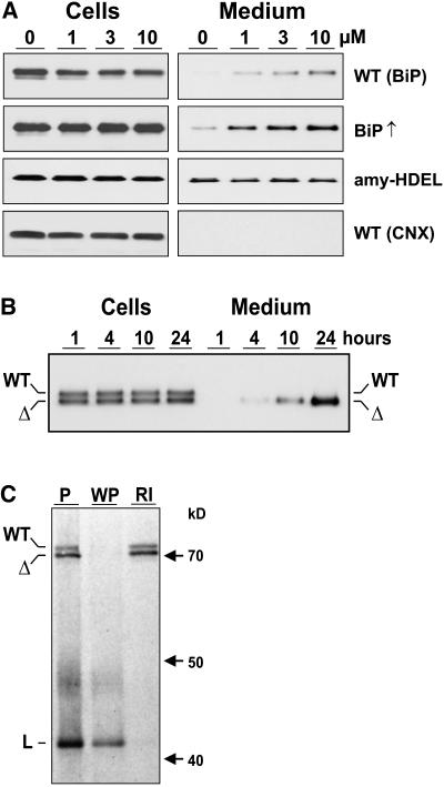
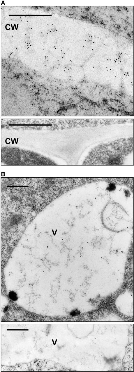
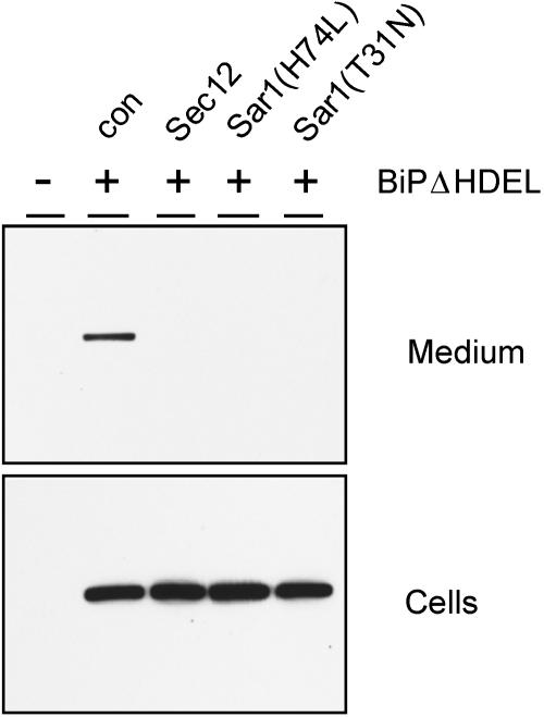
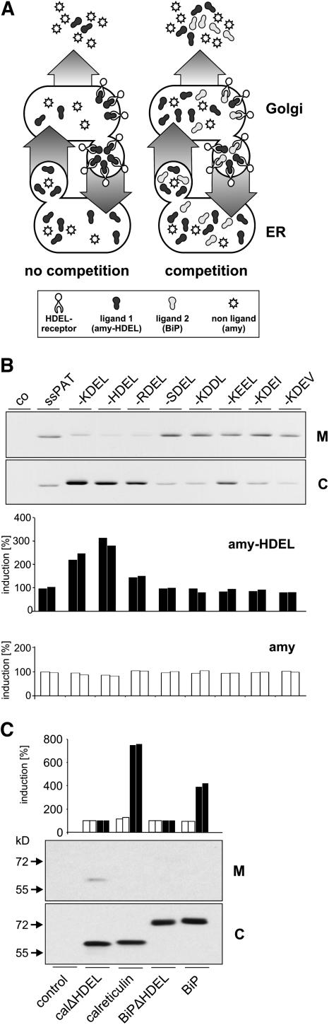
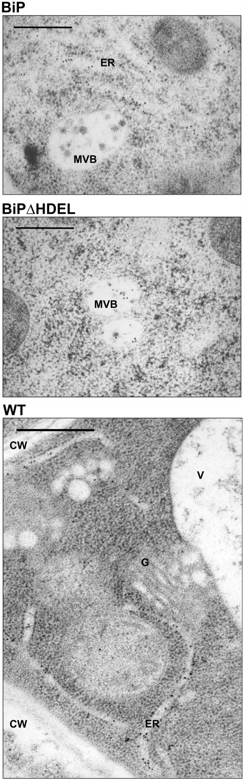
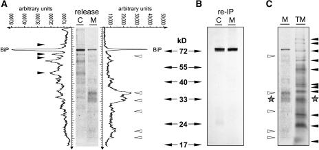
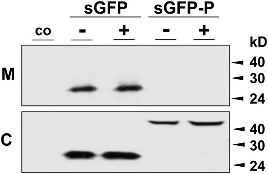
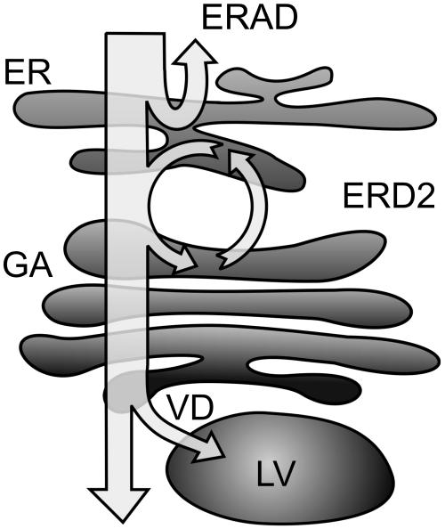
Quality control in the endoplasmic reticulum (ER) prevents the arrival of incorrectly or incompletely folded proteins at their final destinations and targets permanently misfolded proteins for degradation. Such proteins have a high affinity for the ER chaperone BiP and are finally degraded via retrograde translocation from the ER lumen back to the cytosol. This ER-associated protein degradation (ERAD) is currently thought to constitute the main disposal route, but there is growing evidence for a vacuolar role in quality control. We show that BiP is transported to the vacuole in a wortmannin-sensitive manner in tobacco (Nicotiana tabacum) and that it could play an active role in this second disposal route. ER export of BiP occurs via COPII-dependent transport to the Golgi apparatus, where it competes with other HDEL receptor ligands. When HDEL-mediated retrieval from the Golgi fails, BiP is transported to the lytic vacuole via multivesicular bodies, which represent the plant prevacuolar compartment. We also demonstrate that a subset of BiP-ligand complexes is destined to the vacuole and differs from those likely to be disposed of via the ERAD pathway. Vacuolar disposal could act in addition to ERAD to maximize the efficiency of quality control in the secretory pathway.


Figures

Figure 1.
Figure 1.
Wortmannin Induces BiP Secretion. (A) Immunodetection of BiP in cells and 10-fold concentrated medium from tobacco wild-type protoplasts (WT BiP) incubated with an increasing concentration of wortmannin given in micromoles above the lanes. The second panel shows the result of the same type of experiment but from protoplasts prepared from stable BiP overproducing transgenic plants (BiP↑) (Leborgne-Castel et al., 1999). The third panel shows results from protoplasts prepared from transgenic plants producing recombinant α-amylase-HDEL (amy-HDEL). The bottom panel shows samples from the top panel but probed with anticalnexin antiserum (WT CNX). (B) Secretion as a function of time in protoplast suspensions prepared from transgenic plants producing BiP (WT) and BiPΔHDEL (Δ) (Leborgne-Castel et al., 1999) incubated with 10 μM wortmannin. Sampling was performed as in (A) except that equal levels of cells and medium were loaded. Numbers above the lanes refer to time of protoplast incubation until harvesting. (C) Coimmunoprecipitation and ATP-mediated release of BiP and BiPΔHDEL from a model ligand (L). Protoplasts prepared from a transgenic plant producing both wild-type BiP (WT) and truncated BiPΔHDEL (Δ) were transfected with a plasmid encoding a secreted form of green fluorescent protein (GFP) fused to the P-domain of calreticulin (sGFP-P), previously known to be a strong BiP-ligand (Brandizzi et al., 2003). After 5 h of expression, cells were labeled for 2 h and cell extracts immunoprecipitated with anti-GFP serum. One pellet was washed with ATP release buffer, followed by a reimmunoprecipitation of the released material with anti-BiP serum. Shown is the unwashed pellet (P), the washed pellet (WP), and the reimmunoprecipitation from the washing medium (RI). Molecular mass markers are given in kilodaltons. Note that both BiP and BiPΔHDEL bind to the ligand in an ATP-sensitive manner typical for this chaperone.
Figure 2.
Figure 2.
Immunolocalization of BiP. (A) Wild-type tobacco roots were incubated with 10 μM wortmannin for 24 h, fixed with formaldehyde/glutaraldehyde, and embedded in Lowicryl HM20 at −35°C (top panel). Sections were exposed to BiP antibodies (1:200 dilution) then 10 nm gold-coupled secondary antibodies (Biocell GAR10 1:50). Gold particles are seen throughout the cell wall (CW). The control without wortmannin (bottom panel) does not show immunogold labeling for BiP in the cell wall. Bars = 0.5 μm. (B) Untreated tobacco cell from a transgenic BiP-overproducing plant (Leborgne-Castel et al., 1999). Immunogold labeling for BiP is observed in the vacuole (V). The control panel below is from a wild-type tobacco plant and does not exhibit labeling of the vacuole. Bars = 0.5 μm.
Figure 3.
Figure 3.
Inhibition of BiPΔHDEL Secretion by Blocking COPII-Mediated ER-to-Golgi Traffic. Protoplasts were transfected with 30 μg of BiPΔHDEL-encoding plasmid (+) alone (con) or with plasmids (30 μg) encoding Sec12p, Sar1(H74L), or Sar1(T31N) and incubated for 48 h in the presence of wortmannin (10 μM), followed by separation of cells and medium. Mock transfected cells (−) were used as a negative control to illustrate the specificity of the anti-C-myc serum. Note the clear presence of secreted BiPΔHDEL in the transfected cells only when effector molecules were absent.
Figure 4.
Figure 4.
Cargo Competition Assay for the HDEL Receptor. (A) The principle of the cargo competition assay in the absence (no competition) or presence (competition) of competing ligands for binding to the HDEL receptor in the Golgi apparatus. The model illustrates how a second ligand will lead to increased secretion of itself and the monitoring reporter amy-HDEL and has no effect on the nonligand (amy). (B) Cotransfection of tobacco protoplasts with 5 μg plasmid DNA encoding either amy or amy-HDEL with 30 μg plasmid DNA encoding PAT derivatives. The top panel shows the immunodetection of PAT derivatives, using anti-PAT serum in culture medium (M) and cellular fractions (C). The constructs were described previously (Denecke et al., 1992) and represent chimeric genes composed of a signal peptide fused to phosphinothricine acetyl transferase (ssPAT) and additionally fused tetrapeptides to the C terminus, indicated in single-letter codes above the lanes. As a control (co) for protein gel blots and amy assays, protoplasts were mock transfected. The bar graphs below show the increase in the secretion index in percent for amy-HDEL (closed bars) and amy (open bars). The secretion index is defined as the ratio between amy activity in the culture medium compared with the activity in the cellular fraction (Phillipson et al., 2001). To facilitate comparison, the secretion index was set to 100% for the samples in which secretory PAT (ssPAT) was cotransfected. (C) Cotransfection of tobacco protoplasts with 5 μg plasmid DNA encoding for either amy or amy-HDEL with 30 μg plasmid DNA encoding for myc-tagged calreticulinΔHDEL, wild-type calreticulin, BiPΔHDEL, and wild-type BiP. The bar graph shows the increase in the secretion index in percent for amy (open bars) and amy-HDEL (closed bars). The bottom panel shows the immunodetection of myc-tagged ER chaperones in the medium (M) and the cells (C). Molecular mass markers are indicated at the left hand side of the panel. Note that only calreticulinΔHDEL is detected in the medium but that both chaperones compete with amy-HDEL for the HDEL receptor.
Figure 5.
Figure 5.
Specific Localization of BiPΔHDEL to Multivesicular Bodies. Roots from transgenic plants overproducing wild-type BiP or BiPΔHDEL were analyzed by high-pressure freezing followed by freeze substitution and Lowicryl HM20 embedding. Thin sections were stained with anti-BiP serum and secondary antibodies linked to 10-nm gold particles. Note the 10-nm gold labeling in the ER of BiP-overproducing plants and the absence in the multivesicular body (MVB). Out of 25 multivesicular bodies, none exhibited labeling in BiP-overproducing plants. By contrast, sections from BiPΔHDEL-overproducing plants revealed an average multivesicular body labeling density of 5.2 out of a total of 25 multivesicular bodies analyzed. The control panel from untransformed wild-type plants shows an overview to illustrate the specificity of the antiserum. Note that only the ER is labeled in this section, and no labeling was seen in the vacuole (V), the cell wall (CW), and the Golgi apparatus (G). Bars = 20 μm.
Figure 6.
Figure 6.
Demonstration of BiP-Ligand Complexes Exiting the ER. (A) Immunoprecipitation of BiP-ligands from cell extracts (C) and the culture medium (M) of wortmannin-treated wild-type cells followed by ATP release and subsequent analysis by SDS-PAGE and autoradiography. Line scanning data obtained with AIDA software (Raytest) are given in arbitrary units. Arrowheads indicate specific BiP-ligands exclusively found either in cell extracts (closed) or the culture medium (open). (B) Reimmunoprecipitation (re-IP) of ATP-released polypeptides with anti-BiP serum from (A). Compared with the quantities loaded in (A), fivefold higher amounts were used for the reimmunoprecipitation in order to test the presence of lower molecular weight BiP degradation products. Note that none of the bands identified in (A) (closed and open arrowheads) are precipitated with anti-BiP serum. (C) Comparison between the total labeled secreted fraction in the medium and the ATP-released polypeptides from secreted BiP in (A). Note the different patterns in the two lanes except for one band (stars).
Figure 7.
Figure 7.
Transient Expression Experiment to Measure Wortmannin-Induced Secretion. Wild-type tobacco protoplasts were either mock-transfected (co) or transfected with secreted GFP (sGFP) or sGFP-P. After incubation in the presence (+) and absence (−) of wortmannin (10 μM), cells (C) and medium (M) were harvested 24 h after transfection, and equal quantities were compared by protein gel blots. Molecular mass markers are given in kilodaltons on the right side of the panel. Note the complete lack of secretion of sGFP-P regardless of drug treatment.
Figure 8.
Figure 8.
Model Describing the Possible Fate of Newly Synthesized Proteins within the Secretory Pathway. A first quality control step occurs via ERAD back through the Sec61 pore into the cytosol for disposal by the proteasome. The second mechanism is based on the action of ER chaperones such as BiP. HDEL-mediated recycling from the Golgi apparatus (GA) via the HDEL receptor (ERD2) will either permit further folding, disposal via ERAD, or renewed ER export. If the HDEL retrieval checkpoint is overcome as well, the final quality control leads to vacuolar disposal (VD) in the lytic vacuole (LV).

Similar articles

Cited by

References

    1. Barlowe, C., Orci, L., Yeung, T., Hosobuchi, M., Hamamoto, S., Salama, N., Rexach, M.F., Ravazzola, M., Amherdt, M., and Schekman, R. (1994). COPII: A membrane coat formed by Sec proteins that drive vesicle budding from the endoplasmic reticulum. Cell 77 895–907. - PubMed
    1. Ben-Zvi, A., De Los Rios, P., Dietler, G., and Goloubinoff, P. (2004). Active solubilization and refolding of stable protein aggregates by cooperative unfolding action of individual hsp70 chaperones. J. Biol. Chem. 279 37298–37303. - PubMed
    1. Blond-Elguindi, S., Cwirla, S.E., Dower, W.J., Lipshutz, R.J., Sprang, S.R., Sambrook, J.F., and Gething, M.J. (1993). Affinity panning of a library of peptides displayed on bacteriophages reveals the binding specificity of BiP. Cell 75 717–728. - PubMed
    1. Brandizzi, F., Hanton, S., DaSilva, L.L., Boevink, P., Evans, D., Oparka, K., Denecke, J., and Hawes, C. (2003). ER quality control can lead to retrograde transport from the ER lumen to the cytosol and the nucleoplasm in plants. Plant J. 34 269–281. - PubMed
    1. Caldwell, S.R., Hill, K.J., and Cooper, A.A. (2001). Degradation of endoplasmic reticulum (ER) quality control substrates requires transport between the ER and Golgi. J. Biol. Chem. 276 23296–23303. - PubMed

Publication types

MeSH terms

Associated data

LinkOut - more resources