Comparative Study
. 2005 Dec 7;25(49):11455-67.
doi: 10.1523/JNEUROSCI.3153-05.2005.

Modifying the subunit composition of TASK channels alters the modulation of a leak conductance in cerebellar granule neurons

Affiliations
Comparative Study

Modifying the subunit composition of TASK channels alters the modulation of a leak conductance in cerebellar granule neurons

M Isabel Aller et al. J Neurosci. .

Abstract

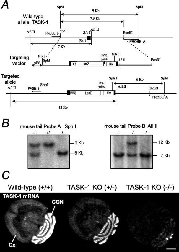
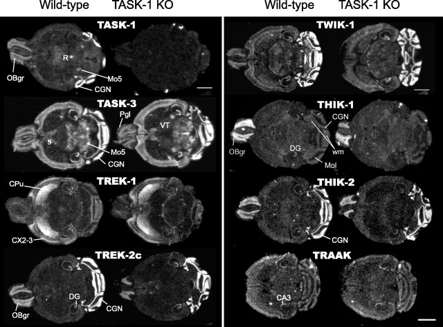
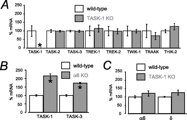
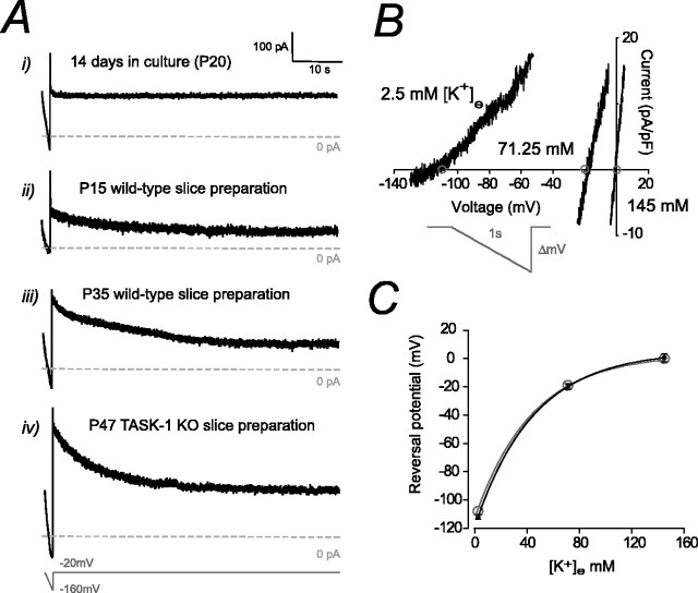
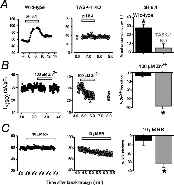
Two-pore domain potassium (K2P) channel expression is believed to underlie the developmental emergence of a potassium leak conductance [IK(SO)] in cerebellar granule neurons (CGNs), suggesting that K2P function is an important determinant of the input conductance and resting membrane potential. To investigate the role that different K2P channels may play in the regulation of CGN excitability, we generated a mouse lacking TASK-1, a K2P channel known to have high expression levels in CGNs. In situ hybridization and real-time PCR studies in wild-type and TASK-1 knock-outs (KOs) demonstrated that the expression of other K2P channels was unaltered in CGNs. TASK-1 knock-out mice were healthy and bred normally but exhibited compromised motor performance consistent with altered cerebellar function. Whole-cell recordings from adult cerebellar slice preparations revealed that the resting excitability of mature CGNs was no different in TASK-1 KO and littermate controls. However, the modulation of IK(SO) by extracellular Zn2+, ruthenium red, and H+ was altered. The IK(SO) recorded from TASK-1 knock-out CGNs was no longer sensitive to alkalization and was blocked by Zn2+ and ruthenium red. These results suggest that a TASK-1-containing channel population has been replaced by a homodimeric TASK-3 population in the TASK-1 knock-out. These data directly demonstrate that TASK-1 channels contribute to the properties of IK(SO) in adult CGNs. However, TASK channel subunit composition does not alter the resting excitability of CGNs but does influence sensitivity to endogenous modulators such as Zn2+ and H+.


Figures

Figure 1.
Figure 1.
TASK-1 gene targeting to generate a knock-out allele. A, Homologous recombination strategy for exon 1 (Ex 1) of the mouse TASK-1 gene showing the structure of the native gene (top), the linearized targeting vector (middle), and the targeted allele (bottom). Exon 1 is marked as a filled box. pBS, pBluescript; SV40 polyA, Simian virus 40 polyadenylation; Neo, neomycin. B, Southern blots confirming germ-line transmission of the targeted TASK-1 allele. After SphI digestion and hybridizing with Probe A, which flanks the 3′ end of the targeting vector, wild-type bands are 9 kb, but a heterozygote mouse (+/-) gives an additional 6 kb band; homozygous mice (-/-) have only the 6 kb band. When tail DNA is digested with AflII, and the blot is hybridized with a 5′ flanking probe (Probe B), a 12 kb band from the targeted allele is found (+/-), whereas the wild-type gene gives a 7 kb band. C, In situ hybridization showing absence of TASK-1 mRNA in adult TASK-1 knock-out mouse brains. In situ hybridization autoradiographs (x-ray film) with a 35S-labeled antisense oligonucleotide complimentary to the nucleotides encoding amino acids 1-15 (i.e., N terminus) of TASK-1. In the wild-type (+/+), the signal is highest in CGNs but is also present in cortex (Cx), thalamus, and inferior colliculus; in heterozygotes (+/-), the signal is partially reduced, and in the knock-out (-/-), no specific signal remains. Scale bar, 2 mm.
Figure 2.
Figure 2.
In situ hybridization of K2P channels. In situ hybridization for eight members of the K2P gene family in wild-type and TASK-1 KO mouse brains. Mol, Molecular layer; wm, white matter; R, raphe nuclei; OBgr, olfactory bulb granule cells; Pgl, periglomerular cells; Mo5, motor nuclei; CX, neocortex; VT, ventral thalamus; CA3, hippocampal pyramidal cells; DG, hippocampal dentate granule cells; IC, inferior colliculi; CPu, caudate-putamen; S, septum. Scale bar, 2 mm.
Figure 3.
Figure 3.
Real-time PCR analysis of K2P gene expression in TASK1 KO cerebellum. A, Comparison of real-time PCR mRNA levels for eight K2P subunits between wild-type and TASK-1 KO cerebellum. B, Comparison of real-time PCR mRNA levels for TASK-1 and TASK-3 K2P subunits between wild-type and GABAA receptor α6 knock-out cerebellum. C, Real-time PCR analysis of α6 and δ GABAA subunit mRNA levels in wild-type and TASK-1 KO cerebellum. Mean levels of gene expression are normalized to cyclophilin expression (n = 3 mice for each subunit). *p < 0.05.
Figure 4.
Figure 4.
Behavioral deficits in the TASK-1 knock-out mice. A, TASK-1 KO (-/-) mice stayed shorter times on the rotating rod than wild-type (+/+) littermates, but both strains improved their performance during a training period. Mice were trained for 6 d (3-6 trials a day). In each 3 min trial, the rotation speed was accelerated from 5 to 40 rpm. Repeated-measures one-way ANOVA confirmed that the genotype effect on the rotarod performance was significant (F(1,112) = 14.92; p < 0.01). B, The latency to walk along the 100-cm-long horizontally placed beam of 1.2 cm diameter back to the home cage. The number of falls from the 100-cm-long horizontally placed beam of 0.8 cm diameter during walk back to the home cage. Data are means ± SEM (n = 7-9); *p < 0.05.
Figure 5.
Figure 5.
Properties of IK(SO) in CGNs. A, Current traces taken from granule cells in the culture preparation (i), the wild-type slice preparation at P15 (ii) and P35 (iii), and a TASK-1 KO (-/-) slice preparation at P47 (iv). In each case, the holding potential was ramped from -20 to -160 mV and then maintained at -20 mV for 1 min. Note the absence of a slowly inactivating component in the data taken from a cultured granule cell (i). In the presence of this slowly inactivating current, IK(SO) was measured after the holding potential had been clamped at -20 mV for at least 1 min to avoid contamination by this conductance. B, Effect of altering external K+ concentration on the reversal potential of IK(SO). The current-voltage plot shows three traces recorded during a voltage-ramp protocol performed on the same adult wild-type granule cell. The predicted reversal potential, calculated from the Nernst equation, is marked with an open circle. C, Plot to compare the observed change in reversal potential (black filled circles) with the predicted values (open gray circles) calculated from the Nernst equation. This experiment illustrates that IK(SO) in CGNs is mediated by a pure K+ conductance. As expected from previous studies, this noninactivating K+ conductance is present in both the culture and the acute slice preparation.
Figure 6.
Figure 6.
Comparison of IK(SO) recorded from CGNs in wild-type and TASK-1 KO mice. A, Left plot is a comparison of IK(SO) measured at -20 mV immediately before the ramp protocol recorded from granule cells at 2, 7, and 14 d in culture, prepared from either wild-type (black) or TASK-1 KO (gray) mice. No significant difference between the magnitude of IK(SO) was observed between these strains at any stage in culture. The right plot illustrates the magnitude of IK(SO) measured at -20 mV from wild-type granule cells (black) at different ages in the acute slice preparation. Once again, there was no difference in the magnitude of IK(SO) recorded from TASK-1 KO (gray) mice. B, Comparison of current-voltage relationships between wild-type (black) and TASK-1 KO (gray) granule cells recorded from the adult slice preparation. The current-voltage plots are averages from each strain with the mean ± SEM superimposed. C, A plot of the resting membrane potential (RMP) of the cells included in the current-voltage plot in B. There was no significant difference between the data obtained from wild-type and TASK-1 KO granule cells. D, Current-clamp recordings from CGNs from wild-type (left) and TASK-1 KO (right) mice. In each case, the response to a hyperpolarizing (8 pA) and depolarizing (30 pA) current injection step have been superimposed. The relationship between injected current and action potential frequency for CGNs in each strain have been plotted in the bar graph. As predicted form the voltage-clamp experiments, there was no significant difference in the data obtained from the two strains.
Figure 7.
Figure 7.
Discrimination between recombinant TASK-1- and TASK-3-mediated currents. A, Effect of altering external pH on the leak current recorded at -20 mV after recombinant expression of mTASK-1 and mTASK-3 cDNA. In control conditions, the external pH was maintained at pH 7.4. During test periods, the pH was shifted to 6.4 and 8.4, as indicated by the filled bars. The left plot illustrates the change recorded from a representative mTASK-1-mediated current. Note how external acidification blocks the current, whereas alkalization enhances the leak current. In contrast, the middle plot illustrates how the mTASK-3-mediated current only responds to external acidification. The right plot is a bar graph comparing the results obtained with mouse (black filled) and human (gray filled) clones. B, Application of 100 μm Zn2+ to the external medium has little effect on mTASK-1-mediated currents (left plot) but has a substantial blocking action on the mTASK-3-mediated leak current (middle plot). The right bar graph compares the effects of external Zn2+ application when using either mouse (black filled) or human (gray filled) cDNA in the recombinant expression studies. C, Similar conventions to A and B but examining the action of 10 μm ruthenium red (RR). Data are means ± SEM (n = 4-16). No significant difference between the results obtained using mouse or human TASK-1 and TASK-3 was observed.
Figure 8.
Figure 8.
Altered properties of IK(SO) in TASK-1 KO adult CGNs. A, Plot of IK(SO) measured at -20 mV from wild-type and TASK-1 KO adult granule cell during a transient change in external pH from 7.4 to 8.4. The graph on the right illustrates the average data from all granule cells exposed to external alkalization. Note the significant increase in IK(SO) recorded from wild-type granule cells but no change in IK(SO) in TASK-1 KO granule cells. B, C, Conventions are the same as in A, but results were obtained after addition of 100 μm Zn2+ and 10 μm ruthenium red (RR). In both cases, a significant reduction in IK(SO) was observed in recordings from TASK-1 KO granule cells, with no change in IK(SO) recorded from wild-type granule cells. Data are mean ± SEM; *p < 0.05.

Similar articles

Cited by

References

    1. Aiba A, Kano M, Chen C, Stanton ME, Fox GD, Herrup K, Zwingman TA, Tonegawa S (1994) Deficient cerebellar long-term depression and impaired motor learning in mGluR1 mutant mice. Cell 79: 377-388. - PubMed
    1. Aller MI, Jones A, Merlo D, Paterlini M, Meyer AH, Amtmann U, Brickley S, Jolin HE, McKenzie ANJ, Monyer H, Farrant M, Wisden W (2003) Cerebellar granule cell Cre recombinase expression. Genesis 36: 97-103. - PubMed
    1. Bayliss DA, Sirois JE, Talley EM (2003) The TASK family: two-pore domain background K+ channels. Mol Interv 3: 205-219. - PubMed
    1. Berg AP, Talley EM, Manger JP, Bayliss DA (2004) Motoneurons express heteromeric TWIK-related acid-sensitive K+ (TASK) channels containing TASK-1 (KCNK3) and TASK-3 (KCNK9) subunits. J Neurosci 24: 6693-6702. - PMC - PubMed
    1. Brickley SG, Cull-Candy SG, Farrant M (1996) Development of a tonic form of synaptic inhibition in rat cerebellar granule cells resulting from persistent activation of GABAA receptors. J Physiol (Lond) 497: 753-759. - PMC - PubMed

Publication types

MeSH terms

Substances

LinkOut - more resources