. 2006 Mar 15;394(Pt 3):693-8.
doi: 10.1042/BJ20050576.

Dimerization is required for SH3PX1 tyrosine phosphorylation in response to epidermal growth factor signalling and interaction with ACK2

Affiliations

Dimerization is required for SH3PX1 tyrosine phosphorylation in response to epidermal growth factor signalling and interaction with ACK2

Chandra Childress et al. Biochem J. .

Abstract

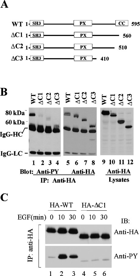
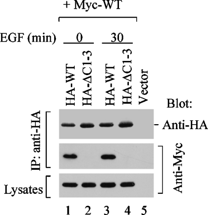
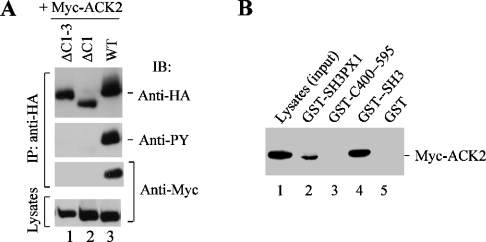
SH3PX1 [SNX9 (sorting nexin 9)] is a member of SNX super-family that is recognized by sharing a PX (phox homology) domain. We have previously shown that SH3PX1, phosphorylated by ACK2 (activated Cdc42-associated tyrosine kinase 2), regulates the degradation of EGF (epidermal growth factor) receptor. In mapping the tyrosine phosphorylation region, we found that the C-terminus of SH3PX1 is required for its tyrosine phosphorylation. Further analysis indicates that this region, known as the coiled-coil domain or the BAR (Bin-amphiphysin-Rvs homology) domain, is the dimerization domain of SH3PX1. Truncation of as little as 13 amino acid residues at the very C-terminus in the coiled-coil/BAR domain of SH3PX1 resulted in no dimerization, no ACK2-catalysed and EGF-stimulated tyrosine phosphorylation and no interaction with ACK2. The intracellular localization of SH3PX1 became dysfunctional upon truncation in the BAR domain. Taken together, our results indicate that the dimerization, which is mediated by the BAR domain, is essential for the intracellular function of SH3PX1.


Figures

Figure 1
Figure 1. The C-terminus of SH3PX1 is required for its tyrosine phosphorylation
(A) A schematic representation of the C-terminus truncation mutants of SH3PX1. CC, the coiled-coil domain that is the region for dimerization of SH3PX1. (B) Deletion of the coiled-coil domain of SH3PX1 eliminated the ACK2-catalysed phosphorylation. The HA-tagged wild-type or truncation mutants of SH3PX1 were co-transfected with Myc-tagged ACK2 (all lanes) into COS7 cells for 48 h. The SH3PX1 and its truncation mutants were immunoprecipitated with anti-HA and immunoblotted with anti-PY (4G10) (lanes 1–4) and anti-HA (lanes 5–8). The expression of the proteins was determined by immunoblotting the cell lysates with anti-HA (lanes 9–12). IgG-HC, IgG heavy chain; IgG-LC, IgG light chain. IP, immunoprecipitation. (C) The C-terminus is required for EGF-stimulated tyrosine phosphorylation of SH3PX1. The COS7 cells were transfected with HA-tagged SH3PX1 or ΔC1 for 36 h followed by 12 h serum starvation and subsequently subjected to EGF stimulation (100 ng/ml EGF) for the indicated times. SH3PX1 or ΔC1 was immunoprecipitated and immunoblotted with anti-HA (upper panel) and anti-PY (4G10) (lower panel). IB, immunoblotting.
Figure 2
Figure 2. Diminution of the tyrosine phosphorylation of SH3PX1 by deletion of the C-terminus did not result from losing the tyrosine phosphorylation sites
(A) HA-tagged SH3PX1, ΔC1 or ΔC1-3 was co-transfected with Myc-tagged ACK2 into COS7 cells. SH3PX1 and the truncation mutants were immunoprecipitated with anti-HA and immunoblotted with anti-HA (the top panel) and anti-PY (the middle panel). The expression of ACK2 was detected by immunoblotting the cell lysates with anti-Myc (the bottom panel). IP, immunoprecipitation; IB, immunoblotting. (B) COS7 cells were transfected with HA-tagged SH3PX1 or its truncation mutants for 36 h followed by serum starvation for 12 h and subsequently stimulated with EGF (100 ng/ml) for the indicated times. SH3PX1 and the truncation mutants were immunoprecipitated with anti-HA, and immunoblotted with anti-HA (the bottom panel) and anti-PY (the top panel).
Figure 3
Figure 3. The C-terminus of SH3PX1 is required for its dimerization
(A) HA-tagged SH3PX1, ΔC1 or ΔC2 was co-transfected with Myc-tagged SH3PX1 into COS7 cells. The HA-tagged SH3PX1 and its truncation mutants were immunoprecipitated and immunoblotted by anti-HA (the top panel). The co-immunoprecipitated Myc-tagged SH3PX1 was detected by immunoblotting with anti-Myc (the middle panel). The expression level of Myc-tagged SH3PX1 was determined by immunoblotting the cell lysates with anti-Myc (the bottom panel). IP, immunoprecipitation. (B) HA-tagged SH3PX1 or ΔC1-3, a deletion mutant that lacks the last 13 amino acid residues at the C-terminus, was co-transfected with Myc-tagged SH3PX1 or ΔC1-3 into COS7 cells. The dimerization between SH3PX1, ΔC1-3 or SH3PX1 and ΔC1-3 was determined by co-immunoprecipitation. The immunoprecipitated Myc–SH3PX1 or Myc–ΔC1-3 and co-immunoprecipitated HA–SH3PX1 or HA–ΔC1-3 were detected by immunoblotting with anti-Myc (the second panel from top) and anti-HA (the top panel) respectively. The expression level of SH3PX1 or ΔC1-3 was determined by immunoblotting the cell lysates with anti-Myc (the bottom panel) or anti-HA (the second panel from bottom).
Figure 4
Figure 4. Dimerization of SH3PX1 is independent of EGF signalling
HA-tagged SH3PX1 or ΔC1-3 was co-transfected with Myc-tagged SH3PX1 into COS7 cells for 36 h. The cells were serum starved for 12 h and subsequently stimulated by EGF for 0 and 30 min. HA-tagged SH3PX1 and the truncation mutants were immunoprecipitated and immunoblotted with anti-HA (the top panel). The co-immunoprecipitated Myc–SH3PX1 was detected by immunoblotting with anti-Myc (the middle panel). The expression level of Myc-tagged SH3PX1 was determined by immunoblotting the cell lysates with anti-Myc (the bottom panel). IP, immunoprecipitation.
Figure 5
Figure 5. Dimerization of SH3PX1 is ACK2-independent
(A) The SH3 domain deletion mutant of SH3PX1 (ΔSH3) is ACK2-binding-defective. HA-tagged SH3PX1 or ΔSH3 was co-transfected with Myc-tagged ACK2 into COS7 cells. ACK2 was immunoprecipitated and immunoblotted with anti-Myc (the top panel). The co-immunoprecipitated SH3PX1 or ΔSH3 was detected by immunoblotting with anti-HA (the middle panel). The expression level of SH3PX1 or ΔSH3 was determined by immunoblotting the cell lysates with anti-HA (the bottom panel). (B) The SH3 domain deletion mutant ΔSH3 is not phosphorylated by ACK2. HA-tagged SH3PX1 or ΔSH3 was co-transfected with Myc-tagged ACK2 into COS7 cells. SH3PX1 or ΔSH3 was immunoprecipitated and immunoblotted with anti-HA antibody. The tyrosine phosphorylation of SH3PX1 or ΔSH3 was detected by immunoblotting with anti-PY (the middle panel). The expression level of ACK2 was determined by immunoblotting the cell lysates with anti-Myc (the bottom panel). (C) The SH3 domain deletion mutant ΔSH3 is capable of dimerization. HA-tagged SH3PX1 or ΔSH3 was co-transfected with Myc-tagged SH3PX1. The HA-tagged SH3PX1 and the SH3 domain deletion mutant were immunoprecipitated and immunoblotted with anti-HA (the top panel). The co-immunoprecipitated Myc-tagged SH3PX1 was detected by immunoblotting with anti-Myc (the middle panel). The expression level of Myc-tagged SH3PX1 was determined by immunoblotting the cell lysates with anti-Myc (the bottom panel). IP, immunoprecipitation.
Figure 6
Figure 6. Dimerization is required for the interaction of SH3PX1 with ACK2
(A) The dimerization-defective mutants do not bind to ACK2. HA-tagged SH3PX1, ΔC1 or ΔC1-3 was co-transfected with Myc-tagged ACK2 into COS7 cells. SH3PX1 and the truncation mutants were immunoprecipitated and immunoblotted with anti-HA (the top panel). The tyrosine phosphorylation of SH3PX1 and the truncation mutants were detected by immunoblotting with anti-PY (the second panel from top). The co-immunoprecipitated Myc-tagged ACK2 was detected by immunoblotting with anti-Myc (the second panel from bottom). The expression of ACK2 was detected by immunoblotting the cell lysates with anti-Myc (the bottom panel). (B) The C-terminus does not contain ACK-binding sites. The GST-fusion proteins that contain full-length SH3PX1, the SH3 domain of SH3PX1 or the C-terminus portion of SH3PX1 (C400–595) were expressed in E. coli and purified by affinity precipitation with glutathione-conjugated agarose beads. The bead-immobilized GST-fusion proteins were incubated with Myc–ACK2 expressed COS7 cell lysates. The co-precipitated ACK2 with the GST-fusion proteins was detected by immunoblotting with anti-Myc. IP, immunoprecipitation; IB, immunoblotting.
Figure 7
Figure 7. The intracellular localization of SH3PX1 is regulated by dimerization
(A) The dimerization domain is required for co-localization of SH3PX1 with ACK2. Myc-tagged SH3PX1 or ΔC1-3 was co-transfected with GFP–ACK2 into COS7 cells. The localization of SH3PX1 (panel a) and ΔC1-3 (panel c) was determined by immunostaining with anti-Myc. ACK2 was visualized by GFP (panels b and d). (B) The dimerization domain is required for EGF-stimulated translocation of SH3PX1 and co-localization with ACK2 upon EGF stimulation. Myc-tagged SH3PX1 or ΔC1-3 was co-transfected with GFP–ACK2 into COS7 cells. After 36 h of transfection, the cells were serum starved for 12 h and then stimulated with EGF (100 ng/ml) for 30 min. The intracellular localization of SH3PX1 and ΔC1-3 was determined by immunostaining with anti-Myc (panels a, c, e and g). The localization of ACK2 was visualized by GFP fluorescence (panels b, d, f and h). No EGF stimulation (panels a–d); EGF stimulation for 30 min (panels e–h).

Similar articles

Cited by

References

    1. Teasdale R. D., Loci D., Houghton F., Karlsson L., Gleeson P. A. A large family of endosome-localized proteins related to sorting nexin 1. Biochem. J. 2001;358:7–16. - PMC - PubMed
    1. Bravo J., Karathanassis D., Pacold C. M., Pacold M. E., Ellson C. D., Anderson K. E., Butler P. J., Lavenir I., Perisic O., Hawkins P. T., et al. The crystal structure of the PX domain from p40phox bound to phosphatidylinositol 3-phosphate. Mol. Cell. 2001;8:829–839. - PubMed
    1. Kanai F., Liu H., Field S. J., Akbary H., Matsuo T., Brown G. E., Cantley L. C., Yaffe M. B. The PX domains of p47phox and p40phox bind to lipid products of PI(3)K. Nat. Cell Biol. 2001;3:675–678. - PubMed
    1. Xu Y., Hortsman H., Seet L., Wong S. H., Hong W. SNX3 regulates endosomal function through its PX-domain-mediated interaction with PtdIns(3)P. Nat. Cell Biol. 2001;3:658–666. - PubMed
    1. Kurten R. C., Cadena D. L., Gill G. N. Enhanced degradation of EGFRs by a sorting nexin, SNX1. Science. 1996;272:1008–1010. - PubMed