. 2005 Dec;25(24):11122-30.
doi: 10.1128/MCB.25.24.11122-11130.2005.

The class II phosphoinositide 3-kinase C2beta is not essential for epidermal differentiation

Affiliations

The class II phosphoinositide 3-kinase C2beta is not essential for epidermal differentiation

Kazutoshi Harada et al. Mol Cell Biol. 2005 Dec.

Abstract

Phosphoinositide 3-kinases (PI3Ks) regulate an array of cellular processes and are comprised of three classes. Class I PI3Ks include the well-studied agonist-sensitive p110 isoforms; however, the functions of class II and III PI3Ks are less well characterized. Of the three class II PI3Ks, C2alpha and C2beta are widely expressed in many tissues, including the epidermis, while C2gamma is confined to the liver. In contrast to the class I PI3K p110alpha, which is expressed throughout the epidermis, C2beta was found to be localized in suprabasal cells, suggesting a potential role for C2beta in epidermal differentiation. Overexpressing C2beta in epidermal cells in vitro induced differentiation markers. To study a role for C2beta in tissue, we generated transgenic mice overexpressing C2beta in both suprabasal and basal epidermal layers. These mice lacked epidermal abnormalities. Mice deficient in C2beta were then generated by targeted gene deletion. C2beta knockout mice were viable and fertile and displayed normal epidermal growth, differentiation, barrier function, and wound healing. To exclude compensation by C2alpha, RNA interference was then used to knock down both C2alpha and C2beta in epidermal cells simultaneously. Induction of differentiation markers was unaffected in the absence of C2alpha and C2beta. These findings indicate that class II PI3Ks are not essential for epidermal differentiation.


Figures

FIG. 1.
FIG. 1.
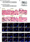
PI3K C2β induces keratinocyte differentiation. (A) Expression of keratinocyte differentiation proteins keratin 1 and involucrin in the presence of low (0.07 mM [−]) or elevated (0.20 mM [+]) calcium in the medium. Cells were coincubated with the PI3K inhibitor LY294002 or transduced with retroviral vectors expressing either the PI3K Δp85 mutant dominant-negative for class Ia PI3K function or a LacZ marker control. Proteins blotted are shown at left. (B) PI3K protein distribution in human epidermis. Note p110α and C2α throughout the epidermis and the restriction of C2β to suprabasal layers. Brackets delineate expression regions: orange, PI3K; green, collagen VII denoting the epidermal basement membrane; and blue, DAPI (4′,6′-diamidino-2-phenylindole) nuclear stain. Bars = 100 μm. (C) Differentiation protein expression in keratinocytes expressing retrovirally introduced LacZ control, p110α, and C2β. Retroviral expression constructs are noted at the top of each lane, and the blotted proteins are indicated at the left of each panel. (D) Differentiation marker protein expression in keratinocytes expressing retrovirally introduced PI3K C2α and LacZ control. (E) Deletion of the C2β PI kinase (PIKΔ), catalytic (CatΔ), or C-terminal C2 (C2Δ) domain abolishes induction of keratinocyte differentiation. Immunoblot verification of expression of the mutants noted at the top of each lane is shown in the top panel, with molecular mass markers to show the size of each mutant. The proteins studied are shown at the left of each panel; retroviral expression of wild-type and mutant C2β proteins invariably produced a doublet on immunoblotting.
FIG.2.
FIG.2.
K14 and HK1-C2β transgenic mice exhibit normal skin. (A) Transgene cassettes used to generate targeted expression of C2β under control of the basal K14 promoter or suprabasal HK1 promoter. Three independent lines were generated for each construct and displayed similar phenotypes. (B) C2β protein expression in keratinocyte extracts isolated from wild-type [tg(−)], singly transgenic [tg(+)], or doubly transgenic [tg(++)] mice. Actin expression is included as a loading control. (C) Histology of wild-type (−) and transgenic skin. Note normal architecture in all cases. Expression of keratin 14 and the differentiation proteins keratin 10, involucrin, filaggrin, and loricrin in (D) wild-type and HK1-C2β and (E) K14-C2β transgenic skin (orange, differentiation marker; blue, DAPI). Bars = 100 μm.
FIG. 3.
FIG. 3.
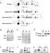
Targeted disruption of the mouse C2β gene. (A) Targeting strategy. Partial restriction enzyme maps and schematic representation of the strategy for ablation of the C2β gene. The PIK3C2B gene of 129Sv mice (wild-type [WT] allele), the targeting vector, the recombinant loxP-flanked locus (floxed allele), and the C2β exon-deleted locus (deleted allele) are shown. Cleavage sites for BamHI (B) and HindIII (H) are marked. The locations of probes 1 and 2 for Southern analysis are indicated by closed bars, and primers for PCR analysis are shown as arrowheads. Exons are symbolized as numbered rectangles. Exon 1 encodes an untranslated region; the start codon is located in exon 2. (B) Southern blot analysis of BamHI-digested genomic DNA and hybridization to probe 1 reveal a 6-kb and an 8-kb band corresponding to wild-type and floxed alleles, respectively. (C) Southern hybridization for detection of the null allele. The HindIII fragments in the wild-type allele and deleted alleles are 8.3 kb and 5.8 kb, respectively, when detected with probe 2. (D) Mice were genotyped by PCR. The amplicon with primer pair 1 and 2 is 251 bp for the wild-type allele. The amplicon with primer pair 3 and 4 is 404 bp for the null allele. (E) Loss of normal C2β mRNA transcript in C2β−/− mice. RNA was isolated from cultured keratinocytes. The forward primer was designed in exon 2, and the reverse primer was designed in exon 7. The wild-type allele yields a 678-bp amplicon, and the deleted allele yields a 301-bp amplicon. (F) Loss of epidermal C2β protein in C2β−/− mice. Western blot analysis showing absence of C2β protein expression in the knockout mice. Proteins were isolated from epidermal extracts and detected with an antiserum to C2β. (G) Loss of C2β protein in visceral tissues of C2β−/− mice.
FIG. 4.
FIG. 4.
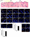
C2β−/− mice display normal epidermal differentiation and proliferation. (A) Histology of back skin from C2β wild-type, heterozygous, and null mice at 8 weeks of age. Note normal tissue architecture in C2β-null skin. (B) Expression of keratin 14 and the differentiation markers keratin 10, involucrin, filaggrin, and loricrin in 8-week-old mice. Bars = 100 μm. (C) Ki-67 (orange) in tissue counterstained with DAPI (blue). Bars = 100 μm. (D) Percentage of Ki-67(+) epidermal cells in skin tissue from C2β wild-type (+/+) and null (−/−) mice (three mice each; values are means ± standard deviations). (E) Filaggrin (orange)-expressing primary keratinocytes in culture from C2β wild-type and null mice when cells were grown in low (0.07 mM)-calcium and higher (0.12 mM)-calcium media. (F) Quantitation of the percent of filaggrin-positive cells in C2β wild-type and null murine keratinocytes after 24 h in 0.12 mM calcium (three independent experiments; values are means ± standard deviations).
FIG. 5.
FIG. 5.
Normal wound healing and barrier function in C2β-deficient mice. (A) C2β−/− mice exhibited normal re-epithelialization. Wound area over a 10-day period following injury (three mice each; values are means ± standard deviations). (B) β-Galactosidase epidermal permeability barrier assay of C2β−/− mice compared to the wild type. Note the lack of increased penetration of X-Gal through the epidermal barrier (as detected by blue staining) of the wild-type and C2β−/− mice. (C) Transepidermal water loss in newborn mice (three mice each; values are means± standard deviations).
FIG. 5.
FIG. 5.
Normal wound healing and barrier function in C2β-deficient mice. (A) C2β−/− mice exhibited normal re-epithelialization. Wound area over a 10-day period following injury (three mice each; values are means ± standard deviations). (B) β-Galactosidase epidermal permeability barrier assay of C2β−/− mice compared to the wild type. Note the lack of increased penetration of X-Gal through the epidermal barrier (as detected by blue staining) of the wild-type and C2β−/− mice. (C) Transepidermal water loss in newborn mice (three mice each; values are means± standard deviations).
FIG. 6.
FIG. 6.
Simultaneous C2α and C2β knockdown fails to prevent keratinocyte differentiation marker expression. Human keratinocytes treated with duplex RNAs to C2α, C2β, or C2α plus C2β were exposed to high and low extracellular calcium concentrations for 2 days. The proteins blotted are shown at the left of each panel, and the RNAi used is at the top. Note the expression of the involucrin differentiation marker and its induction by calcium in the absence of C2α and C2β proteins.

Similar articles

Cited by

References

    1. Anderson, K. E., and S. P. Jackson. 2003. Class I phosphoinositide 3-kinases. Int. J. Biochem. Cell Biol. 35:1028-1033. - PubMed
    1. Arcaro, A., U. K. Khanzada, B. Vanhaesebroeck, T. D. Tetley, M. D. Waterfield, and M. J. Seckl. 2002. Two distinct phosphoinositide 3-kinases mediate polypeptide growth factor-stimulated PKB activation. EMBO J. 21:5097-5108. - PMC - PubMed
    1. Arcaro, A., S. Volinia, M. J. Zvelebil, R. Stein, S. J. Watton, M. J. Layton, I. Gout, K. Ahmadi, J. Downward, and M. D. Waterfield. 1998. Human phosphoinositide 3-kinase C2β, the role of calcium and the C2 domain in enzyme activity. J. Biol. Chem. 273:33082-33090. - PubMed
    1. Arcaro, A., M. J. Zvelebil, C. Wallasch, A. Ullrich, M. D. Waterfield, and J. Domin. 2000. Class II phosphoinositide 3-kinases are downstream targets of activated polypeptide growth factor receptors. Mol. Cell. Biol. 20:3817-3830. - PMC - PubMed
    1. Bedogni, B., M. S. O'Neill, S. M. Welford, D. M. Bouley, A. J. Giaccia, N. C. Denko, and M. B. Powell. 2004. Topical treatment with inhibitors of the phosphatidylinositol 3′-kinase/Akt and Raf/mitogen-activated protein kinase kinase/extracellular signal-regulated kinase pathways reduces melanoma development in severe combined immunodeficient mice. Cancer Res. 64:2552-2560. - PubMed

Publication types

Substances

LinkOut - more resources