. 2005 Dec;115(12):3473-83.
doi: 10.1172/JCI24482. Epub 2005 Nov 23.

Preferential migration of effector CD8+ T cells into the interstitium of the normal lung

Affiliations

Preferential migration of effector CD8+ T cells into the interstitium of the normal lung

Elena Galkina et al. J Clin Invest. 2005 Dec.

Abstract

The respiratory tract is a primary site of infection and exposure to environmental antigens and an important site of memory T cell localization. We analyzed the migration and retention of naive and activated CD8+ T cells within the noninflamed lungs and quantitated the partitioning of adoptively transferred T cells between the pulmonary vascular and interstitial compartments. Activated but not naive T cells were retained within the lungs for a prolonged period. Effector CD8+ T cells preferentially egressed from the pulmonary vascular compartment into the noninflamed pulmonary interstitium. T cell retention within the lung vasculature was leukocyte function antigen-1 dependent, while the egress of effector T cells from the vascular to the interstitium functions through a pertussis toxin-sensitive (PTX-sensitive) mechanism driven in part by constitutive CC chemokine ligand 5 expression in the lungs. These results document a novel mechanism of adhesion receptor- and pulmonary chemokine-dependent regulation of the migration of activated CD8+ T cells into an important nonlymphoid peripheral site (i.e., the normal/noninflamed lung).


Figures

Figure 1
Figure 1
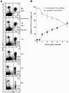
Activated CD8+ T cells but not naive T cells are retained within lungs after adoptive transfer in an antigen-independent manner. (A) Dual modality image (merged image combining γ-ray and x-ray images) following adoptive transfer of 111In-labeled naive, early-activated, or effector CD8+ T cells into WT (non-Tg) recipient mice (top view, prone mouse). In the merged RGB images shown, the red channel indicates γ-counts. (B) Distribution of 111In-labeled CD8+ T cells at the indicated time points after transfer. Data is represented as percentage of radioactivity in ROI defined for the lungs, with the fraction of transferred cells/radioactivity localized to the lungs (∼80%) at 1 minute defined as 100%. Results are representative of 6 independent experiments. (C) Dual modality image and (D) quantitation following adoptive transfer of 111In-labeled effector CTLs into HA-transgenic (HA-Tg) and WT mice.
Figure 2
Figure 2
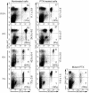
Effector CD8+ T cells migrate into the interstitium of noninflamed lungs. (A) Kinetic analysis of intravascular versus extravascular compartmentalization of effector CD8+ T cells in lungs. We injected 4 × 10⁶ CFSE-labeled CD8+ T cells into recipient mice. Anti–CD8α-APC mAbs were injected at 5 minutes or 1, 2, 3, or 6 hours after transfer; then lungs were harvested 10 minutes later. The percentage of CFSE-labeled cells is shown; the percentage of CFSE-labeled interstitial T cells from total percentage of emigrated CFSE-labeled T cells is shown in parentheses. Ab first indicates that anti–CD8α-APC mAbs were injected 2 minutes prior to T cell transfer. (B) Data from A is expressed as the percentage of transferred CD8+ T cells in the intravascular or extravascular compartment.
Figure 3
Figure 3
Effector CD8+ T cells but not early-activated CD8+ T cells transmigrate to the lung interstitium. (A) Effector CTLs and early-activated (48 hours) CD8+ T cells (labeled with CFSE and CMTMR, respectively) were injected into WT recipient mice. Anti–CD8α-APC mAbs were injected at 6 hours after transfer; lungs were harvested 10 minutes later. The percentages of CFSE and CMTMR-labeled cells from individual recipients are shown in dot plots. (B and C) Analysis of T cell partitioning between pulmonary vascular and interstitial compartments. The numbers in parentheses reflect the percentages of CFSE-labeled (effector CD8+ T cells) or CMTMR-labeled (early-activated CD8+ T cells) cells from individual recipients that emigrated into the interstitium from the total number of CFSE- or CMTMR-labeled cells remaining within the lungs. Results represent the mean ± SEM of 4 recipients in a total of 4 independent experiments.
Figure 4
Figure 4
Regulation of the retention time within the lungs but not transmigration into the interstitium by LFA-1. (A) Pretreated with anti–LFA-1 mAbs, CFSE-labeled effector CD8+ T cells were mixed with CMTMR-labeled–nontreated effector CD8+ T cells and injected into WT recipient mice. Anti–CD8α-APC mAbs were injected at 6 hours after transfer; lungs were harvested 10 minutes later. The percentages of labeled CD8+ T cells from individual mice are shown in dot plots, the percentages of interstitial cells in parentheses. Results are representative of 6 recipients from a total of 6 independent experiments. (B) CMTMR-labeled LFA-1–/– activated CD8+ T cells were mixed with equal numbers of CFSE-labeled activated (LFA-1+/+) CD8+ T cells and injected into WT recipient mice. The percentages of labeled CD8+ T cells from LFA-1–/– and LFA-1+/+ donors localized to the lungs over time are shown. CD8+ T cells were activated by plate-bound anti-CD3/CD28 Abs. Results are representative of 4 recipient mice in a total of 4 independent experiments.
Figure 5
Figure 5
Migration of effector CD8+ T cells into the lung interstitium is PTX-sensitive. PTX-pretreated effector CD8+ T cells or nontreated effector CD8+ T cells were labeled with CFSE and CMTMR, mixed equally, and injected into WT recipient mice. Anti–CD8α-APC mAbs were injected at indicated time points after transfer, and lungs were harvested 10 minutes later. The percentages of total labeled, PTX-treated or control T cells retained in the lungs over time from individual recipient are indicated. The percentages of cells localized to the interstitium (CFSE+/CD8α-APC, CMTMR+/CD8α-APC) are shown in parentheses. Results represent the mean ± SEM from 3 to 9 recipient mice for each time point. In control experiments, cells were treated with mutant PTX (PT9K 129G, provided by E. Hewlett, University of Virginia, Charlottesville, Virginia, USA).
Figure 6
Figure 6
RANTES/CCR5-dependent transmigration of activated effector CD8+ T cells to alveolar interstitium. (A) Naive, early-activated, and effector CD8+ T cells were analyzed for expression of chemokine receptors. Results show the percentages (± SEM) of chemokine receptor expression for naive (white bars), early-activated (gray bars), and effector (black bars) CD8+ T cells from total expression of β-actin as a control. (B) WT recipients were injected with 1 of the following: polyclonal goat anti-RANTES Ab, polyclonal goat Ig, or PBS. After 1 hour, equal numbers of PTX-treated and nontreated effector CD8+ T cells labeled with CMTMR and CFSE were injected into recipient mice. After 4 hours, anti–CD8α-APC mAbs were injected, and 10 minutes later, lungs were harvested. Counterplots represent cells gated on CFSE+ and CMTMR+ T cells. The percentages of emigrated T cells in vascular (CFSE+/CD8-APC+, CMTMR+/CD8-APC+) and interstitial (CFSE+/CD8-APC, CMTMR+/CD8-APC) compartments from individual mice are shown. The percentage of emigrated interstitial labeled cells is shown in parentheses. The results are representative of 6 independent experiments. (C) Percentage of interstitial PTX-treated and control CD8+ T cells in the indicated recipients. *P < 0.05, **P < 0.001 between the percentages of interstitial nontreated T cells in nontreated mice and all other groups. (D) Effector CD8+ T cells from CCR5–/– mice and CCR5+/+ littermates were labeled with CFSE and CMTMR, respectively, mixed in ratio 1:1, and injected i.v. into recipient mice. After 7 hours, anti–CD8α-APC mAbs were injected, and 10 minutes later, lungs were harvested. Flow cytometry plots represent cells gated on CD45+ dye-labeled (CD45+/CFSE+ or CD45+/CMTMR+) cells. The percentages of lung-resident transferred CD8+ T cells in vascular (CFSE+/CD8-APC+, CMTMR+/CD8-APC+) and interstitial (CFSE+/CD8-APC, CMTMR+/CD8-APC) compartments are shown.

Similar articles

Cited by

References

    1. Butcher EC, Picker LJ. Lymphocyte homing and homeostasis. Science. 1996;272:60–66. - PubMed
    1. Weninger W, Crowley MA, Manjunath N, Von Andrian UH. Migratory properties of naive, effector, and memory CD8(+) T-cells. J. Exp. Med. 2001;194:953–966. - PMC - PubMed
    1. Masopust D, Vezys V, Marzo AL, Lefrancois L. Preferential localization of effector memory cells in nonlymphoid tissue. Science. 2001;291:2413–2417. - PubMed
    1. Sallusto F, Lenig D, Forster R, Lipp M, Lanzavecchia A. Two subsets of memory T lymphocytes with distinct homing potentials and effector functions. Nature. 1999;401:708–712. - PubMed
    1. Springer TA. Traffic signals for lymphocyte recirculation and leukocyte emigration: the multistep paradigm. Cell. 1994;76:301–314. - PubMed

Publication types

MeSH terms