Comparative Study
. 2005 Dec 7;24(23):4154-65.
doi: 10.1038/sj.emboj.7600876. Epub 2005 Nov 24.

P-TEFb is not an essential elongation factor for the intronless human U2 snRNA and histone H2b genes

Affiliations
Comparative Study

P-TEFb is not an essential elongation factor for the intronless human U2 snRNA and histone H2b genes

Joanne Medlin et al. EMBO J. .

Abstract

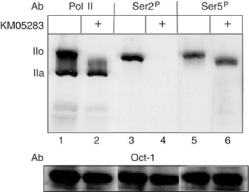
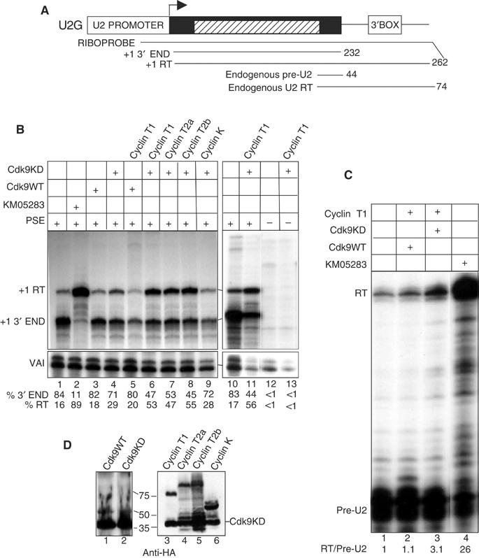
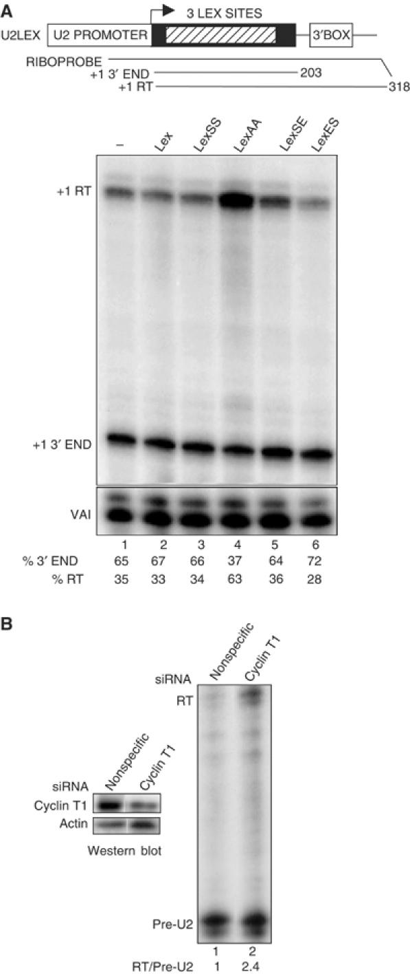
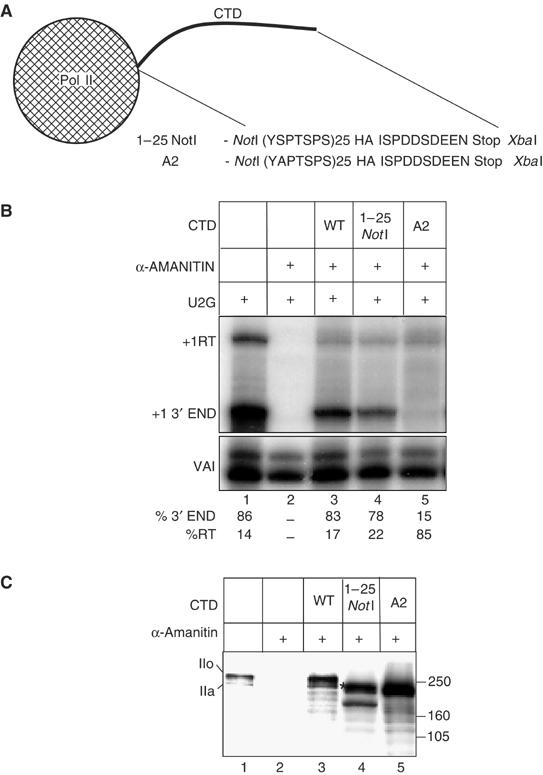
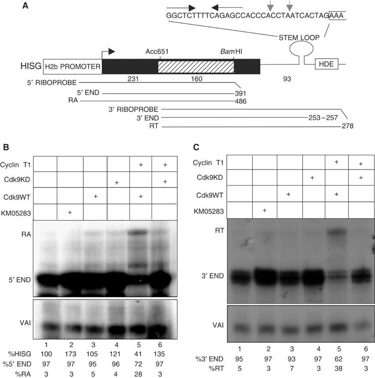
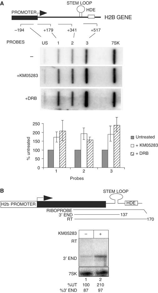
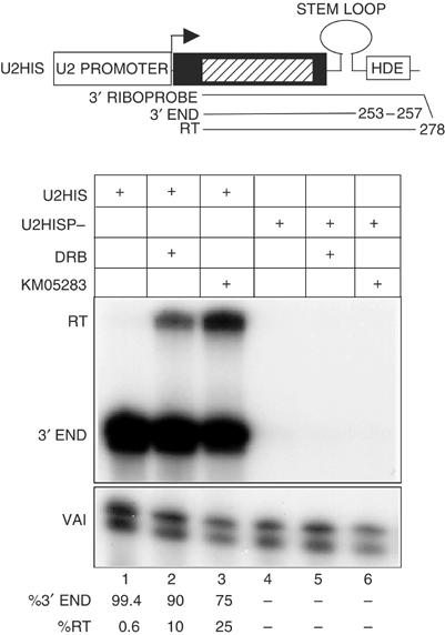
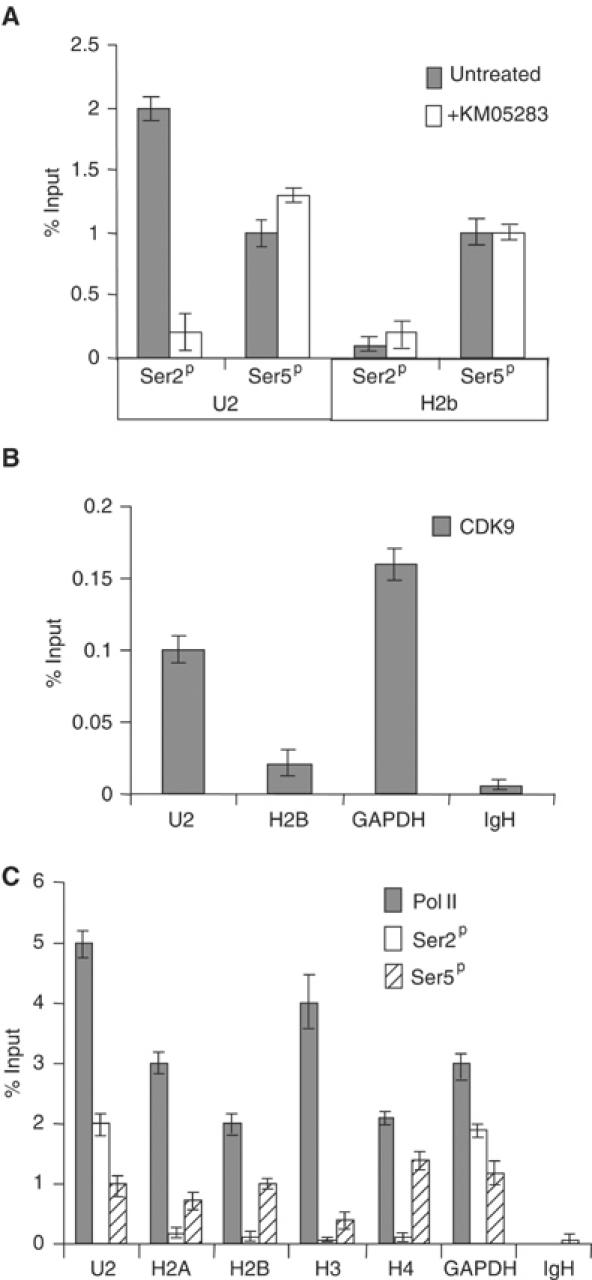
Phosphorylation of Ser2 of the heptapeptide repeat of the CTD of mammalian pol II by P-TEFb is associated with productive elongation of transcription of protein-coding genes. Here, we show that the CTD of pol II transcribing the human U2 snRNA genes is phosphorylated on Ser2 in vivo and that both the CDK9 kinase and cyclin T components of P-TEFb are required for cotranscriptional recognition of the 3' box RNA 3' end processing signal. However, inhibitors of CDK9 do not affect transcription of the U2 genes, indicating that P-TEFb functions exclusively as an RNA processing factor in expression of these relatively short, intronless genes. We also show that inhibition of CDK9 does not adversely affect either transcription of an intron-less, replication-activated histone H2b gene or recognition of the histone gene-specific U7-dependent RNA 3' end formation signal. These results emphasize that the role of P-TEFb as an activator of transcription elongation can be separated from its role in RNA processing and that neither function is universally required for expression of mammalian pol II-dependent genes.


Figures

Figure 1
Figure 1
KM05283 inhibits phosphorylation of Ser2 of the pol II CTD. Western blot analysis of protein from HeLa cells before and after treatment with 100 μM KM05283 using antibodies against the N-terminus of the large subunit of Pol II (Santa Cruz, H-224) (lanes 1, 2), or the CTD repeats phosphorylated on Ser2 (Ser2P) (Covance, H5), lanes 3 and 4, or Ser5 (Ser5P) (Covance, H14), lanes 5 and 6. The positions of the hyperphosphorylated CTD form (IIo) and the hypophosphorylated CTD form (IIa) are indicated on the left. The level of the transcription factor Oct-1 (Santa Cruz, C-21 antibody) serves as a loading control.
Figure 2
Figure 2
P-TEFb is required for proper 3′ end formation of U2 transcripts. (A) Structure of the U2G construct (Medlin et al, 2003) with the relative position of the riboprobe and expected RNase protection products. The hatched box corresponds to β-globin gene sequences. The size of the expected products is noted at the right. RT denotes readthrough transcripts that mismatch to the riboprobe at the 3′ end in this and subsequent figures. (B) RNAse protection analysis of RNA transcribed from U2G after ectopic expression of the wild-type CDK9 (CDK9WT) or the D167N kinase dead mutant of CDK9 (CDK9KD) together with the CDK9 cyclin partners. VAI serves as a transfection control in this and subsequent experiments. The CDK9 and cyclin construct transfected, the addition of KM05283 and the presence or absence of a functional PSE in the U2G construct is noted above each lane. The positions of the protected products are noted on the left. The relative amount of correct 3′ end and readthrough is noted below each lane. (C) RNase protection analysis of endogenous U2 gene transcripts after ectopic expression of the CDK9WT or KD together with cyclin T1. The ratio of readthrough (RT) to pre-U2 3′ end is noted below each lane. (D) Western blot analysis of cells transfected with templates encoding CDK9WT or KD and the four cyclins using an anti-HA antibody (Santa Cruz, F-7). The transfected construct is shown above each lane and the positions of size markers are noted at the left. For lanes 3–6, CDK9DN was cotransfected and the position of this protein is noted at the right.
Figure 3
Figure 3
Inhibition of P-TEFb by CTD analogues or RNAi-mediated knockdown affects 3′ box-dependent processing. (A) Diagram of the U2LEX construct with the 3 lexA binding sites denoted as a hatched box (top). The relative position of the riboprobe and size of the expected RNase protection products are shown. RNase protection analysis of U2LEX transcripts after ectopic expression of the CTD analogue noted above each lane is shown below. The positions of the protected products are noted on the left. The relative amount of correct 3′ end and readthrough is shown beneath each lane. (B) RNase protection analysis of transcripts from endogenous U2 genes after transfection of a nonspecific siRNA (lane 1) or an siRNA specific for cyclin T1 (lane 2). The ratio of readthrough (RT) to pre-U2 3′ end is noted below each lane. Western blot analysis of cells treated with the same siRNAs using antibodies against cyclin T1 (Santa Cruz H245) or α-actin (Santa Cruz C-11) is shown at the left.
Figure 4
Figure 4
Mutation of Ser2 of the pol II CTD repeats causes readthrough of the U2 3′ box. (A) Diagram of the CTD region of the 1–25 NotI and A2 cassette constructs of the pol II large subunit showing the amino acids encoded by the region between the NotI and XbaI sites. HA and Stop denote the HA epitope tag and a stop codon respectively. (B) RNase protection analysis of RNA transcribed from U2G after ectopic expression of the α-amanitin resistant pol II large subunit constructs indicated above the lanes and treatment of cells with α-amanitin. The positions of the protected products are shown on the left. The relative amount of correct 3′ end and readthrough is noted below each lane. (C) Western blot analysis of the proteins encoded by the pol II large subunit constructs using an antibody against the large subunit of pol II (Santa Cruz, H-224). The construct transfected is shown above each lane. The position of the hyperphosphorylated form (IIo) and the hypophosphorylated form (IIa) of the endogenous and ectopically expressed pol II large subunits with a full-length CTD are noted on the left. The putative hyperphosphorylated form of 1–25 NotI is marked with an *.
Figure 5
Figure 5
P-TEFb is not required for elongation of transcription of a transfected H2b gene or 3′ processing of the transcripts. (A) Diagram of the HISG construct with the relative position of the riboprobes and expected RNase protection products. The size of the expected products is noted at the right. The hatched box corresponds to β-globin gene sequences that allow transcripts to be distinguished from endogenous H2b mRNA and the sizes of the histone and globin sequences are noted below the diagram. The sequence of the processing signal is shown above the diagram with the stem loop denoted by convergent arrows, the start of the HDE bracketed and the positions of two potential 3′ ends marked with grey arrows. (B) 5′ RNase protection analysis of RNA transcribed from HISG after treatment of cells with KM05283 and after ectopic expression of CDK9WT or CDK9KD together with cyclin T1. The CDK9 and cyclin construct transfected and the addition of KM05283 is noted above each lane and the positions of the protected products are noted on the left in this and subsequent panels. The relative amount of expression and correct 5′ end and readaround transcription (RA) is noted below each lane. For lanes 2, 4 and 6 the average of three experiments and standard deviations were 193% and 19, 116% and 10, 129 and 16 respectively. (C) 3′ RNase protection analysis of RNA transcribed from HISG after treatment of cells with KM05283 and after ectopic expression of CDK9WT or CDK9KD together with cyclin T1. The relative amount of correct 3′ end and readthrough is noted below each lane.
Figure 6
Figure 6
P-TEFb is not required for elongation of transcription of an endogenous H2b gene or 3′ processing of the transcripts. (A) Nuclear run-on analysis of an endogenous replication-activated H2b gene after treatment of cells with CTD kinase inhibitors. The structure of the human H2b gene analysed is shown with the relative positions of the run-on probes marked below. The numbers next to the probes indicate the 3′ boundary of the probes relative to the site of initiation. The results of run-on analysis in the absence and presence of 100 μM KM05283 or DRB are shown below each probe. The 7SK probe serves as a negative control for the effect of the kinase inhibitors. Quantitation of the results for each probe is shown below as an average of three experiments, with the signals after CTD kinase inhibitor treatment represented as a percentage of the untreated controls. The error bars indicate standard deviations. (B) RNase protection analysis of the 3′ ends of transcripts from an endogenous replication-activated H2b gene after treatment of cells with KM05283. The position of the RNase protection probe relative to the H2b gene and the sizes of the expected products are shown. The addition of inhibitor is noted above the lane and the % of the level of transcript in lane 1 and % 3′ end is noted below each lane. The 7SK RNA serves as a control for the level of RNA.
Figure 7
Figure 7
CDK9 inhibitors affect recognition of the histone processing signal in transcripts from the U2 promoter. RNase protection analysis of the 3′ ends of transcripts from constructs where the H2b promoter has been replaced by either the wild-type U2 promoter (U2HIS) or the P-mutant (U2HISP−). The constructs transfected and CDK9 inhibitor used are shown above each lane. The relative amount of correct 3′ end and readthrough is noted below each lane.
Figure 8
Figure 8
CTD Ser2 phosphorylation and CDK9 are differentially associated with U2 genes and H2b genes. (A) Real-time PCR analysis of the output from ChIP using antibodies against the pol II CTD phosphorylated on Ser2 (Ser2P) (H5, Covance) or Ser5 (Ser5P) (H14, Covance) before and after treatment of cells with KM05283. The graph shows the values obtained using primers against the U2 and H2b genes. Error bars indicate the standard deviation of real-time triplicates in this and subsequent panels. Immunoprecipitation is given as a percentage of the amount of the input DNA after subtraction of the no antibody control in this and subsequent panels. The no antibody controls were less than 0.1% of input DNA. (B) Real-time PCR analysis of the output from ChIP using antibodies against CDK9 (Santa Cruz, L-19). The graph shows the values obtained using primers against the U2, H2b, GAPDH and IgH genes. The no antibody controls were less than 0.008% of input DNA. (C) Real-time PCR analysis of the output from ChIP using antibodies against the large subunit of pol II (Santa Cruz, H-224) and the pol II CTD phosphorylated on Ser2 or Ser5. The graph shows the values obtained using primers against the U2, H2a, H2b, H3, H4, GAPDH and IgH genes. The no antibody controls were less than 0.008% of input DNA for the pol II antibody.

Similar articles

Cited by

References

    1. Ahn SH, Kim M, Buratowski S (2004) Phosphorylation of serine 2 within the RNA polymerase II C-terminal domain couples transcription and 3′ end processing. Mol Cell 13: 67–76 - PubMed
    1. Barilla D, Lee BA, Proudfoot NJ (2001) Cleavage/polyadenylation factor IA associates with the carboxyl-terminal domain of RNA polymerase II in Saccharomyces cerevisiae. Proc Natl Acad Sci USA 98: 445–450 - PMC - PubMed
    1. Bird G, Zorio DA, Bentley DL (2004) RNA polymerase II carboxy-terminal domain phosphorylation is required for cotranscriptional pre-mRNA splicing and 3′-end formation. Mol Cell Biol 24: 8963–8969 - PMC - PubMed
    1. Boyd KE, Wells J, Gutman J, Bartley SM, Farnham PJ (1998) c-Myc target gene specificity is determined by a post-DNA binding mechanism. Proc Natl Acad Sci USA 95: 13887–13892 - PMC - PubMed
    1. Chao SH, Price DH (2001) Flavopiridol inactivates P-TEFb and blocks most RNA polymerase II transcription in vivo. J Biol Chem 276: 31793–31799 - PubMed

Publication types

MeSH terms

LinkOut - more resources