. 2005 Nov 23;25(47):10930-40.
doi: 10.1523/JNEUROSCI.2029-05.2005.

The small GTPase Rab7 controls the endosomal trafficking and neuritogenic signaling of the nerve growth factor receptor TrkA

Affiliations

The small GTPase Rab7 controls the endosomal trafficking and neuritogenic signaling of the nerve growth factor receptor TrkA

Smita Saxena et al. J Neurosci. .

Abstract

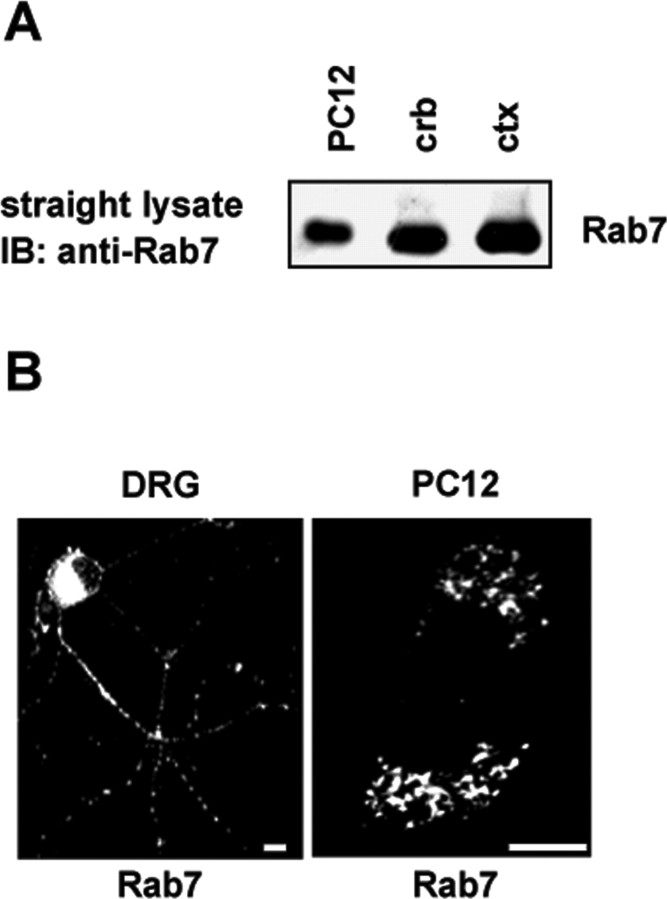
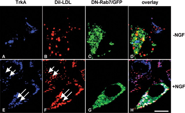
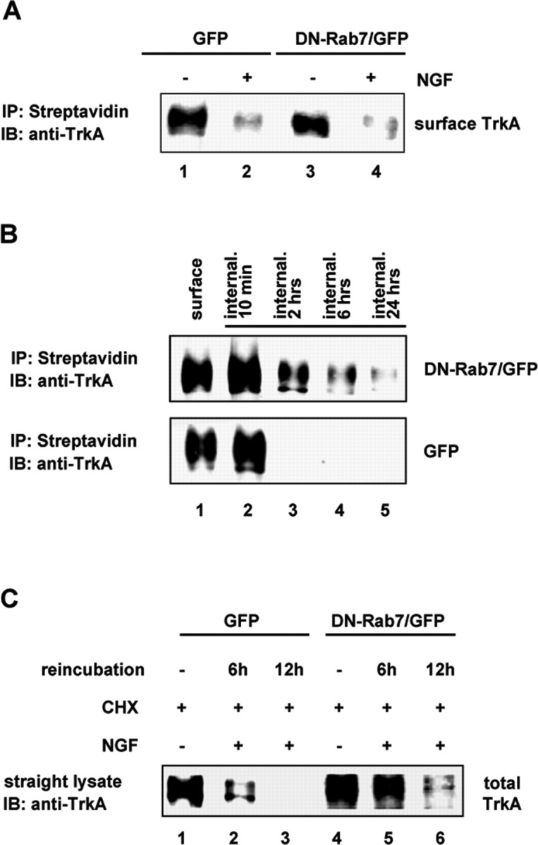
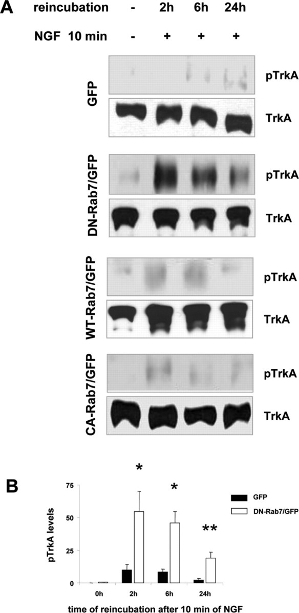
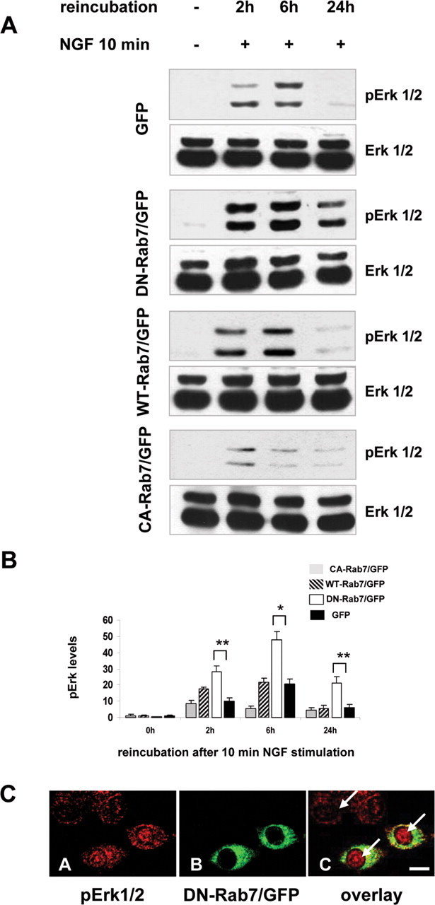
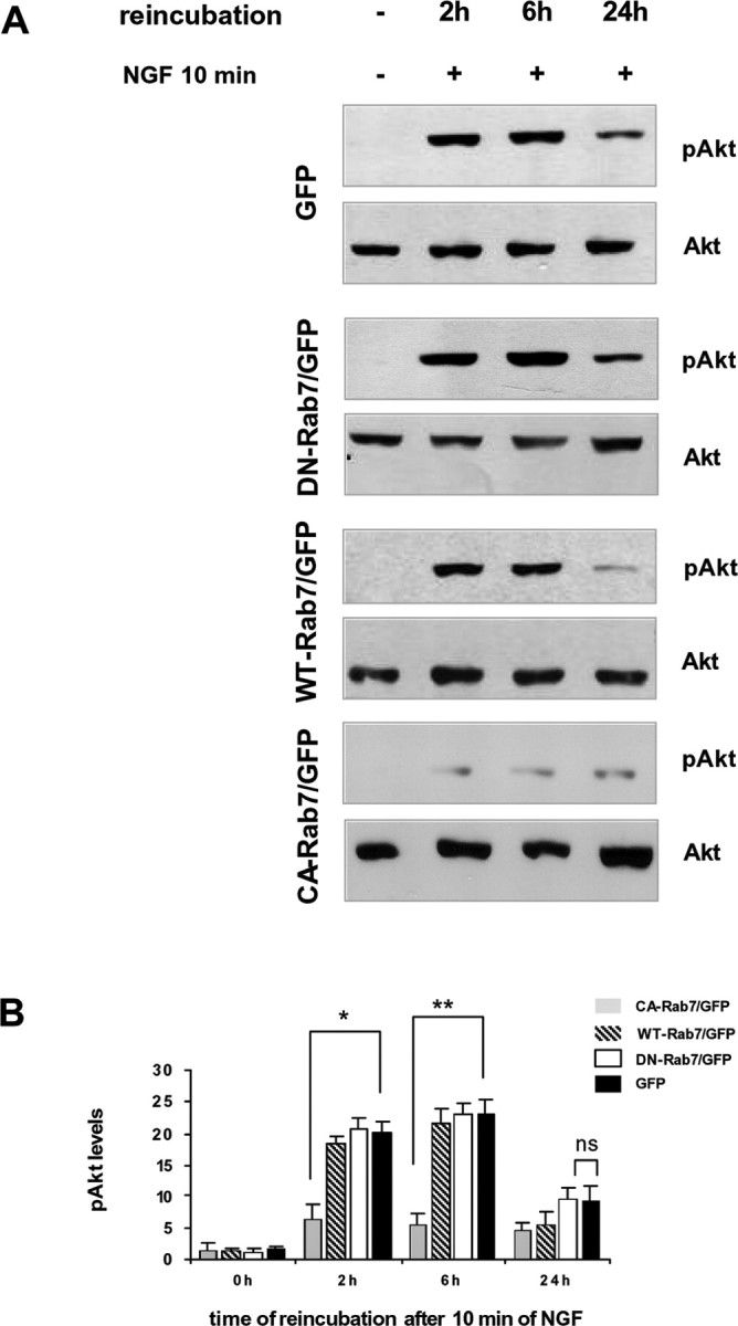
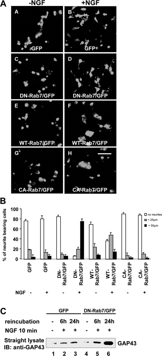
Nerve growth factor (NGF) and its TrkA receptor exert important bioactivities on neuronal cells such as promoting survival and neurite outgrowth. Activated TrkA receptors are not only localized on the cell surface but also in signaling endosomes, and internalized TrkA receptors are important for the mediation of neurite outgrowth. The regulation of the endosomal trafficking of TrkA is so far unknown. Because the endosome-associated GTPase Rab7 coimmunoprecipitated with TrkA, we examined whether the endosomal trafficking of TrkA might be under the control of Rab7. Inhibiting Rab7 by expression of a green fluorescent protein-tagged, dominant-negative Rab7 variant resulted in endosomal accumulation of TrkA and pronounced enhancement of TrkA signaling in response to limited stimulations with NGF, such as increased activation of Erk1/2 (extracellular signal-regulated kinase 1/2), neurite outgrowth, and expression of GAP-43 (growth-associated protein 43). Our studies show that the endosomal GTPase Rab7 controls the endosomal trafficking and neurite outgrowth signaling of TrkA. Because mutations of Rab7 are found in patients suffering from hereditary polyneuropathies, dysfunction of Rab7 might contribute to neurodegenerative conditions by affecting the trafficking of neurotrophins. Moreover, strategies aimed at controlling Rab7 activity might be useful for the treatment of neurodegenerative diseases.


Figures

Figure 1.
Figure 1.
Expression of Rab7 protein in PC12 cells and neurons. A, Western blot analysis of equal amounts of protein lysates from embryonicrat cerebellum (crb) and cortex (ctx), as well as from undifferentiated PC12 cells. A 23 kDa band corresponding to Rab7 was observed in all lanes. A representative blot is shown. IB, Immunoblot. B, Confocal immunofluorescence analysis of Rab7 in fixed and permeabilized DRGs and PC12 cells. Vesicular staining for Rab7 in PC12 cells and within neuronal processes and cell bodies of DRG neurons. A representative experiment is shown. Scale bars, 10 μm.
Figure 2.
Figure 2.
Rab7 inhibition by transient expression of DN-Rab7/GFP results in endosomal accumulation of TrkA. DN-Rab7/GFP-transfected PC12 cells were stimulated without NGF (-NGF; A-D) or with NGF (+NGF; E-H) in the presence of fluorescently labeled LDL (DiI-LDL) to allow for endocytosis of LDL and TrkA, followed by surface strip and reincubation in ligand-free DMEM/BSA for 2 h. Subsequently, cells were fixed, permeabilized, and stained for TrkA. The arrows indicate vesicles containing TrkA and LDL. Colocalization of LDL (red) and TrkA (blue) in enlarged vesicles in DN-Rab7/GFP-expressing cells (green) is shown. A representative experiment is shown. Scale bar, 8 μm.
Figure 3.
Figure 3.
Intracellular persistence of internalized TrkA depends on Rab7 activity. A, Surface biotinylation of PC12 cells transfected with DN-Rab7/GFP or GFP (control). Cells were stimulated with NGF for 30 min, chilled on ice and washed, and surface receptors were biotinylated. Then, cells were lysed, and lysates were subjected to precipitation with streptavidin beads, followed by immunoblot with an antibody against TrkA. Surface TrkA was internalized to a similar extent in both control cells expressing GFP (lanes 1, 2) and cells transfected with DN-Rab7/GFP (lanes 3, 4). A representative blot is shown. B, Surface biotinylation-based internalization assay of PC12 cells transfected with DN-Rab7/GFP or GFP (control). Surface proteins of PC12 cells were first biotinylated on ice with a cleavable membrane-impermeable cross-linker. Cells were then washed and incubated with NGF at 37°C for 10 min. This step drives the internalization of biotinylated surface receptors. Afterward, cells were cooled on ice and washed, and the biotin of remaining surface-exposed biotinylated receptors was cleaved. This step leaves only internalized biotinylated receptors. Then, cells were rewarmed in NGF-free medium for different time points, as indicated. Afterward, cells were lysed, lysates were subjected to precipitation with streptavidin beads, and precipitates were immunoblotted with anti-TrkA. Both GFP and DN-Rab7/GFP samples were run on the same gel and probed on the same nitrocellulose membrane. Lane 1 (surface) reflects surface levels of TrkA before stimulation with NGF, whereas lanes 2-5 reflect the intracellular levels of internalized TrkA after different incubation periods in NGF-free medium. Prolonged intracellular persistence of TrkA in DN-Rab7/GFP-expressing cells (top, lanes 3-5) after 2 and 6 h of reincubation in NGF-free medium compared with GFP transfected control cells (bottom, lanes 3-5) is shown. A representative blot is shown. C, Same numbers of PC12 cells transiently transfected with either GFP (control) or DN-Rab7/GFP were pretreated for 30 min with the translation blocker CHX, followed by a 10 min NGF stimulation, and subsequently surface stripped for ligand removal. Cells were reincubated in serum-free medium in the presence of CHX. Then, cells were lysed, and lysates were immunoblotted for TrkA. The intracellular persistence of internalized TrkA is prolonged in DN-Rab7/GFP-expressing cells (lanes 5, 6) compared with GFP-expressing cells (lanes 2, 3). A representative blot is shown. IP, Immunoprecipitate; IB, immunoblot.
Figure 4.
Figure 4.
Association of TrkA with Rab7. A, PC12 cells were stimulated with NGF for different time points, and equal amounts of lysates were immunoprecipitated with antibodies against Rab7, followed by immunoblotting with antibodies directed against TrkA. Subsequently, the membrane was stripped and reprobed with antibodies against Rab7 to confirm that similar amounts of Rab7 were pulled down in each lane. To confirm that our lysates contain similar amounts of Rab7 or TrkA, equal amounts of lysates were immunoblotted with antibodies against TrkA or Rab7. Blots from a representative experiment are shown. B, PC12 cells were transfected with WT-Rab7/GFP and stimulated with NGF for different time points. Equal amounts of lysates were immunoprecipitated with antibodies against the GFP tag to pull down Rab7/GFP, followed by immunoblotting with antibodies directed against TrkA. Subsequently, the membrane was stripped and reprobed with antibodies against GFP to confirm that similar amounts of Rab7/GFP were pulled down in each lane. Blots from a representative experiment are shown. C, PC12 cells were transfected with WT-Rab7/GFP and stimulated with or without NGF for 30 min. Cells were fixed, permeabilized, and stained for TrkA (red). Increased colocalization of WT-Rab7/GFP (green) with TrkA (red) appears as yellow in C and F. Scale bar, 10 μm. IP, Immunoprecipitate; IB, immunoblot.
Figure 5.
Figure 5.
Enhanced TrkA phosphorylation in PC12 cells expressing DN-Rab7/GFP. A, PC12 cells transfected with GFP, DN-Rab7/GFP, WT-Rab7/GFP, or CA-Rab7/GFP were stimulated for 10 min with NGF, followed by ligand strip and reincubation in NGF-free medium for the indicated time points. Subsequently, cells were lysed, and equal amounts of protein were subjected to immunoblotting with antibodies directed against tyrosine-phosphorylated TrkA (pTrkA). Higher levels of pTrkA were found even after 24 h of reincubation in serum-free medium compared with GFP-expressing cells. Subsequently, membranes were stripped and reprobed with antibodies against unphosphorylated TrkA as loading control (bottom). Representative blots are shown. B, Films derived from four separate experiments were quantified by densitometry. Values of p TrkA are compared with control values (unstimulated). Error bars represent SD. *p < 0.05; **p < 0.01.
Figure 6.
Figure 6.
Enhanced pErk1/2 levels in PC12 cells expressing DN-Rab7/GFP. A, The same lysates as those in Figure 5 were probed for tyrosine-phosphorylated Erk1/2 (pErk1/2). We found persistent enhanced activation of Erk 1/2 in DN-Rab7/GFP-expressing cells compared with GFP-expressing control cells. Decreased pErk1/2 levels are shown in cells transfected with CA-Rab7/GFP. Subsequently, membranes were stripped and reprobed with antibodies against unphosphorylated Erk1/2 as a loading control (bottom). Representative blots are shown. B, Films derived from four separate experiments were quantified by densitometry. Values of pErk are compared with control values (unstimulated). Error bars represent SD. *p < 0.05; **p < 0.01. C, Confocal immunofluorescence analysis of pErk1/2 in DN-Rab7/GFP cells stimulated with NGF. Increased nuclear localization of pErk1/2 (red, arrow) in cells expressing DN-Rab7/GFP (green) are compared with untransfected control cells. Scale bar, 10 μm.
Figure 7.
Figure 7.
Unaltered Akt phosphorylation in PC12 cells expressing DN-Rab7/GFP. A, The same lysates as those in Figures 5 and 6 were probed for phosphorylated Akt (pAkt). Subsequently, membranes were stripped and reprobed with antibodies against unphosphorylated Akt as a loading control (bottom). Representative blots are shown. B, Films derived from four separate experiments were quantified by densitometry. Values of pAkt are compared with control values (unstimulated). Error bars represent SD. ns, Not significant. *p < 0.05; **p < 0.01.
Figure 8.
Figure 8.
Rab7 inhibition results in potentiated neurite outgrowth in response to brief stimulation with NGF. A, Epifluorescence images of PC12 cells expressing GFP (control; A, B) or DN-Rab7/GFP (C, D), WT-Rab7/GFP (E, F), or CA-Rab7/GFP (G, H) stimulated for 10 min with (+NGF) or without (-NGF) NGF, followed by surface strip and reincubation in NGF-free medium for 24 h. DN-Rab7/GFP cells differentiated in response to a 10 min NGF stimulus, whereas control cells failed to do so. Scale bar, 100 μm. B, Graphical representation of A from three independent experiments. The number of cells without neurites (no neurites) or with neurites <25 μm or >50 μm were determined. p < 0.00001, DN-Rab7/GFP-transfected cells and GFP-transfected control cells. C, Western blotting with an antibody directed against a neuronal differentiation marker. PC12 cells transfected with GFP (control) or DN-Rab7/GFP were stimulated for 10 min with NGF, followed by ligand strip and reincubation in NGF-free medium for the indicated time points. Subsequently, cells were lysed, and equal amounts of protein were subjected to immunoblotting with antibodies directed against GAP-43. A representative blot is shown. IB, immunoblot.

Similar articles

Cited by

References

    1. Bananis E, Nath S, Gordon K, Satir P, Stockert RJ, Murray JW, Wolkoff AW (2004) Microtubule-dependent movement of late endocytic vesicles in vitro: requirements for dynein and kinesin. Mol Biol Cell 15: 3688-3697. - PMC - PubMed
    1. Barbieri MA, Fernandez-Pol S, Hunker C, Horazdovsky BH, Stahl PD (2004) Role of rab5 in EGF receptor-mediated signal transduction. Eur J Cell Biol 83: 305-314. - PubMed
    1. Bernd P, Greene LA (1984) Association of 125I-nerve growth factor with PC12 pheochromocytoma cells. Evidence for internalization via high-affinity receptors only and for long-term regulation by nerve growth factor of both high- and low-affinity receptors. J Biol Chem 259: 15509-15516. - PubMed
    1. Bronfman FC, Tcherpakov M, Jovin TM, Fainzilber M (2003) Ligand-induced internalization of the p75 neurotrophin receptor: a slow route to the signaling endosome. J Neurosci 23: 3209-3220. - PMC - PubMed
    1. Bucci C, Thomsen P, Nicoziani P, McCarthy J, van Deurs B (2000) Rab7: a key to lysosome biogenesis. Mol Biol Cell 11: 467-480. - PMC - PubMed

Publication types

MeSH terms

LinkOut - more resources